Wartlick, O., Julicher, F. and Gonzalez-Gaitan, M. (2014). Growth control by a moving morphogen gradient during Drosophila eye development. Development 141: 1884-1893. PubMed ID: 24757005
Summary:
During morphogenesis, organs grow to stereotyped sizes, but growth control mechanisms are poorly understood. This study measured the signaling dynamics of the morphogen Dpp, one of several Drosophila factors controlling morphogenetic growth, in the developing eye. In this tissue, the Dpp expression domain advances from the posterior to the anterior tissue edge. In front of this moving morphogen source, signaling inputs, including Dpp, activate the target gene hairy in a gradient that scales with tissue size. Proliferation, in turn, occurs in a mitotic wave in front of the source, whereas behind it, cells arrest and differentiate. This study found that cells divide when their signaling levels have increased by around 60%. This simple mechanism quantitatively explains the proliferation and differentiation waves in wild type and mutants. Furthermore, this mechanism may be a common feature of different growth factors, because a Dpp-independent growth input also follows this growth rule.
Herrera, S. C. and Morata, G. (2014). Transgressions of compartment boundaries and cell reprogramming during regeneration in Drosophila. Elife 3: e01831. PubMed ID: 24755288
Summary:
Animals have developed mechanisms to reconstruct lost or damaged tissues. To regenerate those tissues the cells implicated have to undergo developmental reprogramming. The imaginal discs of Drosophila are subdivided into distinct compartments, which derive from different genetic programs. This feature makes them a convenient system to study reprogramming during regeneration. This study found that massive damage inflicted to the posterior or the dorsal compartment of the wing disc causes a transient breakdown of compartment boundaries, which are quickly reconstructed. The cells involved in the reconstruction often modify their original identity, visualized by changes in the expression of developmental genes like engrailed or cubitus interruptus. This reprogramming is mediated by up regulation of the JNK pathway and transient debilitation of the epigenetic control mechanism. These results also show that the local developmental context plays a role in the acquisition of new cell identities: cells expressing engrailed induce engrailed expression in neighbor cells.
Zschatzsch, M., Oliva, C., Langen, M., De Geest, N., Ozel, M. N., Williamson, W. R., Lemon, W. C., Soldano, A., Munck, S., Hiesinger, P. R., Sanchez-Soriano, N. and Hassan, B. A. (2014). Regulation of branching dynamics by axon-intrinsic asymmetries in Tyrosine Kinase Receptor signaling. Elife 3: e01699. PubMed ID: 24755286
Summary:
Axonal branching allows a neuron to connect to several targets, increasing neuronal circuit complexity. While axonal branching is well described, the mechanisms that control it remain largely unknown. This study found that in the Drosophila CNS branches develop through a process of excessive growth followed by pruning. In vivo high-resolution live imaging of developing brains as well as loss and gain of function experiments show that activation of Epidermal Growth Factor Receptor (EGFR) is necessary for branch dynamics and the final branching pattern. Live imaging also reveals that intrinsic asymmetry in EGFR localization regulates the balance between dynamic and static filopodia. Elimination of signaling asymmetry by either loss or gain of EGFR function results in reduced dynamics leading to excessive branch formation. In summary, it is proposed that the dynamic process of axon branch development is mediated by differential local distribution of signaling receptors.
Valentine, M., Hogan, J. and Collier, S. (2014). The Drosophila Chmp1 protein determines wing cell fate through regulation of Epidermal Growth Factor Receptor signaling. Dev Dyn [Epub ahead of print]. PubMed ID: 24753138
Summary:
Receptor down-regulation by the multivesicular body (MVB) pathway is critical for many cellular signaling events. MVB generation is mediated by the highly conserved ESCRT (0, I, II, and III) protein complexes. Chmp1 is an ESCRT-III component and a putative tumor suppressor in humans. However, published data on Chmp1 activity are conflicting and its role during tissue development is not well defined. This study investigated the function of Drosophila Chmp1 and found that it is an essential gene. In the wing, loss of Chmp1 activity causes a cell fate change from intervein to vein, and interactions between Chmp1 and Drosophila Epidermal Growth Factor Receptor (DER) regulators suggest that Chmp1 negatively regulates DER signaling. Chmp1 knockdown also decreases Blistered expression, which is repressed by DER signaling. Chmp1 protein was found to localize to the late endosome in Drosophila embryos, which is consistent with its effects on DER signaling resulting from its function in the ESCRT-III complex. It is concluded that Chmp1 negatively regulates DER signaling, likely through its role in MVB formation. Loss of Chmp1 activity in the Drosophila wing induces a cell fate change from intervein to vein that should provide a useful tool for future studies of ESCRT protein activity.
Tuesday, April 29th
Eun, S. H., Shi, Z., Cui, K., Zhao, K. and Chen, X. (2014). A Non-Cell Autonomous Role of E(z) to Prevent Germ Cells from Turning on a Somatic Cell Marker. Science 343: 1513-1516. PubMed ID: 24675960
Summary:
In many metazoans, germ cells are separated from somatic lineages early in development and maintain their identity throughout life. This study shows that a Polycomb group (PcG) component, Enhancer of Zeste [E(z)], a histone transferase that generates trimethylation at lysine 27 of histone H3, maintains germline identity in Drosophila adult testes. Excessive early-stage somatic gonadal cells in E(z) mutant testes, which originate from both overproliferative cyst stem cells and germ cells turning on an early-stage somatic cell marker. Using complementary lineage-tracing experiments in E(z) mutant testes, a portion of excessive early-stage somatic gonadal cells are found to originate from early-stage germ cells, including germline stem cells. Moreover, knocking down E(z) specifically in somatic cells caused this change, which suggests a non-cell autonomous role of E(z) to antagonize somatic identity in germ cells.
Salzmann, V., Inaba, M., Cheng, J. and Yamashita, Y. M. (2013). Lineage tracing quantification reveals symmetric stem cell division in Drosophila male germline stem cells. Cell Mol Bioeng 6: 441-448. PubMed ID: 24465278
Summary:
In the homeostatic state, adult stem cells divide either symmetrically to increase the stem cell number to compensate stem cell loss, or asymmetrically to maintain the population while producing differentiated cells. This study investigated the mode of stem cell division in the testes of Drosophila melanogaster by lineage tracing and confirm the presence of symmetric stem cell division in this system. The rate of symmetric division was found to be limited to 1-2% of total germline stem cell (GSC) divisions, but it increases with expression of a cell adhesion molecule, E-cadherin, or a regulator of the actin cytoskeleton, Moesin, which may modulate adhesiveness of germ cells to the stem cell niche. The results indicate that the decision regarding asymmetric vs. symmetric division is a dynamically regulated process that contributes to tissue homeostasis, responding to the needs of the tissue.
Eliazer, S., Palacios, V., Wang, Z., Kollipara, R. K., Kittler, R. and Buszczak, M. (2014). Lsd1 restricts the number of germline stem cells by regulating multiple targets in escort cells. PLoS Genet 10: e1004200. PubMed ID: 24625679
Summary:
Specialized microenvironments called niches regulate tissue homeostasis by controlling the balance between stem cell self-renewal and the differentiation of stem cell daughters. However the mechanisms that govern the formation, size and signaling of in vivo niches remain poorly understood. Loss of the highly conserved histone demethylase Lsd1 in Drosophila ovarian escort cells results in increased BMP signaling outside the cap cell niche and an expanded germline stem cell (GSC) phenotype. This study presents evidence that loss of Lsd1 also results in gradual changes in escort cell morphology and their eventual death. To better characterize the function of Lsd1 in different cell populations within the ovary, Chromatin immunoprecipitation was performed coupled with massive parallel sequencing (ChIP-seq). This analysis shows that Lsd1 associates with a surprisingly limited number of sites in escort cells and fewer, and often, different sites in cap cells. These findings indicate that Lsd1 exhibits highly selective binding that depends greatly on specific cellular contexts. Lsd1 does not directly target the dpp locus in escort cells. Instead, Lsd1 regulates engrailed expression and disruption of engrailed and its putative downstream target hedgehog suppress the Lsd1 mutant phenotype. Interestingly, over-expression of engrailed, but not hedgehog, results in an expansion of GSC cells, marked by the expansion of BMP signaling. Knockdown of other potential direct Lsd1 target genes, not obviously linked to BMP signaling, also partially suppresses the Lsd1 mutant phenotype. These results suggest that Lsd1 restricts the number of GSC-like cells by regulating a diverse group of genes and provide further evidence that escort cell function must be carefully controlled during development and adulthood to ensure proper germline differentiation.
Vazquez-Pianzola, P., Adam, J., Haldemann, D., Hain, D., Urlaub, H. and Suter, B. (2014). Clathrin heavy chain plays multiple roles in polarizing the Drosophila oocyte downstream of Bic-D. Development 141(9): 1915-26. PubMed ID: 24718986
Summary:
Bicaudal-D (Bic-D), Egalitarian (Egl), microtubules and their motors form a transport machinery that localizes a remarkable diversity of mRNAs to specific cellular regions during oogenesis and embryogenesis. Bic-D family proteins also promote dynein-dependent transport of Golgi vesicles, lipid droplets, synaptic vesicles and nuclei. However, the transport of these different cargoes is still poorly understood. This study sought novel proteins that either mediate Bic-D-dependent transport processes or are transported by them. Clathrin heavy chain (Chc) co-immunopurifies with Bic-D in embryos and ovaries, and a fraction of Chc colocalizes with Bic-D. Both proteins control posterior patterning of the Drosophila oocyte and endocytosis. Although the role of Chc in endocytosis is well established, the results show that Bic-D is also needed for the elevated endocytic activity at the posterior of the oocyte. Apart from affecting endocytosis indirectly by its role in osk mRNA localization, Bic-D is also required to transport Chc mRNA into the oocyte and for transport and proper localization of Chc protein to the oocyte cortex, pointing to an additional, more direct role of Bic-D in the endocytic pathway. Furthermore, similar to Bic-D, Chc also contributes to proper localization of osk mRNA and to oocyte growth. However, in contrast to other endocytic components and factors of the endocytic recycling pathway, such as Rabenosyn-5 (Rbsn-5) and Rab11, Chc is needed during early stages of oogenesis (from stage 6 onwards) to localize osk mRNA correctly. Moreover, a novel, presumably endocytosis-independent, role of Chc was uncovered in the establishment of microtubule polarity in stage 6 oocytes.
Monday, April 28th
Chen, J., Zhang, Z., Li, L., Chen, B. C., Revyakin, A., Hajj, B., Legant, W., Dahan, M., Lionnet, T., Betzig, E., Tjian, R. and Liu, Z. (2014). Single-molecule dynamics of enhanceosome assembly in embryonic stem cells. Cell 156: 1274-1285. PubMed ID: 24630727
Summary:
Enhancer-binding pluripotency regulators (Sox2 and Oct4) play a seminal role in embryonic stem (ES) cell-specific gene regulation. This study combined in vivo and in vitro single-molecule imaging, transcription factor (TF) mutagenesis, and ChIP-exo mapping to determine how TFs dynamically search for and assemble on their cognate DNA target sites. Enhanceosome assembly was found to be hierarchically ordered with kinetically favored Sox2 engaging the target DNA first, followed by assisted binding of Oct4. Sox2/Oct4 follow a trial-and-error sampling mechanism involving 84-97 events of 3D diffusion (3.3-3.7 s) interspersed with brief nonspecific collisions (0.75-0.9 s) before acquiring and dwelling at specific target DNA (12.0-14.6 s). Sox2 employs a 3D diffusion-dominated search mode facilitated by 1D sliding along open DNA to efficiently locate targets. These findings also reveal fundamental aspects of gene and developmental regulation by fine-tuning TF dynamics and influence of the epigenome on target search parameters.
Huff, J. T. and Zilberman, D. (2014). Dnmt1-Independent CG Methylation Contributes to Nucleosome Positioning in Diverse Eukaryotes.. Cell 156: 1286-1297. PubMed ID: 24630728
Summary:
Dnmt1 epigenetically propagates symmetrical CG methylation in many eukaryotes. Their genomes are typically depleted of CG dinucleotides because of imperfect repair of deaminated methylcytosines. This study extensively surveyed diverse species lacking Dnmt1 and showed that, surprisingly, symmetrical CG methylation is nonetheless frequently present and catalyzed by a different DNA methyltransferase family, Dnmt5. Numerous Dnmt5-containing organisms that diverged more than a billion years ago exhibit clustered methylation, specifically in nucleosome linkers. Clustered methylation occurs at unprecedented densities and directly disfavors nucleosomes, contributing to nucleosome positioning between clusters. Dense methylation is enabled by a regime of genomic sequence evolution that enriches CG dinucleotides and drives the highest CG frequencies known. Species with linker methylation have small, transcriptionally active nuclei that approach the physical limits of chromatin compaction. These features constitute a previously unappreciated genome architecture, in which dense methylation influences nucleosome positions, likely facilitating nuclear processes under extreme spatial constraints.
Brewster, R. C., Weinert, F. M., Garcia, H. G., Song, D., Rydenfelt, M. and Phillips, R. (2014). The transcription factor titration effect dictates level of gene expression. Cell 156: 1312-1323. PubMed ID: 24612990
Summary:
Models of transcription are often built around a picture of RNA polymerase and transcription factors (TFs) acting on a single copy of a promoter. However, most TFs are shared between multiple genes with varying binding affinities. Beyond that, genes often exist at high copy number-in multiple identical copies on the chromosome or on plasmids or viral vectors with copy numbers in the hundreds. Using a thermodynamic model, this study characterized the interplay between TF copy number and the demand for that TF. The parameter-free predictive power of this model was demonstrated as a function of the copy number of the TF and the number and affinities of the available specific binding sites; such predictive control is important for the understanding of transcription and the desire to quantitatively design the output of genetic circuits. Finally, these experiments were used to dynamically measure plasmid copy number through the cell cycle.
Cai, H., Katoh-Kurasawa, M., Muramoto, T., Santhanam, B., Long, Y., Li, L., Ueda, M., Iglesias, P. A., Shaulsky, G. and Devreotes, P. N. (2014). Nucleocytoplasmic shuttling of a GATA transcription factor functions as a development timer. Science 343: 1249531. PubMed ID: 24653039
Summary:
Biological oscillations are observed at many levels of cellular organization. In the social amoeba Dictyostelium discoideum, starvation-triggered multicellular development is organized by periodic cyclic adenosine 3',5'-monophosphate (cAMP) waves, which provide both chemoattractant gradients and developmental signals. GtaC, a GATA transcription factor, was shown to exhibit rapid nucleocytoplasmic shuttling in response to cAMP waves. This behavior requires coordinated action of a nuclear localization signal and reversible G protein (heterotrimeric guanine nucleotide-binding protein)-coupled receptor-mediated phosphorylation. Although both are required for developmental gene expression, receptor occupancy promotes nuclear exit of GtaC, which leads to a transient burst of transcription at each cAMP cycle. This biological circuit filters out high-frequency signals and counts those admitted, thereby enabling cells to modulate gene expression according to the dynamic pattern of the external stimuli.
Sunday, April 27th
Barreira, M., Fabbiano, S., Couceiro, J. R., Torreira, E., Martinez-Torrecuadrada, J. L., Montoya, G., Llorca, O. and Bustelo, X. R. (2014). The C-Terminal SH3 Domain Contributes to the Intramolecular Inhibition of Vav Family Proteins. Sci Signal 7: ra35. PubMed ID: 24736456
Summary:
Vav (See Drosophila Vav) proteins are phosphorylation-dependent guanine nucleotide exchange factors (GEFs) that catalyze the activation of members of the Rho family of guanosine triphosphatases (GTPases). The current regulatory model holds that the nonphosphorylated, catalytically inactive state of these GEFs is maintained by intramolecular interactions among the amino-terminal domains and the central catalytic core, which block the binding of Vav proteins to GTPases. This autoinhibition is shown to be mechanistically more complex, also involving the bivalent association of the carboxyl-terminal Src homology 3 (SH3) region of Vav with its catalytic and pleckstrin homology (PH) domains. Such interactions occurred through proline-rich region-independent mechanisms. Full release from this double-locked state required synergistic weakening effects from multiple phosphorylated tyrosine residues, thus providing an optimized system to generate gradients of Vav GEF activity depending on upstream signaling inputs. This mechanism is shared by mammalian and Drosophila melanogaster Vav proteins, suggesting that it may be a common regulatory feature for this protein family.
Dave, A., Cooley, C., Garg, M. and Bianchi, A. (2014). Protein phosphatase 1 recruitment by Rif1 regulates DNA replication origin firing by counteracting DDK activity. Cell Rep 7: 53-61. PubMed ID: 24656819
Summary:
The firing of eukaryotic origins of DNA replication requires CDK (see Drosophila Cdc2) and DDK [Cdc7-Dbf4 (also called Dbf4-dependent kinase; see Drosophila Chiffon] kinase activities. DDK, in particular, is involved in setting the temporal program of origin activation, a conserved feature of eukaryotes. Rif1 (see Drosophila Rif1), originally identified as a telomeric protein, was recently implicated in specifying replication timing in yeast and mammals. This function of Rif1 is shown to depend on its interaction with PP1 phosphatases (see for example Drosophila Flap wing). Mutations of two PP1 docking motifs in Rif1 lead to early replication of telomeres in budding yeast and misregulation of origin firing in fission yeast. Several lines of evidence indicate that Rif1/PP1 counteract DDK activity on the replicative MCM helicase. These data suggest that the PP1/Rif1 interaction is downregulated by the phosphorylation of Rif1, most likely by CDK/DDK. These findings elucidate the mechanism of action of Rif1 in the control of DNA replication and demonstrate a role of PP1 phosphatases in the regulation of origin firing.
Pan, H., Qin, K., Guo, Z., Ma, Y., April, C., Gao, X., Andrews, T. G., Bokov, A., Zhang, J., Chen, Y., Weintraub, S. T., Fan, J. B., Wang, D., Hu, Y., Aune, G. J., Lindsey, M. L. and Li, R. (2014). Negative elongation factor controls energy homeostasis in cardiomyocytes. Cell Rep 7: 79-85. PubMed ID: 24656816
Summary:
Negative elongation factor (see Drosophila Nelf-E) is known to enforce promoter-proximal pausing of RNA polymerase II (Pol II), a pervasive phenomenon observed across multicellular genomes. However, the physiological impact of NELF on tissue homeostasis remains unclear. This study shows that whole-body conditional deletion of the B subunit of NELF (NELF-B; see Drosophila NELF-B) in adult mice results in cardiomyopathy and impaired response to cardiac stress. Tissue-specific knockout of NELF-B confirms its cell-autonomous function in cardiomyocytes. NELF directly supports transcription of those genes encoding rate-limiting enzymes in fatty acid oxidation (FAO) and the tricarboxylic acid (TCA) cycle. NELF also shares extensively transcriptional target genes with peroxisome proliferator-activated receptor alpha (PPARalpha), a master regulator of energy metabolism in the myocardium. Mechanistically, NELF helps stabilize the transcription initiation complex at the metabolism-related genes. These findings strongly indicate that NELF is part of the PPARalpha-mediated transcription regulatory network that maintains metabolic homeostasis in cardiomyocytes.
Skibinski, A., Breindel, J. L., Prat, A., Galvan, P., Smith, E., Rolfs, A., Gupta, P. B., Labaer, J. and Kuperwasser, C. (2014). The Hippo Transducer TAZ Interacts with the SWI/SNF Complex to Regulate Breast Epithelial Lineage Commitment. Cell Rep 6: 1059-1072. PubMed ID: 24613358
Summary:
Lineage-committed cells of many tissues exhibit substantial plasticity in contexts such as wound healing and tumorigenesis, but the regulation of this process is not well understood. This study has identified the Hippo transducer WWTR1/TAZ (see Drosophila Yorkie) in a screen of transcription factors that are able to prompt lineage switching of mammary epithelial cells. Forced expression of TAZ in luminal cells induces them to adopt basal characteristics, and depletion of TAZ in basal and/or myoepithelial cells leads to luminal differentiation. In human and mouse tissues, TAZ is active only in basal cells and is critical for basal cell maintenance during homeostasis. Accordingly, loss of TAZ affects mammary gland development, leading to an imbalance of luminal and basal populations as well as branching defects. Mechanistically, TAZ interacts with components of the SWI/SNF complex (see Drosophila Brahma) to modulate lineage-specific gene expression. Collectively, these findings uncover a new role for Hippo signaling in the determination of lineage identity through recruitment of chromatin-remodeling complexes.
Saturday, April 26th
Ghosh, A. C. and O'Connor, M. B. (2014). Systemic Activin signaling independently regulates sugar homeostasis, cellular metabolism, and pH balance in Drosophila melanogaster. Proc Natl Acad Sci U S A. 111(15): 5729-34. PubMed ID: 24706779
Summary:
The ability to maintain cellular and physiological metabolic homeostasis is key for the survival of multicellular organisms in changing environmental conditions. However, understanding of extracellular signaling pathways that modulate metabolic processes remains limited. This study shows that the Activin-like ligand Dawdle (Daw) is a major regulator of systemic metabolic homeostasis and cellular metabolism in Drosophila. Loss of canonical Smad signaling downstream of Daw leads to defects in sugar and systemic pH homeostasis. Although Daw regulates sugar homeostasis by positively influencing insulin release, the effect of Daw on pH balance was found to be independent of its role in insulin signaling and is caused by accumulation of organic acids that are primarily tricarboxylic acid (TCA) cycle intermediates. RNA sequencing reveals that a number of TCA cycle enzymes and nuclear-encoded mitochondrial genes including genes involved in oxidative phosphorylation and beta-oxidation are up-regulated in the daw mutants, indicating either a direct or indirect role of Daw in regulating these genes. These findings establish Activin signaling as a major metabolic regulator and uncover a functional link between TGF-beta signaling, insulin signaling, and metabolism in Drosophila.
Xu, P. F., Houssin, N., Ferri-Lagneau, K. F., Thisse, B. and Thisse, C. (2014). Construction of a vertebrate embryo from two opposing morphogen gradients. Science 344: 87-89. PubMed ID: 24700857
Summary:
Development of vertebrate embryos involves tightly regulated molecular and cellular processes that progressively instruct proliferating embryonic cells about their identity and behavior. Whereas numerous gene activities have been found to be essential during early embryogenesis, little is known about the minimal conditions and factors that would be sufficient to instruct pluripotent cells to organize the embryo. This study shows that opposing gradients of bone morphogenetic protein (BMP; see Drosophila Dpp) and Nodal, two transforming growth factor family members that act as morphogens, are sufficient to induce molecular and cellular mechanisms required to organize, in vivo or in vitro, uncommitted cells of the zebrafish blastula animal pole into a well-developed embryo.
Weissmueller, S., Manchado, E., Saborowski, M., Morris, J. P. t., Wagenblast, E., Davis, C. A., Moon, S. H., Pfister, N. T., Tschaharganeh, D. F., Kitzing, T., Aust, D., Markert, E. K., Wu, J., Grimmond, S. M., Pilarsky, C., Prives, C., Biankin, A. V. and Lowe, S. W. (2014). Mutant p53 Drives Pancreatic Cancer Metastasis through Cell-Autonomous PDGF Receptor beta Signaling. Cell 157: 382-394. PubMed ID: 24725405
Summary:
Missense mutations in the p53 tumor suppressor (see Drosophila p53) inactivate its antiproliferative properties but can also promote metastasis through a gain-of-function activity. This study shows that sustained expression of mutant p53 is required to maintain the prometastatic phenotype of a murine model of pancreatic cancer, a highly metastatic disease that frequently displays p53 mutations. Transcriptional profiling and functional screening identified the platelet-derived growth factor receptor b (PDGFRb; see Drosophila Pvr) as both necessary and sufficient to mediate these effects. Mutant p53 induced PDGFRb through a cell-autonomous mechanism involving inhibition of a p73/NF-Y complex that represses PDGFRb expression in p53-deficient, noninvasive cells. Blocking PDGFRb signaling by RNA interference or by small molecule inhibitors prevented pancreatic cancer cell invasion in vitro and metastasis formation in vivo. Finally, high PDGFRb expression correlates with poor disease-free survival in pancreatic, colon, and ovarian cancer patients, implicating PDGFRb as a prognostic marker and possible target for attenuating metastasis in p53 mutant tumors.
Seo, J., Giusti-Rodriguez, P., Zhou, Y., Rudenko, A., Cho, S., Ota, K. T., Park, C., Patzke, H., Madabhushi, R., Pan, L., Mungenast, A. E., Guan, J. S., Delalle, I. and Tsai, L. H. (2014). Activity-Dependent p25 Generation Regulates Synaptic Plasticity and Abeta-Induced Cognitive Impairment. Cell 157: 486-498. PubMed ID: 24725413
Summary:
Cyclin-dependent kinase 5 (see Drosophila Cdk5) regulates numerous neuronal functions with its activator, p35. Under neurotoxic conditions, p35 undergoes proteolytic cleavage to liberate p25, which has been implicated in various neurodegenerative diseases. This study shows that p25 is generated following neuronal activity under physiological conditions in an NMDA receptor subunit GluN2B- and CaMKIIalpha-dependent manner (see Drosophila Nmdar1 and Nmdar2 and CaMKII). Moreover, a knockin mouse model was developed in which endogenous p35 is replaced with a calpain-resistant mutant p35 (Deltap35KI) to prevent p25 generation. The Deltap35KI mice exhibit impaired long-term depression and defective memory extinction, likely mediated through persistent GluA1 (see Drosophila Glu-RIIA and Glu-RIIB) phosphorylation at Ser845. Finally, crossing the Deltap35KI mice with the 5XFAD mouse model of Alzheimer's disease (AD) resulted in an amelioration of beta-amyloid (Abeta)-induced synaptic depression and cognitive impairment (see Drosophila Appl). Together, these results reveal a physiological role of p25 production in synaptic plasticity and memory and provide new insights into the function of p25 in Abeta-associated neurotoxicity and AD-like pathology.
Friday, April 25th
Martin, I., Kim, J. W., Lee, B. D., Kang, H. C., Xu, J. C., Jia, H., Stankowski, J., Kim, M. S., Zhong, J., Kumar, M., Andrabi, S. A., Xiong, Y., Dickson, D. W., Wszolek, Z. K., Pandey, A., Dawson, T. M. and Dawson, V. L. (2014). Ribosomal Protein s15 Phosphorylation Mediates LRRK2 Neurodegeneration in Parkinson's Disease. Cell 157: 472-485. PubMed ID: 24725412
Summary:
Mutations in leucine-rich repeat kinase 2 (LRRK2) are a common cause of familial and sporadic Parkinson's disease (PD). Elevated LRRK2 kinase activity and neurodegeneration are linked, but the phosphosubstrate that connects LRRK2 kinase activity to neurodegeneration is not known. This study showed that ribosomal protein s15 is a key pathogenic LRRK2 substrate in Drosophila and human neuron PD models. Phosphodeficient s15 carrying a threonine 136 to alanine substitution rescues dopamine neuron degeneration and age-related locomotor deficits in G2019S LRRK2 transgenic Drosophila and substantially reduces G2019S LRRK2-mediated neurite loss and cell death in human dopamine and cortical neurons. Remarkably, pathogenic LRRK2 stimulates both cap-dependent and cap-independent mRNA translation and induces a bulk increase in protein synthesis in Drosophila, which can be prevented by phosphodeficient T136A s15. These results reveal a novel mechanism of PD pathogenesis linked to elevated LRRK2 kinase activity and aberrant protein synthesis in vivo.
Bolterstein, E., Rivero, R., Marquez, M. and McVey, M. (2014). The Drosophila Werner Exonuclease Participates in an Exonuclease-Independent Response to Replication Stress. Genetics [Epub ahead of print]. PubMed ID: 24709634
Summary:
Members of the RecQ family of helicases are known for their roles in DNA repair, replication and recombination. Mutations in the human RecQ helicases, WRN and BLM, cause Werner and Bloom Syndrome, which are diseases characterized by genome instability and an increased risk of cancer. While WRN contains both a helicase and exonuclease domain, the Drosophila melanogaster homolog, WRNexo, contains only the exonuclease domain. Therefore the Drosophila model system provides a unique opportunity to study the exonuclease functions of WRN separate from the helicase. A null allele of WRNexo was created via imprecise P-element excision. The null WRNexo mutants are not sensitive to double strand break-inducing reagents, suggesting that the exonuclease does not play a key role in homologous recombination-mediated repair of DSBs. However, WRNexo mutant embryos have a reduced hatching frequency and larvae are sensitive to the replication fork-stalling reagent, hydroxyurea (HU), suggesting that WRNexo is important in responding to replication stress. The role of WRNexo in the HU-induced stress response is independent of Rad51. Interestingly, the hatching defect and HU sensitivity of WRNexo mutants do not occur in flies containing an exonuclease-dead copy of WRNexo, suggesting that the role of WRNexo in replication is independent of exonuclease activity. Additionally, WRNexo and Blm mutants exhibit similar sensitivity to HU and synthetic lethality in combination with mutations in structure-selective endonucleases. It is proposed that WRNexo and BLM interact to promote fork reversal following replication fork stalling and in their absence regressed forks are restarted through a Rad51-mediated process.
Takayama, Y., Itoh, R. E., Tsuyama, T. and Uemura, T. (2014). Age-dependent deterioration of locomotion in Drosophila melanogaster deficient in the homologue of amyotrophic lateral sclerosis 2. Genes Cells [Epub ahead of print]. PubMed ID: 24702731
Summary:
Recessive mutations in the amyotrophic lateral sclerosis 2 (ALS2) gene have been linked to juvenile-onset ALS2. Although one of the molecular functions of the ALS2 protein is clearly the activation of Rab5, the mechanisms underlying the selective dysfunction and degeneration of motor neurons in vivo remain to be fully understood. This study focused on the ALS2 homologue (CG7158) of Drosophila melanogaster, isolated two independent deletions, and systematically compared phenotypes of the mutants with those of animals in which Rab5 function in identified neurons was abrogated. In the dALS2 mutant flies, the stereotypic axonal and dendritic morphologies of neurons were found to share some features with those in Rab5-deficient flies, but the dALS2 mutant phenotypes were much milder. It was also found that the abrogation of Rab5 function in motor neurons strongly depressed the locomotion activity of adults, resembling the behavior of aged dALS2 mutants. Importantly, this age-dependent locomotion deficit of dALS2 mutants was restored to normal by expressing the dALS2 transgene in a wide range of tissues. This finding provided a platform where particular cell types responsible for the phenotype could potentially be identified by tissue-specific rescue experiments.
Wacker, J., Ronicke, R., Westermann, M., Wulff, M., Reymann, K. G., Dobson, C. M., Horn, U., Crowther, D. C., Luheshi, L. M. and Fandrich, M. (2014). Oligomer-targeting with a conformational antibody fragment promotes toxicity in Abeta-expressing flies. Acta Neuropathol Commun 2: 43. PubMed ID: 24725347
Summary:
The self-assembly of Aβ peptides (see Drosophila βamyloid protein precursor-like) into a range of conformationally heterogeneous amyloid states represents a fundamental event in Alzheimer's disease. Within these structures oligomeric intermediates are considered to be particularly pathogenic. To test this hypothesis a conformational targeting approach was used where particular conformational states, such as oligomers or fibrils, are recognized in vivo by state-specific antibody fragments. This study shows that oligomer targeting with the KW1 antibody fragment, but not fibril targeting with the B10 antibody fragment, affects toxicity in Aβ-expressing Drosophila melanogaster. The effect of KW1 is observed to occur selectively with flies expressing Aβ(1-40) and not with those expressing Aβ(1-42) or the arctic variant (E22G) of Aβ(1-42). This finding is consistent with the binding preference of KW1 for Aβ(1-40) oligomers that has been established in vitro. Strikingly, and in contrast to the previously demonstrated in vitro ability of this antibody fragment to block oligomeric toxicity in long-term potentiation measurements, KW1 promotes toxicity in the flies rather than preventing it. This result shows the crucial importance of the environment in determining the influence of antibody binding on the nature and consequences of the protein misfolding and aggregation. While these data support to the pathological relevance of oligomers, they highlight the issues to be addressed when developing inhibitory strategies that aim to neutralize these states by means of antagonistic binding agents.
Thursday, April 24th
Chen, B., Brinkmann, K., Chen, Z., Pak, C. W., Liao, Y., Shi, S., Henry, L., Grishin, N. V., Bogdan, S. and Rosen, M. K. (2014). The WAVE regulatory complex links diverse receptors to the actin cytoskeleton. Cell 156: 195-207. PubMed ID: 24439376
Summary:
The WAVE regulatory complex (WRC) controls actin cytoskeletal dynamics throughout the cell by stimulating the actin-nucleating activity of the Arp2/3 complex at distinct membrane sites. However, the factors that recruit the WRC to specific locations remain poorly understood. This study has identified a large family of potential WRC ligands, consisting of approximately 120 diverse membrane proteins, including protocadherins, ROBOs, netrin receptors, neuroligins, GPCRs, and channels. Structural, biochemical, and cellular studies reveal that a sequence motif that defines these ligands binds to a highly conserved interaction surface of the WRC formed by the Sra and Abi subunits. Mutating this binding surface in flies resulted in defects in actin cytoskeletal organization and egg morphology during oogenesis, leading to female sterility. These findings directly link diverse membrane proteins to the WRC and actin cytoskeleton and have broad physiological and pathological ramifications in metazoans.
Zimdahl, B., Ito, T., Blevins, A., Bajaj, J., Konuma, T., Weeks, J., Koechlein, C. S., Kwon, H. Y., Arami, O., Rizzieri, D., Broome, H. E., Chuah, C., Oehler, V. G., Sasik, R., Hardiman, G. and Reya, T. (2014). Lis1 regulates asymmetric division in hematopoietic stem cells and in leukemia. Nat Genet 46: 245-252. PubMed ID: 24487275
Summary:
Cell fate can be controlled through asymmetric division and segregation of protein determinants, but the regulation of this process in the hematopoietic system is poorly understood. This study shows that the dynein-binding protein Lis1 (see Drosophila Lissencephaly-1) is critically required for hematopoietic stem cell function and leukemogenesis. Conditional deletion of Lis1 (also known as Pafah1b1) in the hematopoietic system led to a severe bloodless phenotype, depletion of the stem cell pool and embryonic lethality. Further, real-time imaging revealed that loss of Lis1 caused defects in spindle positioning and inheritance of cell fate determinants, triggering accelerated differentiation. Finally, deletion of Lis1 blocked the propagation of myeloid leukemia and led to a marked improvement in survival, suggesting that Lis1 is also required for oncogenic growth. These data identify a key role for Lis1 in hematopoietic stem cells and mark its directed control of asymmetric division as a critical regulator of normal and malignant hematopoietic development.
Soundararajan, H. C. and Bullock, S. L. (2014). The influence of dynein processivity control, MAPs, and microtubule ends on directional movement of a localising mRNA. Elife 3: e01596. PubMed ID: 24737859
Summary:
Many cellular constituents travel along microtubules in association with multiple copies of motor proteins. How the activity of these motors is regulated during cargo sorting is poorly understood. This study addressed this issue using a novel in vitro assay for the motility of localising Drosophila mRNAs bound to native dynein-dynactin complexes. High precision tracking reveals that individual RNPs within a population undergo either diffusive, or highly processive, minus end-directed movements along microtubules. RNA localisation signals stimulate the processive movements, with regulation of dynein-dynactin's activity rather than its total copy number per RNP, responsible for this effect. These data support a novel mechanism for multi-motor translocation based on the regulation of dynein processivity by discrete cargo-associated features. Studying the in vitro responses of RNPs to microtubule-associated proteins (MAPs) and microtubule ends provides insights into how an RNA population could navigate the cytoskeletal network and become anchored at its destination in cells.
Forth, S., Hsia, K. C., Shimamoto, Y. and Kapoor, T. M. (2014). Asymmetric Friction of Nonmotor MAPs Can Lead to Their Directional Motion in Active Microtubule Networks. Cell 157: 420-432. PubMed ID: 24725408
Summary:
Diverse cellular processes require microtubules to be organized into distinct structures, such as asters or bundles. Within these dynamic motifs, microtubule-associated proteins (MAPs) are frequently under load, but how force modulates these proteins' function is poorly understood. This study combined optical trapping with TIRF-based microscopy to measure the force dependence of microtubule interaction for three nonmotor MAPs [NuMA (see Drosophila mushroom body defect), PRC1 (see Drosophila Fascetto), and EB1 (see Drosophila Eb1)] required for cell division. Frictional forces were found to increase nonlinearly with MAP velocity across microtubules and depend on filament polarity, with NuMA's friction being lower when moving toward minus ends, EB1's lower toward plus ends, and PRC1's exhibiting no directional preference. Mathematical models predict, and experiments confirm, that MAPs with asymmetric friction can move directionally within actively moving microtubule pairs they crosslink. These findings reveal how nonmotor MAPs can generate frictional resistance in dynamic cytoskeletal networks via micromechanical adaptations whose anisotropy may be optimized for MAP localization and function within cellular structures.
Wednesday, April 23rd
Li, Y., Guo, F., Shen, J. and Rosbash, M. (2014). PDF and cAMP enhance PER stability in Drosophila clock neurons. Proc Natl Acad Sci U S A 111: E1284-1290. PubMed ID: 24707054
Summary:
The neuropeptide PDF is important for Drosophila circadian rhythms: pdf01 (pdf-null) animals are mostly arrhythmic or short period in constant darkness and have an advanced activity peak in light-dark conditions. PDF contributes to the amplitude, synchrony, as well as the pace of circadian rhythms within clock neurons. PDF is known to increase cAMP levels in PDR receptor (PDFR)-containing neurons. However, there is no known connection of PDF or of cAMP with the Drosophila molecular clockworks. This study discovered that the mutant period gene perS ameliorates the phenotypes of pdf-null flies. The Period protein (PER) is a well-studied repressor of clock gene transcription, and the perS protein (PERS) has a markedly short half-life. The result therefore suggests that the PDF-mediated increase in cAMP might lengthen circadian period by directly enhancing PER stability. Indeed, increasing cAMP levels and cAMP-mediated protein kinase A (PKA) activity stabilizes PER, in S2 tissue culture cells and in fly circadian neurons. Adding PDF to fly brains in vitro has a similar effect. Consistent with these relationships, a light pulse causes more prominent PER degradation in pdf01 circadian neurons than in wild-type neurons. The results indicate that PDF contributes to clock neuron synchrony by increasing cAMP and PKA, which enhance PER stability and decrease clock speed in intrinsically fast-paced PDFR-containing clock neurons. It is further suggested that the more rapid degradation of PERS bypasses PKA regulation and makes the pace of clock neurons more uniform, allowing them to avoid much of the asynchrony caused by the absence of PDF.
Tran, D. H., Meissner, G. W., French, R. L. and Baker, B. S. (2014). A small subset of fruitless subesophageal neurons modulate early courtship in Drosophila. PLoS One 9: e95472. PubMed ID: 24740138
Summary:
A small subset of two to six subesophageal neurons, expressing the male products of the male courtship master regulator gene products fruitlessMale (fruM), are required in the early stages of the Drosophila melanogaster male courtship behavioral program. Loss of fruM expression or inhibition of synaptic transmission in these fruM(+) neurons results in delayed courtship initiation and a failure to progress to copulation primarily under visually-deficient conditions. A fruM-dependent sexually dimorphic arborization was identified in the tritocerebrum made by two of these neurons. Furthermore, these SOG neurons extend descending projections to the thorax and abdominal ganglia. These anatomical and functional characteristics place these neurons in the position to integrate gustatory and higher-order signals in order to properly initiate and progress through early courtship.
Muijres, F. T., Elzinga, M. J., Melis, J. M. and Dickinson, M. H. (2014). Flies evade looming targets by executing rapid visually directed banked turns. Science 344: 172-177. PubMed ID: 24723606
Summary:
Avoiding predators is an essential behavior in which animals must quickly transform sensory cues into evasive actions. Sensory reflexes are particularly fast in flying insects such as flies, but the means by which they evade aerial predators is not known. Using high-speed videography and automated tracking of flies in combination with aerodynamic measurements on flapping robots, this study showed that flying flies react to looming stimuli with directed banked turns. The maneuver consists of a rapid body rotation followed immediately by an active counter-rotation and is enacted by remarkably subtle changes in wing motion. These evasive maneuvers of flies are substantially faster than steering maneuvers measured previously and indicate the existence of sensory-motor circuitry that can reorient the fly's flight path within a few wingbeats.
Farine, J. P., Cortot, J. and Ferveur, J. F. (2014). Drosophila adult and larval pheromones modulate larval food choice. Proc Biol Sci 281: 20140043. PubMed ID: 24741012
Summary:
Insects use chemosensory cues to feed and mate. In Drosophila, the effect of pheromones has been extensively investigated in adults, but rarely in larvae. The colonization of natural food sources by Drosophila buzzatii and Drosophila simulans species may depend on species-specific chemical cues left in the food by larvae and adults. Such chemicals were identified in both species, and their influence on larval food preference and puparation behaviour was measured. Compounds were tested that varied between these species: (2) two larval volatile compounds: hydroxy-3-butanone-2 and phenol (predominant in D. simulans and D. buzzatii, respectively), and (2) adult cuticular hydrocarbons (CHs). Drosophila buzzatii larvae were rapidly attracted to non-CH adult conspecific cues, whereas D. simulans larvae were strongly repulsed by CHs of the two species and also by phenol. Larval cues from both species generally reduced larval attraction and pupariation on food, which was generally-but not always-low, and rarely reflected larval response. As these larval and adult pheromones specifically influence larval food search and the choice of a pupariation site, they may greatly affect the dispersion and survival of Drosophila species in nature.
Tuesday, April 22nd
Kerr, K. S., Fuentes-Medel, Y., Brewer, C., Barria, R., Ashley, J., Abruzzi, K. C., Sheehan, A., Tasdemir-Yilmaz, O. E., Freeman, M. R. and Budnik, V. (2014). Glial wingless/wnt regulates glutamate receptor clustering and synaptic physiology at the Drosophila neuromuscular junction. J Neurosci 34: 2910-2920. PubMed ID: 24553932
Summary:
Glial cells are emerging as important regulators of synapse formation, maturation, and plasticity through the release of secreted signaling molecules. This study used chromatin immunoprecipitation along with Drosophila genomic tiling arrays to define potential targets of the glial transcription factor Reversed polarity (Repo). Unexpectedly, wingless (wg), encoding a secreted morphogen that regulates synaptic growth at the Drosophila larval neuromuscular junction (NMJ), was identified as a potential Repo target gene. Repo regulates wg expression in vivo, and local glial cells secrete Wg at the NMJ to regulate glutamate receptor clustering and synaptic function. This work identifies Wg as a novel in vivo glial-secreted factor that specifically modulates assembly of the postsynaptic signaling machinery at the Drosophila NMJ.
Piccioli, Z. D. and Littleton, J. T. (2014). Retrograde BMP Signaling Modulates Rapid Activity-Dependent Synaptic Growth via Presynaptic LIM Kinase Regulation of Cofilin. J Neurosci 34: 4371-4381. PubMed ID: 24647957
Summary:
The Drosophila neuromuscular junction (NMJ) is capable of rapidly budding new presynaptic varicosities over the course of minutes in response to elevated neuronal activity. Using live imaging of synaptic growth, this dynamic process was characterized, and it was demonstrated that rapid bouton budding requires retrograde bone morphogenic protein (BMP) signaling and local alteration in the presynaptic actin cytoskeleton. BMP acts during development to provide competence for rapid synaptic growth by regulating the levels of the Rho-type guanine nucleotide exchange factor Trio, a transcriptional output of BMP-Smad signaling. In a parallel pathway, it was found that the BMP type II receptor Wit signals through the effector protein LIM domain kinase 1 (Limk) to regulate bouton budding. Limk interfaces with structural plasticity by controlling the activity of the actin depolymerizing protein Cofilin. Expression of constitutively active or inactive Cofilin in motor neurons demonstrates that increased Cofilin activity promotes rapid bouton formation in response to elevated synaptic activity. Correspondingly, the overexpression of Limk, which inhibits Cofilin, inhibits bouton budding. Live imaging of the presynaptic F-actin cytoskeleton reveals that activity-dependent bouton addition is accompanied by the formation of new F-actin puncta at sites of synaptic growth. Pharmacological disruption of actin turnover inhibits bouton budding, indicating that local changes in the actin cytoskeleton at pre-existing boutons precede new budding events. It is proposed that developmental BMP signaling potentiates NMJs for rapid activity-dependent structural plasticity that is achieved by muscle release of retrograde signals that regulate local presynaptic actin cytoskeletal dynamics.
Seo, J., Giusti-Rodriguez, P., Zhou, Y., Rudenko, A., Cho, S., Ota, K. T., Park, C., Patzke, H., Madabhushi, R., Pan, L., Mungenast, A. E., Guan, J. S., Delalle, I. and Tsai, L. H. (2014). Activity-Dependent p25 Generation Regulates Synaptic Plasticity and Abeta-Induced Cognitive Impairment. Cell 157: 486-498. PubMed ID: 24725413
Summary:
Cyclin-dependent kinase 5 (see Drosophila Cdk5) regulates numerous neuronal functions with its activator, p35. Under neurotoxic conditions, p35 undergoes proteolytic cleavage to liberate p25, which has been implicated in various neurodegenerative diseases. This study shows that p25 is generated following neuronal activity under physiological conditions in a GluN2B- and CaMKIIalpha-dependent manner. Moreover, a knockin mouse model was developed in which endogenous p35 is replaced with a calpain-resistant mutant p35 (Deltap35KI) to prevent p25 generation. The Deltap35KI mice exhibit impaired long-term depression and defective memory extinction, likely mediated through persistent GluA1 phosphorylation at Ser845. Finally, crossing the Deltap35KI mice with the 5XFAD mouse model of Alzheimer's disease (AD) resulted in an amelioration of beta-amyloid (Abeta)-induced synaptic depression and cognitive impairment. Together, these results reveal a physiological role of p25 production in synaptic plasticity and memory and provide new insights into the function of p25 in Abeta-associated neurotoxicity and AD-like pathology.
Gertner, D. M., Desai, S. and Lnenicka, G. A. (2014). Synaptic excitation is regulated by the postsynaptic dSK channel at the Drosophila larval NMJ. J Neurophysiol. PubMed ID: 24671529
Summary:
In the mammalian CNS, the postsynaptic small-conductance Ca2+-dependent K+ (SK) channel has been shown to reduce postsynaptic depolarization and limit Ca2+ influx through NMDA receptors. To examine further the role of the postsynaptic SK channel in synaptic transmission, its action was studied at the Drosophila larval NMJ. Repetitive synaptic stimulation produced an increase in postsynaptic membrane conductance leading to depression of EPSP amplitude and hyperpolarization of the resting membrane potential (RMP). This reduction in synaptic excitation was due to the postsynaptic Drosophila SK (dSK) channel; synaptic depression, increased membrane conductance and RMP hyperpolarization were reduced in dSK mutants or after expressing a Ca2+ buffer in the muscle. Ca2+ entering at the postsynaptic membrane was sufficient to activate dSK channels based upon studies in which the muscle membrane was voltage clamped to prevent opening voltage-dependent Ca2+ channels. Increasing external Ca2+ produced an increase in resting membrane conductance and RMP that was not seen in dSK mutants or after adding the glutamate-receptor blocker philanthotoxin. Thus, it appeared that dSK channels were also activated by spontaneous transmitter release and played a role in setting membrane conductance and RMP. In mammals, dephosphorylation by protein phosphatase 2A (PP2A) increased the Ca2+ sensitivity of the SK channel; PP2A appeared to increase the sensitivity of the dSK channel since PP2A inhibitors reduced activation of the dSK channel by evoked synaptic activity or increased external Ca2+. It is proposed that spontaneous and evoked transmitter release activate the postsynaptic dSK channel to limit synaptic excitation and stabilize synapses.
Monday, April 21st
Zuccaro, E., Bergami, M., Vignoli, B., Bony, G., Pierchala, B. A., Santi, S., Cancedda, L. and Canossa, M. (2014). Polarized Expression of p75NTR Specifies Axons during Development and Adult Neurogenesis. Cell Rep 7: 138-152. PubMed ID: 24685135
Summary:
Newly generated neurons initiate polarizing signals that specify a single axon and multiple dendrites, a process critical for patterning neuronal circuits in vivo. This study reports that the pan-neurotrophin receptor p75NTR (see Drosophila Toll and Toll-6 & Toll-7) is a polarity regulator that localizes asymmetrically in differentiating neurons in response to neurotrophins and is required for specification of the future axon. In cultured hippocampal neurons, local exposure to neurotrophins causes early accumulation of p75NTR) into one undifferentiated neurite to specify axon fate. Moreover, knockout or knockdown of p75NTRb results in failure to initiate an axon in newborn neurons upon cell-cycle exit in vitro and in the developing cortex, as well as during adult hippocampal neurogenesis in vivo. Hence, p75NTR governs neuronal polarity, determining pattern and assembly of neuronal circuits in adult hippocampus and cortical development (Zuccaro, 2014).
Orr, B. O., Borgen, M. A., Caruccio, P. M. and Murphey, R. K. (2014). Netrin and frazzled regulate presynaptic gap junctions at a Drosophila giant synapse. J Neurosci 34: 5416-5430. PubMed ID: 24741033
Summary:
Netrin and its receptor, Frazzled, dictate the strength of synaptic connections in the giant fiber system (GFS) of Drosophila melanogaster by regulating gap junction localization in the presynaptic terminal. In Netrin mutant animals, the synaptic coupling between a giant interneuron and the 'jump' motor neuron was weakened and dye coupling between these two neurons was severely compromised or absent. In cases in which Netrin mutants displayed apparently normal synaptic anatomy, half of the specimens exhibited physiologically defective synapses and dye coupling between the giant fiber (GF) and the motor neuron was reduced or eliminated, suggesting that gap junctions were disrupted in the Netrin mutants. When the gap junctions were examined with antibodies to Shaking-B (ShakB) Innexin, they were significantly decreased or absent in the presynaptic terminal of the mutant GF. Frazzled loss of function mutants exhibited similar defects in synaptic transmission, dye coupling, and gap junction localization. These data are the first to show that Netrin and Frazzled regulate the placement of gap junctions presynaptically at a synapse.
Hadziselimovic, N., Vukojevic, V., Peter, F., Milnik, A., Fastenrath, M., Fenyves, B. G., Hieber, P., Demougin, P., Vogler, C., de Quervain, D. J., Papassotiropoulos, A. and Stetak, A. (2014). Forgetting Is Regulated via Musashi-Mediated Translational Control of the Arp2/3 Complex. Cell 156: 1153-1166. PubMed ID: 24630719
Summary:
A plastic nervous system requires the ability not only to acquire and store but also to forget. This study reports that RNA-binding protein musashi (msi-1) (see Drosophila Musashi) is necessary for time-dependent memory loss in C. elegans. Tissue-specific rescue demonstrates that MSI-1 function is necessary in the AVA interneuron. Using RNA-binding protein immunoprecipitation (IP), it was found that MSI-1 binds to mRNAs of three subunits of the Arp2/3 actin branching regulator complex in vivo and downregulates translation of actin related protein ARX-1, ARX-2, and ARX-3 upon associative learning. The role of msi-1 in forgetting is also reflected by the persistence of learning-induced GLR-1 synaptic size increase in msi-1 mutants. Memory length is regulated cooperatively through the activation of adducin (add-1) (see Drosophila Hu-li tai shao) and by the inhibitory effect of msi-1. Thus, a GLR-1/MSI-1/Arp2/3 pathway induces forgetting and represents a novel mechanism of memory decay by linking translational control to the structure of the actin cytoskeleton in neurons.
Ma, L., Wu, Y., Qiu, Q., Scheerer, H., Moran, A. and Yu, C. R. (2014). A developmental switch of axon targeting in the continuously regenerating mouse olfactory system. Science 344: 194-197. PubMed ID: 24723610
Summary:
The mammalian olfactory system has the natural capacity to regenerate throughout the animal's life span. Despite constant neurogenesis, olfactory sensory neurons project to precise, stereotypical positions in the brain. This study identified a critical period of olfactory sensory axon targeting during postnatal development in mouse. Perturbing axon projection beyond postnatal day 7 permanently disrupts targeting specificity of the sensory neurons. In addition, the establishment of the convergence map requires perinatal sensory neurons. Late-born neurons appear to connect with prospective glomeruli based on homotypic interactions among neurons expressing the same odorant receptor. The results reveal a developmental switch in axon guidance and a mechanism of circuit integration of adult-born neurons.
Tsai, L. and Barnea, G. (2014). A critical period defined by axon-targeting mechanisms in the murine olfactory bulb. Science 344: 197-200. PubMed ID: 24723611
Summary:
The olfactory system remains plastic throughout life because of continuous neurogenesis of sensory neurons in the nose and inhibitory interneurons in the olfactory bulb. This study reveals that transgenic expression of an odorant receptor has non-cell autonomous effects on axons expressing this receptor from the endogenous gene. Perinatal expression of transgenic odorant receptor causes rerouting of like axons to new glomeruli, whereas expression after the sensory map is established does not lead to rerouting. Further, chemical ablation of the map after rerouting does not restore the normal map, even when the transgenic receptor is no longer expressed. These results reveal that glomeruli are designated as targets for sensory neurons expressing specific odorant receptors during a critical period in the formation of the olfactory sensory map.
Sunday, April 20th
Liu, F. and Posakony, J. W. (2014). An enhancer composed of interlocking submodules controls transcriptional autoregulation of Suppressor of Hairless. Dev Cell 29: 88-101. PubMed ID: 24735880
Summary:
Positive autoregulation is an effective mechanism for the long-term maintenance of a transcription factor's expression. This strategy is widely deployed in cell lineages, where the autoregulatory factor controls the activity of a battery of genes that constitute the differentiation program of a postmitotic cell type. In Drosophila, the Notch pathway transcription factor Suppressor of Hairless activates its own expression, specifically in the socket cell of external sensory organs, via an autoregulatory enhancer called the ASE. This study shows that the ASE is composed of several enhancer submodules, each of which can independently initiate weak Su(H) autoregulation. Cross-activation by these submodules is critical to ensure that Su(H) rises above a threshold level necessary to activate a maintenance submodule, which then sustains long-term Su(H) autoregulation. This study reveals the use of interlinked positive-feedback loops to control autoregulation dynamically and provides mechanistic insight into initiation, establishment, and maintenance of the autoregulatory state.
Shlyueva, D., Stelzer, C., Gerlach, D., Yanez-Cuna, J. O., Rath, M., Boryn, L. M., Arnold, C. D. and Stark, A. (2014). Hormone-Responsive Enhancer-Activity Maps Reveal Predictive Motifs, Indirect Repression, and Targeting of Closed Chromatin. Mol Cell [Epub ahead of print]. PubMed ID: 24685159
Summary:
Steroid hormones act as important developmental switches, and their nuclear receptors regulate many genes. However, few hormone-dependent enhancers have been characterized, and important aspects of their sequence architecture, cell-type-specific activating and repressing functions, or the regulatory roles of their chromatin structure have remained unclear. This study used STARR-seq, a recently developed enhancer-screening assay, and ecdysone signaling in two different Drosophila cell types to derive genome-wide hormone-dependent enhancer-activity maps. Enhancer activation was shown to depend on cis-regulatory motif combinations that differ between cell types, and cell-type-specific ecdysone targeting can be predicted. Activated enhancers are often not accessible prior to induction. Enhancer repression following hormone treatment seems independent of receptor motifs and receptor binding to the enhancer, as was shown using ChIP-seq, but appears to rely on motifs for other factors, including Eip74. This strategy is applicable to study signal-dependent enhancers for different pathways and across organisms.
Yanez-Cuna, J. O., Arnold, C. D., Stampfel, G., Boryn, L. M., Gerlach, D., Rath, M. and Stark, A. (2014). Dissection of thousands of cell type-specific enhancers identifies dinucleotide repeat motifs as general enhancer features. Genome Res [Epub ahead of print]. PubMed ID: 24714811
Summary:
Gene expression is determined by genomic elements called enhancers, which contain short motifs bound by different transcription factors (TFs). However, how enhancer sequences and TF motifs relate to enhancer activity is unknown and general sequence requirements for enhancers or comprehensive sets of important enhancer sequence elements have remained elusive. This study computationally dissected thousands of functional enhancer sequences from three different Drosophila cell lines. It was found that the enhancers display distinct cis-regulatory sequence signatures, which are predictive of the enhancers' cell type-specific or broad activities. These signatures contain transcription factor motifs and a novel class of enhancer sequence elements, dinucleotide repeat motifs (DRMs). DRMs are highly enriched in enhancers, particularly in enhancers that are broadly active across different cell types. The importance of the identified TF motifs and DRMs for enhancer function was experimentally validated, and they were shown to be sufficient to create an active enhancer de novo from non-functional sequence. The function of DRMs as a novel class of general enhancer features that are also enriched in human regulatory regions might explain their implication in several diseases and provides important insights into gene regulation.
Groschel, S., et al. (2014). A Single Oncogenic Enhancer Rearrangement Causes Concomitant EVI1 and GATA2 Deregulation in Leukemia. Cell 157: 369-381. PubMed ID: 24703711
Summary:
Chromosomal rearrangements without gene fusions have been implicated in leukemogenesis by causing deregulation of proto-oncogenes via relocation of cryptic regulatory DNA elements. AML with inv(3)/t(3;3) is associated with aberrant expression of the stem-cell regulator EVI1 (see Drosophila Hamlet). Applying functional genomics and genome-engineering, this study demonstrates that both 3q rearrangements reposition a distal GATA2 (Drosophila homolog: Serpent) enhancer to ectopically activate EVI1 and simultaneously confer GATA2 functional haploinsufficiency, previously identified as the cause of sporadic familial AML/MDS and MonoMac/Emberger syndromes. Genomic excision of the ectopic enhancer restored EVI1 silencing and led to growth inhibition and differentiation of AML cells, which could be replicated by pharmacologic BET inhibition. These data show that structural rearrangements involving the chromosomal repositioning of a single enhancer can cause deregulation of two unrelated distal genes, with cancer as the outcome.
Saturday, April 19th
Mathew, V., Pauleau, A. L., Steffen, N., Bergner, A., Becker, P. B. and Erhardt, S. (2014). The Histone-Fold Protein CHRAC14 Influences Chromatin Composition in Response to DNA Damage. Cell Rep [Epub ahead of print]. PubMed ID: 24703848
Summary:
Chromatin reorganization and the incorporation of specific histone modifications during DNA damage response are essential steps for the successful repair of any DNA lesion. This study shows that the histone-fold protein CHRAC14 plays an essential role in response to DNA damage in Drosophila. Chrac14 mutants are hypersensitive to genotoxic stress and do not activate the G2/M cell-cycle checkpoint after damage induction. Even though the DNA damage repair process is activated in the absence of CHRAC14, lesions are not repaired efficiently. In the absence of CHRAC14, the centromere-specific histone H3 variant CENP-A localizes to sites of DNA damage, causing ectopic kinetochore formation and genome instability. CENP-A and CHRAC14 are able to interact upon damage. These data suggest that CHRAC14 modulates chromatin composition in response to DNA damage, which is required for efficient DNA damage repair in Drosophila.
Barton, L. J., Wilmington, S. R., Martin, M. J., Skopec, H. M., Lovander, K. E., Pinto, B. S. and Geyer, P. K. (2014). Unique and Shared Functions of Nuclear Lamina LEM Domain Proteins in Drosophila. Genetics [Epub ahead of print]. PubMed ID: 24700158
Summary:
The nuclear lamina is an extensive protein network that contributes to nuclear structure and function. LEM domain (LEM-D) proteins are components of the nuclear lamina, identified by a shared ~45 amino acid motif that binds Barrier to Autointegration Factor (BAF), a chromatin interacting protein. Drosophila melanogaster has three nuclear lamina LEM-D proteins, named Otefin (Ote), Bocksbeutel (Bocks) and dMAN1. Although these LEM-D proteins are globally expressed, loss of either Ote or dMAN1 causes tissue-specific defects in adult flies that differ from each other. The reason for such distinct tissue-restricted defects is unknown. This study shows that null alleles of bocks cause no overt adult phenotypes. Phenotypes associated with lem-d double mutants were studied. Although the absence of individual LEM-D proteins does not affect viability, loss of any two proteins causes lethality. Mutant phenotypes displayed by lem-d double mutants differ from baf mutants, indicating that BAF function is retained in animals with a single nuclear lamina LEM-D protein. Interestingly, lem-d double mutants displayed distinct developmental and cellular mutant phenotypes, suggesting that Drosophila LEM-D proteins have developmental functions that are differentially shared with other LEM-D family members. This conclusion is supported by studies showing that ectopically produced LEM-D proteins have distinct capacities to rescue the tissue-specific phenotypes found in single lem-d mutants. These findings predict that cell-specific mutant phenotypes caused by loss of LEM-D proteins reflect both the constellation of LEM-D proteins within the nuclear lamina and the capacity of functional compensation of the remaining LEM-D proteins.
Greer, E. L., Beese-Sims, S. E., Brookes, E., Spadafora, R., Zhu, Y., Rothbart, S. B., Aristizabal-Corrales, D., Chen, S., Badeaux, A. I., Jin, Q., Wang, W., Strahl, B. D., Colaiacovo, M. P. and Shi, Y. (2014). A Histone Methylation Network Regulates Transgenerational Epigenetic Memory in C. elegans. Cell Rep 7: 113-126. PubMed ID: 24685137
Summary:
How epigenetic information is transmitted from generation to generation remains largely unknown (reviewed in Transgenerational epigenetics in the germline cycle of Caenorhabditis elegans). Deletion of the C. elegans histone H3 lysine 4 dimethyl (H3K4me2) demethylase spr-5 leads to inherited accumulation of the euchromatic H3K4me2 mark and progressive decline in fertility. This study identified multiple chromatin-modifying factors, including H3K4me1/me2 and H3K9me3 methyltransferases, an H3K9me3 demethylase, and an H3K9me reader, which either suppress or accelerate the progressive transgenerational phenotypes of spr-5 mutant worms. These findings uncover a network of chromatin regulators that control the transgenerational flow of epigenetic information and suggest that the balance between euchromatic H3K4 and heterochromatic H3K9 methylation regulates transgenerational effects on fertility.
Soares, L. M., Radman-Livaja, M., Lin, S. G., Rando, O. J. and Buratowski, S. (2014). Feedback Control of Set1 Protein Levels Is Important for Proper H3K4 Methylation Patterns. Cell Rep 6: 961-972. PubMed ID: 24613354
Summary:
Methylation of histone H3 lysine 4 by the Set1 subunit of COMPASS (see Drosophila Set1) correlates with active transcription. This study shows that Set1 levels are regulated in yeast by protein degradation in response to multiple signals. Set1 levels are greatly reduced when COMPASS recruitment to genes, H3K4 methylation, or transcription is blocked. The degradation sequences map to N-terminal regions that overlap a previously identified autoinhibitory domain, as well as the catalytic domain. Truncation mutants of Set1 that cause under- or overexpression produce abnormal H3K4 methylation patterns on transcribed genes. Surprisingly, SAGA-dependent genes are more strongly affected than TFIID-dependent genes, reflecting differences in their chromatin dynamics. It is proposed that careful tuning of Set1 levels by regulated degradation is critical for the establishment and maintenance of proper H3K4 methylation patterns.
Friday, April 18th
Claudius, A. K., Romani, P., Lamkemeyer, T., Jindra, M. and Uhlirova, M. (2014). Unexpected Role of the Steroid-Deficiency Protein Ecdysoneless in Pre-mRNA Splicing. PLoS Genet 10: e1004287. PubMed ID: 24722212
Summary:
The steroid hormone ecdysone coordinates insect growth and development, directing the major postembryonic transition of forms, metamorphosis. The steroid-deficient ecdysoneless1 (ecd1) strain of Drosophila has long served to assess the impact of ecdysone on gene regulation, morphogenesis, or reproduction. However, ecd also exerts cell-autonomous effects independently of the hormone, and mammalian Ecd homologs have been implicated in cell cycle regulation and cancer. Why the Drosophila ecd1 mutants lack ecdysone has not been resolved. This study shows that in Drosophila cells, Ecd directly interacts with core components of the U5 snRNP spliceosomal complex, including the conserved Prp8 protein. In accord with a function in pre-mRNA splicing, Ecd and Prp8 are cell-autonomously required for survival of proliferating cells within the larval imaginal discs. In the steroidogenic prothoracic gland, loss of Ecd or Prp8 prevents splicing of a large intron from CYP307A2/spookier (spok) pre-mRNA, thus eliminating this essential ecdysone-biosynthetic enzyme and blocking the entry to metamorphosis. Human Ecd (hEcd) can substitute for its missing fly ortholog. When expressed in the Ecd-deficient prothoracic gland, hEcd re-establishes spok pre-mRNA splicing and protein expression, restoring ecdysone synthesis and normal development. This work identifies Ecd as a novel pre-mRNA splicing factor whose function has been conserved in its human counterpart. Whether the role of mammalian Ecd in cancer involves pre-mRNA splicing remains to be discovered.
Horiguchi, T., Fuka, M., Fujisawa, K., Tanimura, A., Miyoshi, K., Murakami, R. and Noma, T. (2014). Adenylate kinase 2 deficiency limits survival and regulates various genes during larval stages of Drosophila melanogaster. J Med Invest 61: 137-150. PubMed ID: 24705759
Summary:
Adenylate kinase isozyme 2 (AK2) is located in mitochondrial intermembrane space and regulates energy metabolism by reversibly converting ATP and AMP to 2 ADPs. Disruption of the Drosophila melanogaster AK2 gene (Dak2) resulted in growth arrest during the larval stage and subsequent death. Human AK2 mutations cause reticular dysgenesis, a form of severe combined immunodeficiency (SCID) that is associated with severe hematopoietic defects and sensorineural deafness. However, the mechanisms underlying differential outcomes of AK2 deficiency in Drosophila and human systems remain unknown. In this study, effects of tissue-specific inactivation of the Dak2 gene on Drosophila development were analyzed using RNAi-mediated gene knockdown. In addition, to investigate the roles of AK2 in the regulation of gene expression during development, microarray analysis was performed using RNA from first and second instar larvae of Dak2-deficient mutant and wild-type D. melanogaster. Knockdown of Dak2 in all germ layers caused cessation of growth and subsequent death of flies. Microarray analysis revealed that Dak2 deficiency downregulates various genes, particularly those involved in the proteasomal function and in mitochondrial translation machinery. These data indicate that adenine nucleotide interconversion by Dak2 is crucial for developmental processes of Drosophila melanogaster.
Gros, J. and Tabin, C. J. (2014). Vertebrate limb bud formation is initiated by localized epithelial-to-mesenchymal transition. Science 343: 1253-1256. PubMed ID: 24626928
Summary:
Vertebrate limbs first emerge as small buds at specific locations along the trunk. Although a fair amount is known about the molecular regulation of limb initiation and outgrowth, the cellular events underlying these processes have remained less clear. This study shows that the mesenchymal limb progenitors arise through localized epithelial-to-mesenchymal transition (EMT) of the coelomic epithelium specifically within the presumptive limb fields. This EMT is regulated at least in part by Tbx5 (see Drosophila Optomotor-blind) and Fgf10, two genes known to control limb initiation. This work shows that limb buds initiate earlier than previously thought, as a result of localized EMT rather than differential proliferation rates.
Nishimoto, S., Minguillon, C., Wood, S. and Logan, M. P. (2014). A combination of activation and repression by a colinear hox code controls forelimb-restricted expression of Tbx5 and reveals hox protein specificity. PLoS Genet 10: e1004245. PubMed ID: 24651482
Summary:
Tight control over gene expression is essential for precision in embryonic development and acquisition of the regulatory elements responsible is the predominant driver for evolution of new structures. Tbx5 and Tbx4 (Drosophila Optomotor-blind related genes), two genes expressed in forelimb and hindlimb-forming regions respectively, play crucial roles in the initiation of limb outgrowth. Evolution of regulatory elements that activate Tbx5 in rostral lateral plate mesoderm (LPM) was essential for the acquisition of forelimbs in vertebrates. This study identified such a regulatory element for Tbx5 and demonstrated Hox genes are essential, direct regulators. While the importance of Hox genes in regulating embryonic development is clear, Hox targets and the ways in which each protein executes its specific function are not known. This study reveals how nested Hox expression along the rostro-caudal axis restricts Tbx5 expression to forelimb. Hoxc9, which is expressed in caudal LPM where Tbx5 is not expressed, can form a repressive complex on the Tbx5 forelimb regulatory element. This repressive capacity is limited to Hox proteins expressed in caudal LPM and carried out by two separate protein domains in Hoxc9 (Drosophila homolog: Abdominal-B). Forelimb-restricted expression of Tbx5 and ultimately forelimb formation is therefore achieved through co-option of two characteristics of Hox genes; their colinear expression along the body axis and the functional specificity of different paralogs. Active complexes can be formed by Hox PG proteins present throughout the rostral-caudal LPM while restriction of Tbx5 expression is achieved by superimposing a dominant repressive (Hoxc9) complex that determines the caudal boundary of Tbx5 expression. These results reveal the regulatory mechanism that ensures emergence of the forelimbs at the correct position along the body. Acquisition of this regulatory element would have been critical for the evolution of limbs in vertebrates and modulation of the factors this study has identified can be molecular drivers of the diversity in limb morphology.
Thursday, April 17th
Santiago, C., Labrador, J. P. and Bashaw, G. J. (2014). The Homeodomain Transcription Factor Hb9 Controls Axon Guidance in Drosophila through the Regulation of Robo Receptors. Cell Rep 7(1):153-65. PubMed ID: 24685136
Summary:
Transcription factors establish neural diversity and wiring specificity; however, how they orchestrate changes in cell morphology remains poorly understood. The Drosophila Roundabout (Robo) receptors regulate connectivity in the CNS, but how their precise expression domains are established is unknown. This study shows that the homeodomain transcription factor Hb9 (Exex) acts upstream of Robo2 and Robo3 to regulate axon guidance in the Drosophila embryo. In ventrally projecting motor neurons, hb9 is required for robo2 expression, and restoring Robo2 activity in hb9 mutants rescues motor axon defects. Hb9 requires its conserved repressor domain and functions in parallel with Nkx6 (HGTX) to regulate robo2. Moreover, hb9 can regulate the medio-lateral position of axons through robo2 and robo3, and restoring robo3 expression in hb9 mutants rescues the lateral position defects of a subset of neurons. Altogether, these data identify Robo2 and Robo3 as key effectors of Hb9 in regulating nervous system development.
Loya, C. M., McNeill, E. M., Bao, H., Zhang, B. and Van Vactor, D. (2014). miR-8 controls synapse structure by repression of the actin regulator Enabled. Development [Epub ahead of print]. PubMed ID: 24718988
Summary:
MicroRNAs (miRNAs) are post-transcriptional regulators of gene expression that play important roles in nervous system development and physiology. However, understanding of the strategies by which miRNAs control synapse development is limited. This study found that the highly conserved miRNA miR-8 regulates the morphology of presynaptic arbors at the Drosophila neuromuscular junction (NMJ) through a postsynaptic mechanism. Developmental analysis shows that miR-8 is required for presynaptic expansion that occurs in response to larval growth of the postsynaptic muscle targets. With an in vivo sensor, the hypothesis was confirmed that the founding member of the conserved Ena/VASP (Enabled/Vasodilator Activated Protein) family is regulated by miR-8 through a conserved site in the Ena 3' untranslated region (UTR). Synaptic marker analysis and localization studies suggest that Ena functions within the subsynaptic reticulum (SSR) surrounding presynaptic terminals. Transgenic lines that express forms of a conserved mammalian Ena ortholog further suggest that this localization and function of postsynaptic Ena/VASP family protein is dependent on conserved C-terminal domains known to mediate actin binding and assembly while antagonizing actin-capping proteins. Ultrastructural analysis demonstrates that miR-8 is required for SSR morphogenesis. As predicted by this model, it was found that Ena is both sufficient and necessary to account for miR-8-mediated regulation of SSR architecture, consistent with its localization in this compartment. Finally, electrophysiological analysis shows that miR-8 is important for spontaneous neurotransmitter release frequency and quantal content. However, unlike the structural phenotypes, increased expression of Ena fails to mimic the functional defects observed in miR-8-null animals. Together, these findings suggest that miR-8 limits the expansion of presynaptic terminals during larval synapse development through regulation of postsynaptic actin assembly that is independent of changes in synapse physiology.
Kuert, P. A., Hartenstein, V., Bello, B. C., Lovick, J. K. and Reichert, H. (2014). Neuroblast lineage identification and lineage-specific Hox gene action during postembryonic development of the subesophageal ganglion in the Drosophila central brain. Dev Biol [Epub ahead of print]. PubMed ID: 24713419
Summary:
The central brain of Drosophila consists of the supraesophageal ganglion (SPG) and the subesophageal ganglion (SEG), both of which are generated by neural stem cell-like neuroblasts during embryonic and postembryonic development. Considerable information has been obtained on postembryonic development of the neuroblasts and their lineages in the SPG. In contrast, very little is known about neuroblasts, neural lineages, or any other aspect of the postembryonic development in the SEG. This study characterized the neuroanatomy of the larval SEG in terms of tracts, commissures, and other landmark features as compared to a thoracic ganglion. We then use clonal MARCM labeling to identify all adult-specific neuroblast lineages in the late larval SEG; a surprisingly small number of neuroblast lineages, 13 paired and one unpaired, was found. The Hox genes Dfd, Scr, and Antp are expressed in a lineage-specific manner in these lineages during postembryonic development. Hox gene loss-of-function causes lineage-specific defects in axonal targeting and reduction in neural cell numbers. Moreover, it results in the formation of novel ectopic neuroblast lineages. Apoptosis block also results in ectopic lineages suggesting that Hox genes are required for lineage-specific termination of proliferation through programmed cell death. Taken together, these findings show that postembryonic development in the SEG is mediated by a surprisingly small set of identified lineages and requires lineage-specific Hox gene action to ensure the correct formation of adult-specific neurons in the Drosophila brain.
Luthy, K., Ahrens, B., Rawal, S., Lu, Z., Tarnogorska, D., Meinertzhagen, I. A. and Fischbach, K. F. (2014). The irre Cell Recognition Module (IRM) Protein Kirre Is Required to Form the Reciprocal Synaptic Network of L4 Neurons in the Drosophila Lamina. J Neurogenet [Epub ahead of print]. PubMed ID: 24697410
Summary:
Each neuropil module, or cartridge, in the fly's lamina has a fixed complement of cells. Of five types of monopolar cell interneurons, only L4 has collaterals that invade neighboring cartridges. In the proximal lamina, these collaterals form reciprocal synapses with both the L2 of their own cartridge and the L4 collateral branches from two other neighboring cartridges. During synaptogenesis, L4 collaterals strongly express the cell adhesion protein Kirre, a member of the irre cell recognition module (IRM) group of proteins. Mutant analysis and gene knockdown techniques show that L4 neurons develop their lamina collaterals in the absence of this cell adhesion protein. Using electron microscopy (EM), it was demonstrated, however, that without Kirre protein these L4 collaterals selectively form fewer synapses. The collaterals of L4 neurons of various genotypes reconstructed from serial-section EM revealed that the number of postsynaptic sites was dramatically reduced in the absence of Kirre, almost eliminating any synaptic input to L4 neurons. A significant reduction of presynaptic sites was also detected in kirre0 mutants and gene knockdown flies using RNA interference. L4 neuron reciprocal synapses are thus almost eliminated. A presynaptic marker, Brp-shortGFP confirmed these data using confocal microscopy. This study reveals that removing Kirre protein specifically disrupts the functional L4 synaptic network in the Drosophila lamina.
Wednesday, April 16th
Loedige, I., Stotz, M., Qamar, S., Kramer, K., Hennig, J., Schubert, T., Loffler, P., Langst, G., Merkl, R., Urlaub, H. and Meister, G. (2014). The NHL domain of BRAT is an RNA-binding domain that directly contacts the hunchback mRNA for regulation. Genes Dev 28: 749-764. PubMed ID: 24696456
Summary:
The Drosophila protein Brain tumor (Brat) forms a complex with Pumilio (Pum) and Nanos (Nos) to repress hunchback (hb) mRNA translation at the posterior pole during early embryonic development. It is currently thought that complex formation is initiated by Pum, which directly binds the hb mRNA and subsequently recruits Nos and Brat. This study reports that, in addition to Pum, Brat also directly interacts with the hb mRNA. Brat-binding sites distinct from the Pum consensus motif were identified, and RNA binding and translational repression by Brat were shown not to require Pum, suggesting so far unrecognized Pum-independent Brat functions. Using various biochemical and biophysical methods, it was also demonstrated that the NHL (NCL-1, HT2A, and LIN-41) domain of Brat, a domain previously believed to mediate protein-protein interactions, is a novel, sequence-specific ssRNA-binding domain. The Brat-NHL domain folds into a six-bladed beta propeller, and its positively charged top surface was identified as the RNA-binding site. Brat belongs to the functional diverse TRIM (tripartite motif)-NHL protein family. Using structural homology modeling, it is predicted that the NHL domains of all TRIM-NHL proteins have the potential to bind RNA, indicating that Brat is part of a conserved family of RNA-binding proteins.
Alami, N. H., et al. (2014). Axonal transport of TDP-43 mRNA granules is impaired by ALS-causing mutations. Neuron 81: 536-543. PubMed ID: 24507191
Summary:
The RNA-binding protein TDP-43 (see Drosophila TAR DNA-binding protein-43 homolog) regulates RNA metabolism at multiple levels, including transcription, RNA splicing, and mRNA stability. TDP-43 is a major component of the cytoplasmic inclusions characteristic of amyotrophic lateral sclerosis and some types of frontotemporal lobar degeneration. The importance of TDP-43 in disease is underscored by the fact that dominant missense mutations are sufficient to cause disease, although the role of TDP-43 in pathogenesis is unknown. This study shows that TDP-43 forms cytoplasmic mRNP granules that undergo bidirectional, microtubule-dependent transport in neurons in vitro and in vivo and facilitate delivery of target mRNA to distal neuronal compartments. TDP-43 mutations impair this mRNA transport function, including in stem cell-derived motor neurons from ALS patients bearing any one of three different TDP-43 ALS-causing mutations. Thus, TDP-43 mutations that cause ALS lead to partial loss of a novel cytoplasmic function of TDP-43.
King, I., Yartseva, V., Salas, D., Kumar, A., Heidersbach, A., Ando, D. M., Stallings, N. R., Elliott, J. L., Srivastava, D. and Ivey, K. N. (2014).. The RNA Binding Protein TDP-43 Selectively Disrupts MicroRNA-1/206 Incorporation into the RNA-Induced Silencing Complex. J Biol Chem [Epub ahead of print]. PubMed ID: 24719334
Summary:
microRNA (miRNA) maturation is regulated by interaction of particular miRNA precursors with specific RNA-binding proteins. Following their biogenesis, mature miRNAs are incorporated into the RNA-Induced Silencing Complex (RISC) where they interact with mRNAs to negatively regulate protein production. However, little is known about how mature miRNAs are regulated at the level of their activity. To address this, a screen was carried out for proteins differentially bound to the mature form of the miR-1 or miR-133 miRNA families. These muscle-enriched, co-transcribed miRNA pairs cooperate to suppress smooth muscle gene expression in the heart. However, they also have opposing roles, with the miR-1 family, composed of miR-1 and miR-206, promoting myogenic differentiation, while miR-133 maintains the progenitor state. This study describes a physical interaction between TDP-43, an RNA binding protein that forms aggregates in the neuromuscular disease, amyotrophic lateral sclerosis, and the miR-1, but not miR-133, family. Deficiency of the TDP-43 Drosophila ortholog (TAR DNA-binding protein-43 homolog) enhanced dmiR-1 activity in vivo. In mammalian cells, TDP-43 limited the activity of both miR-1 and miR-206, but not the miR-133 family, by disrupting their RISC association. Consistent with TDP-43 dampening miR-1/206 activity, protein levels of the miR-1/206 targets, IGF-1 and HDAC4, were elevated in TDP-43 transgenic mouse muscle. This occurred without corresponding Igf-1 or Hdac4 mRNA increases and despite higher miR-1 and miR-206 expression. These findings reveal that TDP-43 negatively regulates the activity of the miR-1 family of miRNAs by limiting their bioavailability for RISC loading and suggest a processing-independent mechanism for differential regulation of miRNA activity.
King, M. R., Matzat, L. H., Dale, R. K., Lim, S. J. and Lei, E. P. (2014). The RNA-binding protein Rumpelstiltskin antagonizes gypsy chromatin insulator function in a tissue-specific manner. J Cell Sci [Epub ahead of print]. PubMed ID: 24706949
Summary:
Chromatin insulators are DNA-protein complexes situated throughout the genome that are proposed to contribute to higher order organization and demarcation into distinct transcriptional domains. Mounting evidence in different species implicates RNA and RNA-binding proteins as regulators of chromatin insulator activities. This study identified the Drosophila hnRNP M homolog Rumpelstiltskin (Rump) as an antagonist of gypsy chromatin insulator enhancer-blocking and barrier activities. Despite ubiquitous expression of Rump, decreasing Rump levels leads to improvement of barrier activity only in tissues outside of the central nervous system (CNS). Furthermore, rump mutants restore insulator body localization in an insulator mutant background only in non-CNS tissues. Rump associates physically with core gypsy insulator proteins, and ChIP-Seq analysis of Rump demonstrates extensive colocalization with a subset of insulator sites across the genome. The genome-wide binding profile and tissue-specificity of Rump contrast with that of Shep, a recently identified RNA-binding protein that antagonizes gypsy insulator activity primarily in the CNS. These findings indicate parallel roles for RNA-binding proteins in mediating tissue-specific regulation of chromatin insulator activity.
Tuesday, April 15th
Gurudev, N., Yuan, M. and Knust, E. (2014). chaoptin, prominin, eyes shut and crumbs form a genetic network controlling the apical compartment of Drosophila photoreceptor cells. Biol Open [Epub ahead of print]. PubMed ID: 24705015
Summary:
The apical surface of epithelial cells is often highly specialised to fulfil cell type-specific functions. Many epithelial cells expand their apical surface by forming microvilli, actin-based, finger-like membrane protrusions. The apical surface of Drosophila photoreceptor cells (PRCs) forms tightly packed microvilli, which are organised into the photosensitive rhabdomeres. The GPI-anchored adhesion protein Chaoptin is required for the stability of the microvilli, whereas the transmembrane protein Crumbs is essential for proper rhabdomere morphogenesis. This study shows that chaoptin synergises with crumbs to ensure optimal rhabdomere width. In addition, reduction of crumbs ameliorates morphogenetic defects observed in PRCs mutant for prominin and eyes shut, known antagonists of chaoptin. These results suggest that these four genes provide a balance of adhesion and anti-adhesion to maintain microvilli development and maintenance. Similar to crumbs mutant PRCs, PRCs devoid of prominin or eyes shut undergo light-dependent retinal degeneration. Given the observation that human orthologues of crumbs, prominin and eyes shut result in progressive retinal degeneration and blindness, the Drosophila eye is ideally suited to unravel the genetic and cellular mechanisms that ensure morphogenesis of PRCs and their maintenance under light-mediated stress.
Anderson, A. M., Weasner, B. P., Weasner, B. M. and Kumar, J. P. (2014). The Drosophila Wilms' Tumor 1-Associating Protein (WTAP) Homolog is Required for Eye Development. Dev Biol [Epub ahead of print]. PubMed ID: 24690230
Summary:
Sine Oculis (So), the founding member of the SIX family of homeobox transcription factors, binds to sequence specific DNA elements and regulates transcription of downstream target genes. It does so, in part, through the formation of distinct biochemical complexes with Eyes Absent (Eya) and Groucho (Gro). While these complexes play significant roles during development, they do not account for all So-dependent activities in Drosophila. It is thought that additional So-containing complexes make important contributions as well. This contention is supported by the identification of nearly two-dozen additional proteins that complex with So. However, very little is known about the roles that these additional complexes play in development. This report used yeast two-hybrid screens and co-immunoprecipitation assays from Kc167 cells to identify a biochemical complex consisting of So and Fl(2)d, the Drosophila homolog of human Wilms' Tumor 1-Associating Protein (WTAP). Fl(2)d protein is distributed throughout the entire eye-antennal imaginal disc. Loss-of-function mutations lead to perturbations in retinal development. The eye defects are manifested behind the morphogenetic furrow and result in part from increased levels of the pan-neuronal RNA binding protein Embryonic Lethal Abnormal Vision (Elav) and the RUNX class transcription factor Lozenge (Lz). Evidence is provided that So and Fl(2)d interact genetically in the developing eye. Wilms' tumor-1 (WT1; Drosophila homolog Klumphuss), a binding partner of WTAP, is required for normal eye formation in mammals and loss-of-function mutations are associated with some versions of retinoblastoma. In contrast, WTAP and its homologs have not been implicated in eye development. These results are the first description of a role for WTAP in the retina of any seeing animal.
Vo, N., Taga, A., Inaba, Y., Yoshida, H., Cotterill, S. and Yamaguchi, M. (2014). Drosophila mcm10 is required for DNA replication and differentiation in the compound eye. PLoS One 9: e93450. PubMed ID: 24686397
Summary:
Mini chromosome maintenance 10 (Mcm10) is an essential protein, which is conserved from S. cerevisiae to Drosophila and human, and is required for the initiation of DNA replication. Knockdown of Drosophila Mcm10 (dMcm10) by RNA interference in eye imaginal discs induces abnormal eye morphology (rough eye phenotype), and the number of ommatidia is decreased in adult eyes. A delay was observed in the S phase and M phase in eye discs of dMcm10 knockdown fly lines. These results show important roles for dMcm10 in the progression of S and M phases. Furthermore, genome damage and apoptosis were induced by dMcm10 knockdown in eye imaginal discs. Surprisingly, when deadpan-lacZ and klingon-lacZ enhancer trap lines were used to monitor the photoreceptor cells in eye discs, knockdown of dMcm10 by the GMR-GAL4 driver reduced the signals of R7 photoreceptor cells. These data suggest an involvement of dMcm10 in R7 cell differentiation. This involvement appears to be independent of the apoptosis induced by dMcm10 knockdown. Together, these results suggest that dMcm10 knockdown has an effect on DNA replication and R7 cell differentiation.
Liang, L., Haug, J. S., Seidel, C. W. and Gibson, M. C. (2014). Functional Genomic Analysis of the Periodic Transcriptome in the Developing Drosophila Wing. Dev Cell. PubMed ID: 24684830
Summary:
The eukaryotic cell cycle, driven by both transcriptional and posttranslational mechanisms, is the central molecular oscillator underlying tissue growth throughout animals. Although genome-wide studies have investigated cell-cycle-associated transcription in unicellular systems, global patterns of cell-cycle phased transcription in multicellular tissues remain largely unexplored. This study defines the cell-cycle-associated transcriptome of the developing Drosophila wing epithelium and comparea it with that of cultured Drosophila S2 cells, revealing a core set of periodic genes and a surprising degree of context specificity in periodic transcription. RNAi-mediated phenotypic profiling was employed to define functional requirements for more than 300 periodic genes, with a focus on those required for cell proliferation in vivo. Finally, uncharacterized genes required for interkinetic nuclear migration were characterized. Combined, these findings provide a global perspective on cell-cycle control in vivo, and they highlight a critical need to understand the context-specific regulation of cell proliferation.
Monday, April 14th
Zhang, Y., Kong, D., Reichl, L., Vogt, N., Wolf, F. and Grosshans, J. (2014). The glucosyltransferase Xiantuan of the endoplasmic reticulum specifically affects E-Cadherin expression and is required for gastrulation movements in Drosophila. Dev Biol [Epub ahead of print]. PubMed ID: 24681004
Summary:
The majority of membrane and secreted proteins, including many developmentally important signalling proteins, receptors and adhesion molecules, are cotranslationally N-glycosylated in the endoplasmic reticulum. The structure of the N-glycan is invariant for all substrates and conserved in eukaryotes. Correspondingly, the enzymes are conserved, which successively assemble the glycan precursor from activated monosaccharides prior to transfer to nascent proteins. Despite the well-defined biochemistry, the physiological and developmental role of N-glycosylation and of the responsible enzymes has not been much investigated in metazoa. This study identified a mutation in the Drosophila gene, xiantuan (xit, CG4542), which encodes one of the conserved enzymes involved in addition of the terminal glucose residues to the glycan precursor. xit is required for timely apical constriction of mesoderm precursor cells and ventral furrow formation in early embryogenesis. Furthermore, cell intercalation in the lateral epidermis during germband extension is impaired in xit mutants. xit affects glycosylation and intracellular distribution of E-Cadherin, albeit not the total amount of E-Cadherin protein. As depletion of E-Cadherin by RNAi induces a similar cell intercalation defect, E-Cadherin may be the major xit target that is functionally relevant for germband extension.
Kumichel, A. and Knust, E. (2014). Apical localisation of Crumbs in the boundary cells of the Drosophila hindgut is independent of its canonical interaction partner Stardust. PLoS One 9: e94038. PubMed ID: 24710316
Summary:
The transmembrane protein Crumbs/Crb is a key regulator of apico-basal epithelial cell polarity, both in Drosophila and in vertebrates. In most cases studied so far, the apical localisation of Drosophila Crumbs depends on the interaction of its C-terminal amino acids with the scaffolding protein Stardust. Consequently, embryos lacking either Crumbs or Stardust develop a very similar phenotype, characterised by the loss of epithelial tissue integrity and cell polarity in many epithelia. An exception is the hindgut, which is not affected by the loss of either gene. The hindgut is a single layered epithelial tube composed of two cell populations, the boundary cells and the principal cells. This study shows that Crumbs localisation in the principal cells depends on Stardust, similarly to other embryonic epithelia. In contrast, localisation of Crumbs in the boundary cells does not require Stardust and is independent of its PDZ domain- and FERM-domain binding motifs. In line with this, the considerable upregulation of Crumbs in boundary cells is not followed by a corresponding upregulation of its canonical binding partners. These data are the first to suggest a mechanism controlling apical Crumbs localisation, which is independent of its conserved FERM- and PDZ-domain binding motifs.
Chung, S. and Andrew, D. J. (2014). Cadherin 99C regulates apical expansion and cell rearrangement during epithelial tube elongation. Development [Epub ahead of print]. PubMed ID: 24718992
Summary:
Apical and basolateral determinants specify and maintain membrane domains in epithelia. This study identified new roles for two apical surface proteins - Cadherin 99C (Cad99C) and Stranded at Second (SAS) - in conferring apical character in Drosophila tubular epithelia. Cad99C, the Drosophila ortholog of human Usher protocadherin PCDH15, is expressed in several embryonic tubular epithelial structures. Through loss-of-function and overexpression studies, this study showed that Cad99C is required to regulate cell rearrangement during salivary tube elongation. It was further shown that overexpression of either Cad99C or SAS causes a dramatic increase in apical membrane at the expense of other membrane domains, and that both proteins can do this independently of each other and independently of mislocalization of the apical determinant Crumbs (Crb). Overexpression of Cad99C or SAS results in similar, but distinct effects, suggesting both shared and unique roles for these proteins in conferring apical identity.
Razzell, W., Wood, W. and Martin, P. (2014). Recapitulation of morphogenetic cell shape changes enables wound re-epithelialisation. Development [Epub ahead of print]. PubMed ID: 24718989
Summary:
Wound repair is a fundamental, conserved mechanism for maintaining tissue homeostasis and shares many parallels with embryonic morphogenesis. Small wounds in simple epithelia rapidly assemble a contractile actomyosin cable at their leading edge, as well as dynamic filopodia that finally knit the wound edges together. Most studies of wound re-epithelialisation have focused on the actin machineries that assemble in the leading edge of front row cells and that resemble the contractile mechanisms that drive morphogenetic episodes, including Drosophila dorsal closure, but, clearly, multiple cell rows back must also contribute for efficient repair of the wound. This study examined the role of cells back from the wound edge and shows that they also stretch towards the wound and cells anterior-posterior to the wound edge rearrange their junctions with neighbours to drive cell intercalation events. This process in anterior-posterior cells is active and dependent on pulses of actomyosin that lead to ratcheted shrinkage of junctions; the actomyosin pulses are targeted to breaks in the cell polarity protein Par3 at cell vertices. Inhibiting actomyosin dynamics back from the leading edge prevents junction shrinkage and inhibits the wound edge from advancing. These events recapitulate cell rearrangements that occur during germband extension, in which intercalation events drive the elongation of tissues (Razzellm, 2014).
Sunday, April 13th
Weiss, R., Bartok, O., Mezan, S., Malka, Y. and Kadener, S. (2014). Synergistic interactions between the molecular and neuronal circadian networks drive robust behavioral circadian rhythms in Drosophila melanogaster. PLoS Genet 10: e1004252. PubMed ID: 24698952
Summary:
Most organisms use 24-hr circadian clocks to keep temporal order and anticipate daily environmental changes. In Drosophila melanogaster Clock (Clk) and Cycle (Cyc) initiates the circadian system by promoting rhythmic transcription of hundreds of genes. However, it is still not clear whether high amplitude transcriptional oscillations are essential for circadian timekeeping. In order to address this issue, flies were generated in which the amplitude of Clk-driven transcription can be reduced partially (approx. 60%) or strongly (90%) without affecting the average levels of Clk-target genes. The impaired transcriptional oscillations lead to low amplitude protein oscillations that were not sufficient to drive outputs of peripheral oscillators. However, circadian rhythms in locomotor activity were resistant to partial reduction in transcriptional and protein oscillations. The resilience of the brain oscillator was found to depend on the neuronal communication among circadian neurons in the brain. Indeed, the capacity of the brain oscillator to overcome low amplitude transcriptional oscillations depends on the action of the neuropeptide PDF and on the pdf-expressing cells having equal or higher amplitude of molecular rhythms than the rest of the circadian neuronal groups in the fly brain. Therefore, this work reveals the importance of high amplitude transcriptional oscillations for cell-autonomous circadian timekeeping. Moreover, it was demonstrated that the circadian neuronal network is an essential buffering system that protects against changes in circadian transcription in the brain.
Strother, J. A., Nern, A. and Reiser, M. B. (2014). Direct observation of ON and OFF pathways in the Drosophila visual system. Curr Biol [Epub ahead of print]. PubMed ID: 24704075
Summary:
Visual motion perception is critical to many animal behaviors, and flies have emerged as a powerful model system for exploring this fundamental neural computation. Although numerous studies have suggested that fly motion vision is governed by a simple neural circuit, the implementation of this circuit has remained mysterious for decades. Connectomics and neurogenetics have produced a surge in recent progress, and several studies have shown selectivity for light increments (ON) or decrements (OFF) in key elements associated with this circuit. However, related studies have reached disparate conclusions about where this selectivity emerges and whether it plays a major role in motion vision. To address these questions, this study examined activity in the neuropil thought to be responsible for visual motion detection, the medulla, of Drosophila melanogaster in response to a range of visual stimuli using two-photon calcium imaging. It was confirmed that the input neurons of the medulla, the LMCs, are not responsible for light-on and light-off selectivity. Then the pan-neural response of medulla neurons was examined, and prominent selectivity for light-on and light-off in layers of the medulla was found to be associated with two anatomically derived pathways (L1/L2 associated). Next, the activity of prominent interneurons was examined within each pathway (Mi1 and Tm1), and that these neurons were found to have corresponding selectivity for light-on or light-off. These results provide direct evidence that motion is computed in parallel light-on and light-off pathways, demonstrate that this selectivity emerges in neurons immediately downstream of the LMCs, and specify where crucial elements of motion computation occur.
Bidaye, S. S., Machacek, C., Wu, Y. and Dickson, B. J. (2014). Neuronal control of Drosophila walking direction. Science 344: 97-101. PubMed ID: 24700860
Summary:
Most land animals normally walk forward but switch to backward walking upon sensing an obstacle or danger in the path ahead. A change in walking direction is likely to be triggered by descending 'command' neurons from the brain that act upon local motor circuits to alter the timing of leg muscle activation. This study identified descending neurons for backward walking in Drosophila--the MDN neurons (see Neuronal Control of Drosophila Walking Direction - the fly in which the moonwalker neurons are artificially activated). MDN activity is required for flies to walk backward when they encounter an impassable barrier and is sufficient to trigger backward walking under conditions in which flies would otherwise walk forward. Ascending neurons, MAN, were identified that promote persistent backward walking, possibly by inhibiting forward walking. These findings provide an initial glimpse into the circuits and logic that control walking direction in Drosophila.
Schnell, B., Weir, P. T., Roth, E., Fairhall, A. L. and Dickinson, M. H. (2014). Cellular mechanisms for integral feedback in visually guided behavior. Proc Natl Acad Sci U S A. PubMed ID: 24706794
Summary:
Sensory feedback is a ubiquitous feature of guidance systems in both animals and engineered vehicles. For example, a common strategy for moving along a straight path is to turn such that the measured rate of rotation is zero. This task can be accomplished by using a feedback signal that is proportional to the instantaneous value of the measured sensory signal. In such a system, the addition of an integral term depending on past values of the sensory input is needed to eliminate steady-state error [proportional-integral (PI) control]. However, the means by which nervous systems implement such a computation are poorly understood. This study shows that the optomotor responses of flying Drosophila follow a time course consistent with temporal integration of horizontal motion input. To investigate the cellular basis of this effect, whole-cell patch-clamp recordings were performed from the set of identified visual interneurons [horizontal system (HS) cells] thought to control this reflex during tethered flight. At high stimulus speeds, HS cells exhibit steady-state responses during flight that are absent during quiescence, a state-dependent difference in physiology that is explained by changes in their presynaptic inputs. However, even during flight, the membrane potential of the large-field interneurons exhibits no evidence for integration that could explain the behavioral responses. However, using a genetically encoded indicator, it was found that calcium accumulates in the terminals of the interneurons along a time course consistent with the behavior, and it is proposed that this accumulation provides a mechanism for temporal integration of sensory feedback consistent with PI control.
Saturday, April 12th
Conduit, P. T., Feng, Z., Richens, J. H., Baumbach, J., Wainman, A., Bakshi, S. D., Dobbelaere, J., Johnson, S., Lea, S. M. and Raff, J. W. (2014). The centrosome-specific phosphorylation of Cnn by Polo/Plk1 drives Cnn scaffold assembly and centrosome maturation. Dev Cell 28(6): 659-69. PubMed ID: 24656740
Summary:
Centrosomes are important cell organizers. They consist of a pair of centrioles surrounded by pericentriolar material (PCM) that expands dramatically during mitosis - a process termed centrosome maturation. How centrosomes mature remains mysterious. This study identified a domain in Drosophila Cnn that appears to be phosphorylated by Polo/Plk1 specifically at centrosomes during mitosis. The phosphorylation promotes the assembly of a Cnn scaffold around the centrioles that is in constant flux, with Cnn molecules recruited continuously around the centrioles as the scaffold spreads slowly outward. Mutations that block Cnn phosphorylation strongly inhibit scaffold assembly and centrosome maturation, whereas phosphomimicking mutations allow Cnn to multimerize in vitro and to spontaneously form cytoplasmic scaffolds in vivo that organize microtubules independently of centrosomes. It is concluded that Polo/Plk1 initiates the phosphorylation-dependent assembly of a Cnn scaffold around centrioles that is essential for efficient centrosome maturation in flies.
Mavrakis, M., Azou-Gros, Y., Tsai, F. C., Alvarado, J., Bertin, A., Iv, F., Kress, A., Brasselet, S., Koenderink, G. H. and Lecuit, T. (2014). Septins promote F-actin ring formation by crosslinking actin filaments into curved bundles. Nat Cell Biol 16(4): 322-34. PubMed ID: 24633326
Summary:
Animal cell cytokinesis requires a contractile ring of crosslinked actin filaments and myosin motors. How contractile rings form and are stabilized in dividing cells remains unclear. This problem was addressed by focusing on septins (see Peanut), highly conserved proteins in eukaryotes whose precise contribution to cytokinesis remains elusive. The cleavage of the Drosophila melanogaster embryo was used as a model system, where contractile actin rings drive constriction of invaginating membranes to produce an epithelium in a manner akin to cell division. In vivo functional studies show that septins are required for generating curved and tightly packed actin filament networks. In vitro reconstitution assays show that septins alone bundle actin filaments into rings, accounting for the defects in actin ring formation in septin mutants. The bundling and bending activities are conserved for human septins, and highlight unique functions of septins in the organization of contractile actomyosin rings.
Gallaud, E., Caous, R., Pascal, A., Bazile, F., Gagne, J. P., Huet, S., Poirier, G. G., Chretien, D., Richard-Parpaillon, L. and Giet, R. (2014). Ensconsin/Map7 promotes microtubule growth and centrosome separation in Drosophila neural stem cells. J Cell Biol 204: 1111-1121. PubMed ID: 24687279
Summary:
The mitotic spindle is crucial to achieve segregation of sister chromatids. To identify new mitotic spindle assembly regulators, this study isolated 855 microtubule-associated proteins (MAPs) from Drosophila melanogaster mitotic or interphasic embryos. Using RNAi, 96 poorly characterized genes were screened in the Drosophila central nervous system to establish their possible role during spindle assembly. Ensconsin/MAP7 mutant neuroblasts were found to display shorter metaphase spindles, a defect caused by a reduced microtubule polymerization rate and enhanced by centrosome ablation. In agreement with a direct effect in regulating spindle length, Ensconsin overexpression triggered an increase in spindle length in S2 cells, whereas purified Ensconsin stimulated microtubule polymerization in vitro. Interestingly, ensc-null mutant flies also display defective centrosome separation and positioning during interphase, a phenotype also detected in kinesin-1 mutants. Collectively, these results suggest that Ensconsin cooperates with its binding partner Kinesin-1 during interphase to trigger centrosome separation. In addition, Ensconsin promotes microtubule polymerization during mitosis to control spindle length independent of Kinesin-1.
Pokrywka, N. J., Zhang, H. and Raley-Susman, K. (2014). Distinct roles for hu li tai shao and swallow in cytoskeletal organization during Drosophila oogenesis. Dev Dyn [Epub ahead of print]. PubMed ID: 24677508
Summary:
Cytoskeletal organization is essential for localization of developmentally significant molecules during Drosophila oogenesis. Swallow (Swa) and an isoform of Hu li tai shao (Ovhts-RC) have been implicated in the organization of actin filaments in developing oocytes but their precise roles have been obscured by the dependence of hts RNA localization on swa function. The functional significance of hts RNA localization in the oocyte has not been established. This study examined Ovhts-RC distribution and cytoskeletal organization under conditions in which Swa protein and/or hts RNA localization are perturbed. Swa was found to be required for overall actin organization and for the maintenance of a distinct subset of microtubules in the oocyte. hts RNA localization modulates the distribution of Ovhts-RC in the oocyte and in turn, local actin filament proliferation. These results support separate contributions of Swa and hts RNA localization to actin organization during oogenesis. Swa is crucial for the organization of actin networks that lead to the formation of a specialized microtubule population, while Ovhts-RC acts to modulate spatially restricted actin filament growth at the oocyte cortex. This suggests RNA localization can lead to modifications of both the actin and microtubule cytoskeletons at specific subcellular locales.
Friday, April 11th
Boto, T., Louis, T., Jindachomthong, K., Jalink, K. and Tomchik, S. M. (2014). Dopaminergic Modulation of cAMP Drives Nonlinear Plasticity across the Drosophila Mushroom Body Lobes. Curr Biol. PubMed ID: 24684937
Summary:
Activity of dopaminergic neurons is necessary and sufficient to evoke learning-related plasticity in neuronal networks that modulate learning. During olfactory classical conditioning, large subsets of dopaminergic neurons are activated, releasing dopamine across broad sets of postsynaptic neurons. It is unclear how such diffuse dopamine release generates the highly localized patterns of plasticity required for memory formation. This study has mapped spatial patterns of dopaminergic modulation of intracellular signaling and plasticity in Drosophila mushroom body (MB) neurons, combining presynaptic thermogenetic stimulation of dopaminergic neurons with postsynaptic functional imaging in vivo. Stimulation of dopaminergic neurons generated increases in cyclic AMP (cAMP) across multiple spatial regions in the MB. However, odor presentation paired with stimulation of dopaminergic neurons evoked plasticity in Ca2+ responses in discrete spatial patterns. These patterns of plasticity correlated with behavioral requirements for each set of MB neurons in aversive and appetitive conditioning. Finally, broad elevation of cAMP differentially facilitated responses in the gamma lobe, suggesting that it is more sensitive to elevations of cAMP and that it is recruited first into dopamine-dependent memory traces. These data suggest that the spatial pattern of learning-related plasticity is dependent on the postsynaptic neurons' sensitivity to cAMP signaling. This may represent a mechanism through which single-cycle conditioning allocates short-term memory to a specific subset of eligible neurons (gamma neurons).
Wernet, M. F., Meier, K. M., Baumann-Klausener, F., Dorfman, R., Weihe, U., Labhart, T. and Desplan, C. (2014). Genetic Dissection of Photoreceptor Subtype Specification by the Drosophila melanogaster Zinc Finger Proteins Elbow and No ocelli. PLoS Genet 10: e1004210. PubMed ID: 24625735
Summary:
The elbow/no ocelli (elb/noc) complex of Drosophila melanogaster encodes two paralogs of the evolutionarily conserved NET family of zinc finger proteins. These transcriptional repressors share a conserved domain structure, including a single atypical C2H2 zinc finger. In flies, Elb and Noc are important for the development of legs, eyes and tracheae. Vertebrate NET proteins play an important role in the developing nervous system, and mutations in the homolog ZNF703 human promote luminal breast cancer. However, their interaction with transcriptional regulators is incompletely understood. This study shows that loss of both Elb and Noc causes mis-specification of polarization-sensitive photoreceptors in the 'dorsal rim area' (DRA) of the fly retina. This phenotype is identical to the loss of the homeodomain transcription factor Homothorax (Hth)/dMeis. Development of DRA ommatidia and expression of Hth are induced by the Wingless/Wnt pathway. The current data suggest that Elb/Noc genetically interact with Hth, and two conserved domains crucial for this function were identified. Furthermore, Elb/Noc specifically interact with the transcription factor Orthodenticle (Otd)/Otx, a crucial regulator of rhodopsin gene transcription. Interestingly, different Elb/Noc domains are required to antagonize Otd functions in transcriptional activation, versus transcriptional repression. It is proposed that similar interactions between vertebrate NET proteins and Meis and Otx factors might play a role in development and disease.
Lavado, A. and Oliver, G. (2014). Jagged1 is necessary for postnatal and adult neurogenesis in the dentate gyrus. Dev Biol 388: 11-21. PubMed ID: 24530424
Summary:
Understanding the mechanisms that control the maintenance of neural stem cells is crucial for the study of neurogenesis. In the brain, granule cell neurogenesis occurs during development and adulthood, and the generation of new neurons in the adult subgranular zone of the dentate gyrus contributes to learning. Notch signaling plays an important role during postnatal and adult subgranular zone neurogenesis, and it has been suggested as a potential candidate to couple cell proliferation with stem cell maintenance. This study shows that conditional inactivation of Jagged1 (see Drosophila Delta) affects neural stem cell maintenance and proliferation during postnatal and adult neurogenesis of the subgranular zone. As a result, granule cell production is severely impaired. The results provide additional support to the proposal that Notch/Jagged1 activity is required for neural stem cell maintenance during granule cell neurogenesis and suggest a link between maintenance and proliferation of these cells during the early stages of neurogenesis.
Gibson, D. A., Tymanskyj, S., Yuan, R. C., Leung, H. C., Lefebvre, J. L., Sanes, J. R., Chedotal, A. and Ma, L. (2014). Dendrite self-avoidance requires cell-autonomous slit/robo signaling in cerebellar purkinje cells. Neuron 81: 1040-1056. PubMed ID: 24607227
Summary:
Dendrites from the same neuron usually develop nonoverlapping patterns by self-avoidance, a process requiring contact-dependent recognition and repulsion. Recent studies have implicated homophilic interactions of cell surface molecules, including Dscams and Pcdhgs, in self-recognition, but repulsive molecular mechanisms remain obscure. This study report a role for the secreted molecule Slit2 (see Drosophila Slit) and its receptor Robo2 (see Drosophila Robo) in self-avoidance of cerebellar Purkinje cells (PCs). Both molecules are highly expressed by PCs, and their deletion leads to excessive dendrite self-crossing without affecting arbor size and shape. This cell-autonomous function is supported by the boundary-establishing activity of Slit in culture and the phenotype rescue by membrane-associated Slit2 activities. Furthermore, genetic studies show that they act independently from Pcdhg-mediated recognition. Finally, PC-specific deletion of Robo2 is associated with motor behavior alterations. Thus, this study uncovers a local repulsive mechanism required for self-avoidance and demonstrates the molecular complexity at the cell surface in dendritic patterning.
Thursday, April 10th
Sirajuddin, M., Rice, L. M. and Vale, R. D. (2014). Regulation of microtubule motors by tubulin isotypes and post-translational modifications. Nat Cell Biol 16: 335-344. PubMed ID: 24633327
Summary: The 'tubulin-code' hypothesis proposes that different tubulin genes or post-translational modifications (PTMs), which mainly confer variation in the carboxy-terminal tail (CTT), result in unique interactions with microtubule-associated proteins for specific cellular functions. However, the inability to isolate distinct and homogeneous tubulin species has hindered biochemical testing of this hypothesis. This study engineered 25 alpha/beta-tubulin heterodimers with distinct CTTs and PTMs and tested their interactions with four different molecular motors using single-molecule assays. The results show that tubulin isotypes and PTMs can govern motor velocity, processivity and microtubule depolymerization rates, with substantial changes conferred by even single amino acid variation. Revealing the importance and specificity of PTMs, kinesin-1 motility on neuronal beta-tubulin (TUBB3) was shown to be increased by polyglutamylation, and robust kinesin-2 motility was shown to require detyrosination of alpha-tubulin. The results also show that different molecular motors recognize distinctive tubulin 'signatures', which supports the premise of the tubulin-code hypothesis.
Lammel, U., Bechtold, M., Risse, B., Berh, D., Fleige, A., Bunse, I., Jiang, X., Klambt, C. and Bogdan, S. (2014). The Drosophila FHOD1-like formin Knittrig acts through Rok to promote stress fiber formation and directed macrophage migration during the cellular immune response. Development 141: 1366-1380. PubMed ID: 24553290
Summary:
A tight spatiotemporal control of actin polymerization is important for many cellular processes that shape cells into a multicellular organism. The formation of unbranched F-actin is induced by several members of the formin family. Drosophila encodes six formin genes, representing six of the seven known mammalian subclasses. Knittrig (Formin homology 2 domain containing ortholog), the Drosophila homolog of mammalian FHOD1, is specifically expressed in the developing central nervous system midline glia, the trachea, the wing and in macrophages. knittrig mutants exhibit mild tracheal defects but survive until late pupal stages and mainly die as pharate adult flies. knittrig mutant macrophages are smaller and show reduced cell spreading and cell migration in in vivo wounding experiments. Rescue experiments further demonstrate a cell-autonomous function of Knittrig in regulating actin dynamics and cell migration. Knittrig localizes at the rear of migrating macrophages in vivo, suggesting a cellular requirement of Knittrig in the retraction of the trailing edge. Supporting this notion, this study found that Knittrig is a target of the Rho-dependent kinase Rok. Co-expression with Rok or expression of an activated form of Knittrig induces actin stress fibers in macrophages and in epithelial tissues. Thus, a model is proposed in which Rok-induced phosphorylation of residues within the basic region mediates the activation of Knittrig in controlling macrophage migration.
Banerjee, B., Kestner, C. A. and Stukenberg, P. T. (2014). EB1 enables spindle microtubules to regulate centromeric recruitment of Aurora B. J Cell Biol 204: 947-963. PubMed ID: 24616220
Summary:
The Aurora B kinase (see Drosophila Aurora B) coordinates kinetochore-microtubule attachments with spindle checkpoint signaling on each mitotic chromosome. This study found that EB1 (see Drosophila Eb1) , a microtubule plus end-tracking protein, is required to enrich Aurora B at inner centromeres in a microtubule-dependent manner. This regulates phosphorylation of both kinetochore and chromatin substrates. EB1 regulates the histone phosphorylation marks (histone H2A phospho-Thr120 and histone H3 phospho-Thr3) that localize Aurora B. The chromosomal passenger complex containing Aurora B can be found on a subset of spindle microtubules that exist near prometaphase kinetochores, known as preformed K-fibers (kinetochore fibers). These data suggest that EB1 enables the spindle microtubules to regulate the phosphorylation of kinetochores through recruitment of the Aurora B kinase.
Krieg, M., Dunn, A. R. and Goodman, M. B. (2014). Mechanical control of the sense of touch by beta-spectrin. Nat Cell Biol 16: 224-233. PubMed ID: 24561618
Summary:
The ability to sense and respond to mechanical stimuli emanates from sensory neurons and is shared by most, if not all, animals. Exactly how such neurons receive and distribute mechanical signals during touch sensation remains mysterious. This study shows that sensation of mechanical forces depends on a continuous, pre-stressed spectrin cytoskeleton inside neurons. Mutations in the tetramerization domain of Caenorhabditis elegans beta-spectrin (UNC-70; see Drosophila Karst), an actin-membrane crosslinker, cause defects in sensory neuron morphology under compressive stress in moving animals. Through atomic force spectroscopy experiments on isolated neurons, in vivo laser axotomy and fluorescence resonance energy transfer imaging to measure force across single cells and molecules, this study shows that spectrin is held under constitutive tension in living animals, which contributes to elevated pre-stress in touch receptor neurons. Genetic manipulations that decrease such spectrin-dependent tension also selectively impair touch sensation, suggesting that such pre-tension is essential for efficient responses to external mechanical stimuli.
Wednesday, April 9th
Sorrentino, G., Ruggeri, N., Specchia, V., Cordenonsi, M., Mano, M., Dupont, S., Manfrin, A., Ingallina, E., Sommaggio, R., Piazza, S., Rosato, A., Piccolo, S. and Del Sal, G. (2014). Metabolic control of YAP and TAZ by the mevalonate pathway. Nat Cell Biol 16: 357-366. PubMed ID: 24658687
Summary:
The YAP and TAZ (see Drosophila Yorkie) mediators of the Hippo pathway (hereafter called YAP/TAZ) promote tissue proliferation and organ growth. However, how their biological properties intersect with cellular metabolism remains unexplained. This study shows that YAP/TAZ activity is controlled by the SREBP/mevalonate pathway . Inhibition of the rate-limiting enzyme of this pathway (HMG-CoA reductase) by statins opposes YAP/TAZ nuclear localization and transcriptional responses. Mechanistically, the geranylgeranyl pyrophosphate produced by the mevalonate cascade is required for activation of Rho GTPases that, in turn, activate YAP/TAZ by inhibiting their phosphorylation and promoting their nuclear accumulation. The mevalonate-YAP/TAZ axis is required for proliferation and self-renewal of breast cancer cells. In Drosophila, inhibition of mevalonate biosynthesis and geranylgeranylation blunts the eye overgrowth induced by Yorkie, the YAP/TAZ orthologue. In tumour cells, YAP/TAZ activation is promoted by increased levels of mevalonic acid produced by SREBP transcriptional activity (see Drosophila Helix loop helix protein 106/SREBP), which is induced by its oncogenic cofactor mutant p53. These findings reveal an additional layer of YAP/TAZ regulation by metabolic cues.
Cho, A., Tang, Y., Davila, J., Deng, S., Chen, L., Miller, E., Wernig, M. and Graef, I. A. (2014). Cho, A., Tang, Y., Davila, J., Deng, S., Chen, L., Miller, E., Wernig, M. and Graef, I. A. (2014). Calcineurin Signaling Regulates Neural Induction through Antagonizing the BMP Pathway. Neuron 82: 109-124. PubMed ID: 24698271
Summary:
Development of the nervous system begins with neural induction, which is controlled by complex signaling networks functioning in concert with one another. Fine-tuning of the bone morphogenetic protein (BMP) pathway is essential for neural induction in the developing embryo. However, the molecular mechanisms by which cells integrate the signaling pathways that contribute to neural induction have remained unclear. This study found that neural induction is dependent on the Ca(2+)-activated phosphatase calcineurin (CaN; see Drosophila Calcineurin). Fibroblast growth factor (FGF)-regulated Ca(2+) entry activates CaN, which directly and specifically dephosphorylates BMP-regulated Smad1/5 proteins (see Drosophila Mad). Genetic and biochemical analyses revealed that CaN adjusts the strength and transcriptional output of BMP signaling and that a reduction of CaN activity leads to an increase of Smad1/5-regulated transcription. As a result, FGF-activated CaN signaling opposes BMP signaling during gastrulation, thereby promoting neural induction and the development of anterior structures.
Leitch, C. C., Lodh, S., Prieto-Echague, V., Badano, J. L. and Zaghloul, N. A. (2014). Basal body proteins regulate Notch signaling via endosomal trafficking. J Cell Sci. PubMed ID: 24681783
Summary:
Proteins associated with primary cilia and basal bodies mediate numerous signaling pathways, but little is known about their role in Notch signaling. This study reports that loss of Bardet-Biedl syndrome proteins, BBS1 or BBS4, produced increased Notch-directed transcription in a zebrafish reporter line and in human cell lines. Pathway overactivation was accompanied by reduced localization of Notch receptor at both the plasma membrane and the cilium. In Drosophila mutants, overactivation of Notch can result from receptor accumulation in endosomes and recent studies implicate ciliary proteins in endosomal trafficking, suggesting a possible mechanism by which overactivation occurs in BBS mutants. Consistent with this, genetic interaction was observed of BBS1/4 with the ESCRT gene TSG101 (see Drosophila TSG101) and accumulation of receptor in late endosomes, reduced endosomal recycling and reduced receptor degradation in lysosomes. Similar defects were observed with disruption of BBS3. Loss of another basal body protein, ALMS1, also enhanced Notch activation and accumulation of receptor in late endosomes, but did not disrupt recycling. These findings suggest a role for these proteins in regulation of Notch via endosomal trafficking of the receptor.
Ashton-Beaucage, D., Udell, C. M., Gendron, P., Sahmi, M., Lefrancois, M., Baril, C., Guenier, A. S., Duchaine, J., Lamarre, D., Lemieux, S. and Therrien, M. (2014). A Functional Screen Reveals an Extensive Layer of Transcriptional and Splicing Control Underlying RAS/MAPK Signaling in Drosophila. PLoS Biol 12: e1001809. PubMed ID: 24643257
Summary:
The small GTPase RAS is among the most prevalent oncogenes. The evolutionarily conserved RAF-MEK-MAPK module that lies downstream of RAS is one of the main conduits through which RAS transmits proliferative signals in normal and cancer cells. Genetic and biochemical studies conducted over the last two decades uncovered a small set of factors regulating RAS/MAPK signaling. Interestingly, most of these were found to control RAF activation, thus suggesting a central regulatory role for this event. Whether additional factors are required at this level or further downstream remains an open question. To obtain a comprehensive view of the elements functionally linked to the RAS/MAPK cascade, this study used a quantitative assay in Drosophila S2 cells to conduct a genome-wide RNAi screen for factors impacting RAS-mediated MAPK activation. The screen led to the identification of 101 validated hits, including most of the previously known factors associated to this pathway. Epistasis experiments were then carried out on individual candidates to determine their position relative to core pathway components. While this revealed several new factors acting at different steps along the pathway - including a new protein complex modulating RAF activation - most hits unexpectedly work downstream of MEK and specifically influence MAPK expression. These hits mainly consist of constitutive splicing factors and thereby suggest that splicing plays a specific role in establishing MAPK levels. Two representative members of this group were further characterized and they were found to act by regulating mapk alternative splicing. This study provides an unprecedented assessment of the factors modulating RAS/MAPK signaling in Drosophila. In addition, it suggests that pathway output does not solely rely on classical signaling events, such as those controlling RAF activation, but also on the regulation of MAPK levels. Finally, it indicates that core splicing components can also specifically impact alternative splicing.
Tuesday, April 8th
Yao, Z. and Shafer, O. T. (2014). The Drosophila circadian clock is a variably coupled network of multiple peptidergic units. Science 343: 1516-1520. PubMed ID: 24675961
Summary:
Daily rhythms in behavior emerge from networks of neurons that express molecular clocks. Drosophila's clock neuron network consists of a diversity of cell types, yet is modeled as two hierarchically organized groups, one of which serves as a master pacemaker. This study establishes that the fly's clock neuron network consists of multiple units of independent neuronal oscillators, each unified by its neuropeptide transmitter and mode of coupling to other units. This work reveals that the circadian clock neuron network is not orchestrated by a small group of master pacemakers but rather consists of multiple independent oscillators, each of which drives rhythms in activity. The results reveal that the PDF-negative lateral neurons consist of at least three functionally and neurochemically distinct oscillatory units: two pairs of sNPF+/PDFR+ neurons that are strongly coupled to PDF neurons, two pairs of ITP+/PDFR+ neurons that are less strongly coupled to PDF neurons, and three pairs of PDFR- neurons that are not directly coupled to PDF neurons. Each of these oscillatory units is unified by its neuropeptide output and characterized by a distinct mode of coupling to the other oscillatory units. It is concluded that the clock neuron network consists of multiple independent oscillators, each capable of orchestrating bouts of activity and that behavioral rhythms emerge from the interactions of many independent oscillators rather than from a single group of master pacemakers.
Metaxakis, A., Tain, L. S., Gronke, S., Hendrich, O., Hinze, Y., Birras, U. and Partridge, L. (2014). Lowered insulin signalling ameliorates age-related sleep fragmentation in Drosophila. PLoS Biol 12: e1001824. PubMed ID: 24690889
Summary:
Sleep fragmentation, particularly reduced and interrupted night sleep, impairs the quality of life of older people. Strikingly similar declines in sleep quality are seen during ageing in laboratory animals, including the fruit fly Drosophila. This study investigated whether reduced activity of the nutrient- and stress-sensing insulin/insulin-like growth factor (IIS)/TOR signalling network, which ameliorates ageing in diverse organisms, could rescue the sleep fragmentation of ageing Drosophila. Lowered IIS/TOR network activity improved sleep quality, with increased night sleep and day activity and reduced sleep fragmentation. Reduced TOR activity, even when started for the first time late in life, improved sleep quality. The effects of reduced IIS/TOR network activity on day and night phenotypes were mediated through distinct mechanisms: Day activity was induced by adipokinetic hormone, dFOXO, and enhanced octopaminergic signalling. In contrast, night sleep duration and consolidation were dependent on reduced S6K and dopaminergic signalling. These findings highlight the importance of different IIS/TOR components as potential therapeutic targets for pharmacological treatment of age-related sleep fragmentation in humans.
Fuller, S. B., Straw, A. D., Peek, M. Y., Murray, R. M. and Dickinson, M. H. (2014). Flying Drosophila stabilize their vision-based velocity controller by sensing wind with their antennae. Proc Natl Acad Sci 111(13): E1182-91. PubMed ID: 24639532
Summary:
Flies and other insects use vision to regulate their groundspeed in flight, enabling them to fly in varying wind conditions (see Behavioral paradigms). Compared with mechanosensory modalities, however, vision requires a long processing delay (~100 ms) that might introduce instability if operated at high gain. Flies also sense air motion with their antennae, but how this is used in flight control is unknown. This study manipulated the antennal function of fruit flies by ablating their aristae, forcing them to rely on vision alone to regulate groundspeed. Arista-ablated flies in flight exhibited significantly greater groundspeed variability than intact flies. They were then subjected to a series of controlled impulsive wind gusts delivered by an air piston and experimentally manipulated antennae and visual feedback. The results show that an antenna-mediated response alters wing motion to cause flies to accelerate in the same direction as the gust. This response opposes flying into a headwind, but flies regularly fly upwind. To resolve this discrepancy, a dynamic model of the fly's velocity regulator was obtained by fitting parameters of candidate models to the experimental data. The model suggests that the groundspeed variability of arista-ablated flies is the result of unstable feedback oscillations caused by the delay and high gain of visual feedback. The antenna response drives active damping with a shorter delay (~20 ms) to stabilize this regulator, in exchange for increasing the effect of rapid wind disturbances. This provides insight into flies' multimodal sensory feedback architecture and constitutes a previously unknown role for the antennae.
Mu, L., Bacon, J. P., Ito, K. and Strausfeld, N. J. (2014). Responses of Drosophila giant descending neurons to visual and mechanical stimuli. J Exp Biol [Epub ahead of print]. PubMed ID: 24675562
Summary:
In Drosophila, the paired Giant Descending Neurons (GDN), also known as Giant Fibers (GFs), and the paired Giant Antennal Mechanosensory Descending Neurons (GAMDN), are supplied by visual and mechanosensory inputs. Both neurons have the largest cell bodies in the brain and both supply slender axons to the neck connective. The GDN axon thereafter widens to become the largest axon in the thoracic ganglia, supplying information to leg extensor and wing depressor muscles. The GAMDN axon remains slender, interacting with other DN axons medially. GDN and GAMDN dendrites are partitioned to receive inputs from antennal mechanosensory afferents and inputs from the optic lobes. Although GDN anatomy has been well studied in Musca domestica, less is known about Drosophila homologue, including electrophysiological responses to sensory stimuli. This study provides detailed anatomical comparisons of the GDN and the GAMDN, characterizing their sensory inputs. The GDN showed responses to light-ON and light-OFF stimuli, expanding stimuli that result in luminance decrease, mechanical stimulation of the antennae, and combined mechanical and visual stimulation. Ensembles of lobula columnar neurons (type Col A) and mechanosensory antennal afferents are likely responsible for these responses. The reluctance of the GDN to spike in response to stimulation confirms observations of the Musca GDN. That this reluctance may be a unique property of the GDN is suggested by comparisons with the GAMDN, in which action potentials are readily elicited by mechanical and visual stimuli. The results are discussed in the context of descending pathways involved in multimodal integration and escape responses.
Monday, April 7th
Shamir, E. R., Pappalardo, E., Jorgens, D. M., Coutinho, K., Tsai, W. T., Aziz, K., Auer, M., Tran, P. T., Bader, J. S. and Ewald, A. J. (2014). Twist1-induced dissemination preserves epithelial identity and requires E-cadherin. J Cell Biol 204: 839-856. PubMed ID: 24590176
Summary:
Dissemination of epithelial cells is a critical step in metastatic spread. Molecular models of dissemination focus on loss of E-cadherin (see Drosophila Shotgun) or repression of cell adhesion through an epithelial to mesenchymal transition (EMT). This study sought to define the minimum molecular events necessary to induce dissemination of cells out of primary murine mammary epithelium. Deletion of E-cadherin disrupted epithelial architecture and morphogenesis but only rarely resulted in dissemination. In contrast, expression of the EMT transcription factor Twist1 (see Drosophila Twist) induced rapid dissemination of cytokeratin-positive epithelial cells. Twist1 induced dramatic transcriptional changes in extracellular compartment and cell-matrix adhesion genes but not in cell-cell adhesion genes. Surprisingly, disseminating cells were observed with membrane-localized E-cadherin and beta-catenin (see Drosophila Armadillo), and E-cadherin knockdown strongly inhibited Twist1-induced single cell dissemination. Dissemination can therefore occur with retention of epithelial cell identity. The spread of cancer cells during metastasis could similarly involve activation of an epithelial motility program without requiring a transition from epithelial to mesenchymal character.
Gamblin, C. L., Hardy, E. J., Chartier, F. J., Bisson, N. and Laprise, P. (2014).. A bidirectional antagonism between aPKC and Yurt regulates epithelial cell polarity. J Cell Biol 204: 487-495. PubMed ID: 24515345
Summary:
During epithelial cell polarization, Yurt (Yrt) is initially confined to the lateral membrane and supports the stability of this membrane domain by repressing the Crumbs-containing apical machinery. At late stages of embryogenesis, the apical recruitment of Yrt restricts the size of the apical membrane. However, the molecular basis sustaining the spatiotemporal dynamics of Yrt remains undefined. This paper reports that atypical protein kinase C (aPKC) phosphorylates Yrt to prevent its premature apical localization. A nonphosphorylatable version of Yrt dominantly dismantles the apical domain, showing that its aPKC-mediated exclusion is crucial for epithelial cell polarity. In return, Yrt counteracts aPKC functions to prevent apicalization of the plasma membrane. The ability of Yrt to bind and restrain aPKC signaling is central for its role in polarity, as removal of the aPKC binding site neutralizes Yrt activity. Thus, Yrt and aPKC are involved in a reciprocal antagonistic regulatory loop that contributes to segregation of distinct and mutually exclusive membrane domains in epithelial cells.
Morioka, S., Broglie, P., Omori, E., Ikeda, Y., Takaesu, G., Matsumoto, K. and Ninomiya-Tsuji, J. (2014)S. TAK1 kinase switches cell fate from apoptosis to necrosis following TNF stimulation. J Cell Biol 204: 607-623. PubMed ID: 24535827
Summary:
TNF activates three distinct intracellular signaling cascades leading to cell survival, caspase-8-mediated apoptosis (see Drosophila Death caspase-1), or receptor interacting protein kinase 3 (RIPK3)-dependent necrosis, also called necroptosis. Depending on the cellular context, one of these pathways is activated upon TNF challenge. When caspase-8 is activated, it drives the apoptosis cascade and blocks RIPK3-dependent necrosis. This study reports the biological event switching to activate necrosis over apoptosis. TAK1 kinase (see Drosophila Tak1) is normally transiently activated upon TNF stimulation. Prolonged and hyperactivation of TAK1 induced phosphorylation and activation of RIPK3, leading to necrosis without caspase activation. In addition, it was also demonstrated that activation of RIPK1 and RIPK3 promoted TAK1 activation, suggesting a positive feedforward loop of RIPK1, RIPK3, and TAK1. Conversely, ablation of TAK1 caused caspase-dependent apoptosis, in which Ripk3 deletion did not block cell death either in vivo or in vitro. These results reveal that TAK1 activation drives RIPK3-dependent necrosis and inhibits apoptosis. TAK1 acts as a switch between apoptosis and necrosis.
Mukunda, L., Miazzi, F., Kaltofen, S., Hansson, B. S. and Wicher, D. (2014). Calmodulin modulates insect odorant receptor function. Cell Calcium. PubMed ID: 24661599
Summary:
Insect odorant receptors (ORs) are heteromeric complexes of an odor-specific receptor protein (OrX) and a ubiquitous co-receptor protein (Orco). The ORs operate as non-selective cation channels, also conducting Ca2+ ions. The Orco protein contains a conserved putative calmodulin (CaM)-binding motif indicating a role of CaM in its function. Using Ca2+ imaging to monitor OR activity this study investigated the effect of CaM inhibition on the function of OR proteins. Ca2+ responses elicited in Drosophila olfactory sensory neurons by stimulation with the synthetic OR agonist VUAA1 were reduced and prolonged by CaM inhibition with the potent antagonist W7 but not with the weak antagonist W5. A similar effect was observed for Orco proteins heterologously expressed in CHO cells when CaM was inhibited with W7, trifluoperazine or chlorpromazine, or upon overexpression of CaM-EF-hand mutants. With the Orco CaM mutant bearing a point mutation in the putative CaM site (K339N) the Ca2+ responses were akin to those obtained for wild type Orco in the presence of W7. There was no uniform effect of W7 on Ca2+ responses in CHO cells expressing complete ORs (Or22a/Orco, Or47a/Orco, Or33a/Orco, Or56a/Orco). For Or33a and Or47a no significant effect of W7 was observed, while it caused a reduced response in cells expressing Or22a and a shortened response for Or56a.
Sunday, April 6th
Gavilan, H. S., Kulikauskas, R. M., Gutmann, D. H. and Fehon, R. G. (2014). In Vivo Functional Analysis of the Human NF2 Tumor Suppressor Gene in Drosophila. PLoS One 9: e90853. PubMed ID: 24595234
Summary:
The proper control of tissue growth is essential during normal development and an important problem in human disease. Merlin (see Drosophila Merlin), the product of the Neurofibromatosis 2 tumor suppressor gene, has been extensively studied to understand its functions in growth control. This study describes experiments in which Drosophila was used as an in vivo system to test the functions of the normal human NF2 gene products and patient-derived mutant alleles. Although the predominant NF2 gene isoform, isoform 1, could functionally replace the Drosophila Merlin gene, a second isoform with a distinct C-terminal tail could not. Immunofluorescence studies show that the two isoforms have distinct subcellular localizations when expressed in the polarized imaginal epithelium, and function in genetic rescue assays correlates with apical localization of the NF2 protein. Interestingly, a patient-derived missense allele, NF2L64P, appeared to be temperature sensitive. These studies highlight the utility of Drosophila for in vivo functional analysis of highly conserved human disease genes.
Salado, I. G., Redondo, M., Bello, M. L., Perez, C., Liachko, N. F., Kraemer, B. C., Miguel, L., Lecourtois, M., Gil, C., Martinez, A. and Perez, D. I. (2014). Protein kinase CK-1 inhibitors as new potential drugs for Amyotrophic Lateral Sclerosis. J Med Chem 57(6): 2755-72. PubMed ID: 24592867
Summary:
Amyotrophic lateral sclerosis (ALS) is a neurodegenerative disease where motor neurons in cortex, brainstem and spinal cord die progressively, resulting in muscle wasting, paralysis, and death. Currently, effective therapies for ALS are lacking; however, identification of pathological TAR DNA-binding protein 43 (TDP-43; Drosophila homolog TAR DNA-binding protein-43 homolog) as the hallmark lesion in sporadic ALS suggests new therapeutic targets for pharmacological intervention. Pathological TDP-43 phosphorylation appears to drive the onset and progression of ALS, and may result from upregulation of the protein kinase CK-1 in affected neurons resulting in postranslational TDP-43 modification. Consequently, brain penetrant specific CK-1 inhibitors may provide a new therapeutic strategy for treating ALS and other TDP-43 proteinopathies. Using a chemical genetic approach, this study reports the discovery and further optimization of a number of potent CK-1 inhibitors. Moreover, these small heterocyclic molecules are able to prevent TDP-43 phosphorylation in cell cultures, to increase Drosophila lifespan by reduction of TDP-43 neurotoxicity, and are predicted to cross the blood brain barrier. Thus, N-(benzothiazolyl)-2-phenyl-acetamides are valuable drug candidates for further studies and may be a new therapeutic approach for ALS and others pathologies in where TDP-43 is involved.
Iuso, A., Sibon, O. C., Gorza, M., Heim, K., Organisti, C., Meitinger, T. and Prokisch, H. (2014). Impairment of Drosophila orthologs of the human orphan protein c19orf12 induces bang sensitivity and neurodegeneration. PLoS One 9: e89439. PubMed ID: 24586779
Summary:
Mutations in the orphan gene C19orf12 were identified as a genetic cause in a subgroup of patients with NBIA, a neurodegenerative disorder characterized by deposits of iron in the basal ganglia. C19orf12 was shown to be localized in mitochondria, however, nothing is known about its activity and no functional link exists to the clinical phenotype of the patients. This situation led to an investigation of the effects of C19orf12 down-regulation in the model organism Drosophila melanogaster. Two genes are present in D. melanogaster, which are orthologs of C19orf12, CG3740 and CG11671. This study provides evidence that transgenic flies with impaired C19orf12 homologs reflect the neurodegenerative phenotype and represent a valid tool to further analyze the pathomechanism in C19orf12-associated NBIA.
Morais, V. A., Haddad, D., Craessaerts, K., De Bock, P. J., Swerts, J., Vilain, S., Aerts, L., Overbergh, L., Grunewald, A., Seibler, P., Klein, C., Gevaert, K., Verstreken, P. and De Strooper, B. (2014). PINK1 Loss of Function Mutations Affect Mitochondrial Complex I Activity via NdufA10 Ubiquinone Uncoupling. Science [Epub ahead of print]. PubMed ID: 24652937
Summary:
Under resting conditions, Pink1 (see Drosophila PTEN-induced putative kinase 1) knockout cells and cells derived from patients with PINK1 mutations display a loss of mitochondrial complex I reductive activity causing a decrease in the mitochondrial membrane potential. Analyzing the phosphoproteome of complex I in liver and brain from Pink1-/- mice, this study found specific loss of phosphorylation of Ser250 in complex I subunit NdufA10. Phosphorylation of Ser250 was needed for ubiquinone reduction by complex I. Phosphomimetic NdufA10 reversed Pink1 deficits in mouse knockout cells and rescued mitochondrial depolarization and synaptic transmission defects in pinkB9 null mutant Drosophila. Complex I deficits and ATP synthesis were also rescued in cells derived from PINK1 patients. Thus, this evolutionary conserved pathway may contribute to the pathogenic cascade that eventually leads to Parkinson's disease in patients with PINK1 mutations.
Saturday, April 5th
Ma, X., et al. (2014). Piwi is required in multiple cell types to control germline stem cell lineage development in the Drosophila ovary. PLoS One 9: e90267. PubMed ID: 24658126
Summary:
The piRNA pathway plays an important role in maintaining genome stability in the germ line by silencing transposable elements (TEs) from fly to mammals. As a highly conserved piRNA pathway component, Piwi is widely expressed in both germ cells and somatic cells in the Drosophila ovary and is required for piRNA production in both cell types. In addition to its known role in somatic cap cells to maintain germline stem cells (GSCs), this study has demonstrated that Piwi has novel functions in somatic cells and germ cells of the Drosophila ovary to promote germ cell differentiation. Piwi knockdown in escort cells causes a reduction in escort cell (EC) number and accumulation of undifferentiated germ cells, some of which show active BMP signaling, indicating that Piwi is required to maintain ECs and promote germ cell differentiation. Simultaneous knockdown of dpp, encoding a BMP, in ECs can partially rescue the germ cell differentiation defect, indicating that Piwi is required in ECs to repress dpp. Consistent with its key role in piRNA production, TE transcripts increase significantly and DNA damage is also elevated in the piwi knockdown somatic cells. Germ cell-specific knockdown of piwi surprisingly causes depletion of germ cells before adulthood, suggesting that Piwi might control primordial germ cell maintenance or GSC establishment. Finally, Piwi inactivation in the germ line of the adult ovary leads to gradual GSC loss and germ cell differentiation defects, indicating the intrinsic role of Piwi in adult GSC maintenance and differentiation. This study has revealed new germline requirement of Piwi in controlling GSC maintenance and lineage differentiation as well as its new somatic function in promoting germ cell differentiation. Therefore, Piwi is required in multiple cell types to control GSC lineage development in the Drosophila ovary.
Goriaux, C., Desset, S., Renaud, Y., Vaury, C. and Brasset, E. (2014). Transcriptional properties and splicing of the flamenco piRNA cluster. EMBO Rep 15(4): 411-8. PubMed ID: 24562610
Summary:
In Drosophila, the piRNA cluster, flamenco, produces most of the piRNAs (PIWI-interacting RNAs) that silence transposable elements in the somatic follicle cells during oogenesis. These piRNAs are thought to be processed from a long single-stranded precursor transcript. This study demonstrates that flamenco transcription is initiated from an RNA polymerase II promoter containing an initiator motif (Inr) and downstream promoter element (DPE) and requires the transcription factor, Cubitus interruptus. The flamenco precursor transcript is shown to undergo differential alternative splicing to generate diverse RNA precursors that are processed to piRNAs. These data reveal dynamic processing steps giving rise to piRNA cluster precursors.
Kelly, S. M., Leung, S. W., Pak, C., Banerjee, A., Moberg, K. H. and Corbett, A. H. (2014). A conserved role for the zinc finger polyadenosine RNA binding protein, ZC3H14, in control of poly(A) tail length. RNA [Epub ahead of print]. PubMed ID: 24671764
Summary:
The ZC3H14 gene, which encodes a ubiquitously expressed, evolutionarily conserved, nuclear, zinc finger polyadenosine RNA-binding protein, was recently linked to autosomal recessive, nonsyndromic intellectual disability. Although studies have been carried out to examine the function of putative orthologs of ZC3H14 in Saccharomyces cerevisiae, where the protein is termed Nab2, and Drosophila, where the protein has been designated dNab2, little is known about the function of mammalian ZC3H14. Work from both budding yeast and flies implicates Nab2/dNab2 in poly(A) tail length control, while a role in poly(A) RNA export from the nucleus has been reported only for budding yeast. This study provides the first functional characterization of ZC3H14. Analysis of ZC3H14 function in a neuronal cell line as well as in vivo complementation studies in a Drosophila model identify a role for ZC3H14 in proper control of poly(A) tail length in neuronal cells. Furthermore, this study shows that human ZC3H14 can functionally substitute for dNab2 in fly neurons and can rescue defects in development and locomotion that are present in dNab2 null flies. These rescue experiments provide evidence that this zinc finger-containing class of nuclear polyadenosine RNA-binding proteins plays an evolutionarily conserved role in controlling the length of the poly(A) tail in neurons.
Lin, W. H. and Baines, R. A. (2014). Regulation of membrane excitability: a convergence on voltage-gated sodium conductance. Mol Neurobiol [Epub ahead of print]. PubMed ID: 24677068
Summary:
The voltage-gated sodium channel (Nav) plays a key role in regulation of neuronal excitability. Aberrant regulation of Nav expression and/or function can result in an imbalance in neuronal activity which can progress to epilepsy. Regulation of Nav activity is achieved by coordination of a multitude of mechanisms including RNA alternative splicing and translational repression. Understanding of these regulatory mechanisms is complicated by extensive genetic redundancy: the mammalian genome encodes ten Navs. By contrast, the genome of the fruitfly, Drosophila melanogaster, contains just one Nav homologue, encoded by paralytic (DmNav). Analysis of splicing in DmNav shows variants exhibit distinct gating properties including varying magnitudes of persistent sodium current (INaP). Splicing by Pasilla, an identified RNA splicing factor, alters INaP magnitude as part of an activity-dependent mechanism. Enhanced INaP promotes membrane hyperexcitability that is associated with seizure-like behaviour in Drosophila. Nova-2, a mammalian Pasilla homologue, has also been linked to splicing of Navs and, moreover, mouse gene knockouts display seizure-like behaviour. Expression level of Navs is also regulated through a mechanism of translational repression in both flies and mammals. The translational repressor Pumilio (Pum) can bind to Nav transcripts and repress the normal process of translation, thus regulating sodium current (INa) density in neurons. Pum2-deficient mice exhibit spontaneous EEG abnormalities. Taken together, aberrant regulation of Nav function and/or expression is often epileptogenic. As such, a better understanding of regulation of membrane excitability through RNA alternative splicing and translational repression of Navs should provide new leads to treat epilepsy.
Friday, April 4th
Eroglu, E., Burkard, T. R., Jiang, Y., Saini, N., Homem, C. C., Reichert, H. and Knoblich, J. A. (2014). SWI/SNF Complex Prevents Lineage Reversion and Induces Temporal Patterning in Neural Stem Cells. Cell 156: 1259-1273. PubMed ID: 24630726
Summary:
Members of the SWI/SNF chromatin-remodeling complex are among the most frequently mutated genes in human cancer, but how they suppress tumorigenesis is currently unclear. This study used Drosophila neuroblasts to demonstrate that the SWI/SNF component Osa (ARID1) prevents tumorigenesis by ensuring correct lineage progression in stem cell lineages. Osa induces a transcriptional program in the transit-amplifying population that initiates temporal patterning, limits self-renewal, and prevents dedifferentiation. The Prdm protein Hamlet was identified as a key component of this program. Hamlet is directly induced by Osa and regulates the progression of progenitors through distinct transcriptional states to limit the number of transit-amplifying divisions. These data provide a mechanistic explanation for the widespread tumor suppressor activity of SWI/SNF. Because the Hamlet homologs Evi1 and Prdm16 are frequently mutated in cancer, this mechanism could well be conserved in human stem cell lineages.
Luxenhofer, G., Helmbrecht, M. S., Langhoff, J., Giusti, S. A., Refojo, D. and Huber, A. B. (2014). MicroRNA-9 promotes the switch from early-born to late-born motor neuron populations by regulating Onecut transcription factor expression. Dev Biol 386: 358-370. PubMed ID: 24374159
Summary:
Motor neurons in the vertebrate spinal cord are stereotypically organized along the rostro-caudal axis in discrete columns that specifically innervate peripheral muscle domains. Originating from the same progenitor domain, the generation of spinal motor neurons is orchestrated by a spatially and temporally tightly regulated set of secreted molecules and transcription factors such as retinoic acid and the Lim homeodomain transcription factors Isl1 and Lhx1. However, the molecular interactions between these factors remained unclear. This study examined the role of the microRNA 9 (miR-9) in the specification of spinal motor neurons and identified Onecut1 (OC1; see Drosophila Onecut) as one of its targets. miR-9 and OC1 are expressed in mutually exclusive patterns in the developing chick spinal cord, with high OC1 levels in early-born motor neurons and high miR-9 levels in late-born motor neurons. miR-9 efficiently represses OC1 expression in vitro and in vivo. Overexpression of miR-9 leads to an increase in late-born neurons, while miR-9 loss-of-function induces additional OC1+ motor neurons that display a transcriptional profile typical of early-born neurons. These results demonstrate that regulation of OC1 by miR-9 is a crucial step in the specification of spinal motor neurons and support a model in which miR-9 expression in late-born LMCl neurons downregulates Isl1 expression through inhibition of OC1. In conclusion, this study contributes essential factors to the molecular network specifying spinal motor neurons and emphasizes the importance of microRNAs as key players in the generation of neuronal diversity.
Leyva-Diaz, E., Del Toro, D., Menal, M. J., Cambray, S., Susin, R., Tessier-Lavigne, M., Klein, R., Egea, J. and Lopez-Bendito, G. (2014). FLRT3 Is a Robo1-Interacting Protein that Determines Netrin-1 Attraction in Developing Axons. Curr Biol 24: 494-508. PubMed ID: 24560577
Summary:
Guidance molecules are normally presented to cells in an overlapping fashion; however, little is known about how their signals are integrated to control the formation of neural circuits. In the thalamocortical system, the topographical sorting of distinct axonal subpopulations relies on the emergent cooperation between Slit1 (see Drosophila Slit) and Netrin-1 (see Drosophila Netrins) guidance cues presented by intermediate cellular targets. However, the mechanism by which both cues interact to drive distinct axonal responses remains unknown. This study shows that the attractive response to the guidance cue Netrin-1 is controlled by Slit/Robo1 signaling and by FLRT3, a novel coreceptor for Robo1. While thalamic axons lacking FLRT3 are insensitive to Netrin-1, thalamic axons containing FLRT3 can modulate their Netrin-1 responsiveness in a context-dependent manner. In the presence of Slit1, both Robo1 and FLRT3 receptors are required to induce Netrin-1 attraction by the upregulation of surface DCC (Drosophila homolog Frazzled) through the activation of protein kinase A (see Drosophila Pka). Finally, the absence of FLRT3 produces defects in axon guidance in vivo. These results highlight a novel mechanism by which interactions between limited numbers of axon guidance cues can multiply the responses in developing axons, as required for proper axonal tract formation in the mammalian brain.
Plouhinec, J. L., Roche, D. D., Pegoraro, C., Figueiredo, A. L., Maczkowiak, F., Brunet, L. J., Milet, C., Vert, J. P., Pollet, N., Harland, R. M. and Monsoro-Burq, A. H. (2014). Pax3 and Zic1 trigger the early neural crest gene regulatory network by the direct activation of multiple key neural crest specifiers. Dev Biol 386: 461-472. PubMed ID: 24360906
Summary:
Neural crest development is orchestrated by a complex and still poorly understood gene regulatory network. Premigratory neural crest is induced at the lateral border of the neural plate by the combined action of signaling molecules and transcription factors such as AP2, Gbx2, Pax3 and Zic1. Among them, Pax3 (Drosophila homolog Paired) and Zic1 (Drosophila homolog Odd-paired) are both necessary and sufficient to trigger a complete neural crest developmental program. However, their gene targets in the neural crest regulatory network remain unknown. Through a transcriptome analysis of frog microdissected neural border, this study identified an extended gene signature for the premigratory neural crest, and novel potential members of the regulatory network were defined. This signature includes 34 novel genes, as well as 44 known genes expressed at the neural border. Using another microarray analysis which combined Pax3 and Zic1 gain-of-function and protein translation blockade, 25 Pax3 and Zic1 direct targets within this signature were uncovered. The neural border specifiers Pax3 and Zic1 are direct upstream regulators of neural crest specifiers Snail1/2, Foxd3, Twist1, and Tfap2b. In addition, they may modulate the transcriptional output of multiple signaling pathways involved in neural crest development (Wnt, Retinoic Acid) through the induction of key pathway regulators (Axin2 and Cyp26c1). It was also found that Pax3 could maintain its own expression through a positive autoregulatory feedback loop. These hierarchical inductions, feedback loops, and pathway modulations provide novel tools to understand the neural crest induction network.
Thursday, April 3rd
Lim, H. Y., Wang, W., Chen, J., Ocorr, K. and Bodmer, R. (2014). ROS Regulate Cardiac Function via a Distinct Paracrine Mechanism. Cell Rep [Epub ahead of print]. PubMed ID: 24656823
Summary:
Reactive oxygen species (ROS) can act cell autonomously and in a paracrine manner by diffusing into nearby cells. This study reveals a ROS-mediated paracrine signaling mechanism that does not require entry of ROS into target cells. Under physiological conditions, nonmyocytic pericardial cells (PCs) of the Drosophila heart contain elevated levels of ROS compared to the neighboring cardiomyocytes (CMs). ROS in PCs act in a paracrine manner to regulate normal cardiac function, not by diffusing into the CMs to exert their function, but by eliciting a downstream D-MKK3-D-p38 MAPK signaling cascade in PCs that acts on the CMs to regulate their function. ROS-D-p38 signaling in PCs during development is also important for establishing normal adult cardiac function. These results provide evidence for a previously unrecognized role of ROS in mediating PC/CM interactions that significantly modulates heart function.
Demetriades, C., Doumpas, N. and Teleman, A. A. (2014). Regulation of TORC1 in Response to Amino Acid Starvation via Lysosomal Recruitment of TSC2. Cell 156: 786-799. PubMed ID: 24529380
Summary:
TOR complex 1 (TORC1; see Target of rapamycin) is a potent anabolic regulator of cellular growth and metabolism. When cells have sufficient amino acids, TORC1 is active due to its lysosomal localization mediated via the Rag GTPases. Upon amino acid removal, the Rag GTPases release TORC1, causing it to become cytoplasmic and inactive. This study show that, upon amino acid removal, the Rag GTPases also recruit TSC2 to the lysosome, where it can act on Rheb. Only when both the Rag GTPases and Rheb are inactive is TORC1 fully released from the lysosome. Upon amino acid withdrawal, cells lacking TSC2 fail to completely release TORC1 from the lysosome, fail to completely inactivate TORC1, and fail to adjust physiologically to amino acid starvation. These data suggest that regulation of TSC2 subcellular localization may be a general mechanism to control its activity and place TSC2 in the amino-acid-sensing pathway to TORC1.
Kuang, C., Golden, K. L., Simon, C. R., Damrath, J., Buttitta, L., Gamble, C. E. and Lee, C. Y. (2014). A novel Fizzy/Cdc20-dependent mechanism suppresses necrosis in neural stem cells. Development [Epub ahead of print]. PubMed ID: 24598157
Summary:
Cancer stem cells likely survive chemotherapy or radiotherapy by acquiring mutations that inactivate the endogenous apoptotic machinery or by cycling slowly. Thus, knowledge about the mechanisms linking the activation of an alternative cell death modality and the cell cycle machinery could have a transformative impact on the development of new cancer therapies, but the mechanisms remain completely unknown. The regulation of alternative cell death was investigated in Drosophila larval brain neural stem cells (neuroblasts) in which apoptosis is normally repressed. From a screen, two novel loss-of-function alleles of the Cdc20/fizzy (fzy) gene were identified that lead to premature brain neuroblast loss without perturbing cell proliferation in other diploid cell types. Fzy is an evolutionarily conserved regulator of anaphase promoting complex/cyclosome (APC/C). Neuroblasts carrying the novel fzy allele or exhibiting reduced APC/C function display hallmarks of necrosis. By contrast, neuroblasts overexpressing the non-degradable form of canonical APC/C substrates required for cell cycle progression undergo mitotic catastrophe. These data strongly suggest that Fzy can elicit a novel pro-survival function of APC/C by suppressing necrosis. Neuroblasts experiencing catastrophic cellular stress, or overexpressing p53, lose Fzy expression and undergo necrosis. Co-expression of fzy suppresses the death of these neuroblasts. Consequently, attenuation of the Fzy-dependent survival mechanism functions downstream of catastrophic cellular stress and p53 to eliminate neuroblasts by necrosis. Strategies that target the Fzy-dependent survival mechanism might lead to the discovery of new treatments or complement the pre-existing therapies to eliminate apoptosis-resistant cancer stem cells by necrosis.
Bilak, A., Uyetake, L. and Su, T. T. (2014). Dying cells protect survivors from radiation-induced cell death in Drosophila. PLoS Genet 10: e1004220. PubMed ID: 24675716
Summary:
Induction of cell death by a variety of means in wing imaginal discs of Drosophila larvae resulted in the activation of an anti-apoptotic microRNA, bantam. Cells in the vicinity of dying cells also become harder to kill by ionizing radiation (IR)-induced apoptosis. Both ban activation and increased protection from IR required receptor tyrosine kinase Tie, which was identified in a genetic screen for modifiers of ban. tie mutants are hypersensitive to radiation, and radiation sensitivity of tie mutants was rescued by increased ban gene dosage. It is proposed that dying cells activate ban in surviving cells through Tie to make the latter cells harder to kill, thereby preserving tissues and ensuring organism survival. The protective effect reported in this study differs from classical radiation bystander effect in which neighbors of irradiated cells become more prone to death. The protective effect also differs from the previously described effect of dying cells that results in proliferation of nearby cells in Drosophila larval discs. If conserved in mammals, a phenomenon in which dying cells make the rest harder to kill by IR could have implications for treatments that involve the sequential use of cytotoxic agents and radiation therapy.
Wednesday, April 2nd
Buti, E., Mesquita, D. and Araujo, S. J. (2014). Hedgehog Is a Positive Regulator of FGF Signalling during Embryonic Tracheal Cell Migration. PLoS One 9: e92682. PubMed ID: 24651658
Summary:
Cell migration is a widespread and complex process that is crucial for morphogenesis and for the underlying invasion and metastasis of human cancers. During migration, cells are steered toward target sites by guidance molecules that induce cell direction and movement through complex intracellular mechanisms. The spatio-temporal regulation of the expression of these guidance molecules is of extreme importance for both normal morphogenesis and human disease. One way to achieve this precise regulation is by combinatorial inputs of different transcription factors. This study used Drosophila mutants with migration defects in the ganglionic branches of the tracheal system to further clarify guidance regulation during cell migration. By studying the cellular consequences of overactivated Hh signalling, using ptc mutants, it was found that Hh positively regulates Bnl/FGF levels during embryonic stages. The results show that Hh modulates cell migration non-autonomously in the tissues surrounding the action of its activity. It was further demonstrated that the Hh signalling pathway regulates bnl expression via Stripe (Sr), a zinc-finger transcription factor with homology to the Early Growth Response (EGR) family of vertebrate transcription factors. It is proposed that Hh modulates embryonic cell migration by participating in the spatio-temporal regulation of bnl expression in a permissive mode. By doing so, a molecular link is provided between the activation of Hh signalling and increased chemotactic responses during cell migration.
Tevy, M. F., Seyres, D., Traina, C., Perrin, L. and Capovilla, M. (2014). Ndae1 expression and regulation in Drosophila embryos. PLoS One 9: e92956. PubMed ID: 24676142
Summary:
The construction and prediction of cell fate maps at the whole embryo level require the establishment of an accurate atlas of gene expression patterns throughout development and the identification of the corresponding cis-regulatory sequences. However, while the expression and regulation of genes encoding upstream developmental regulators such as transcription factors or signaling pathway components have been analyzed in detail, up to date the number of cis-regulatory sequences identified for downstream effector genes, like ion channels, pumps and exchangers, is very low. The control and regulation of ion homeostasis in each cell, including at blastoderm stages, are essential for normal embryonic development. This study analyzed in detail the embryonic expression pattern and cis-regulatory modules of the Drosophila Na+-driven anion exchanger 1 (Ndae1) gene, involved in the regulation of pH homeostasis. Ndae1 was shown to be expressed in a tight and complex spatial-temporal pattern. In particular, this downstream effector gene is under the control of the canonical dorsal-ventral patterning cascade through dorsal, Toll, twist and snail at early embryogenesis. Moreover, several cis-regulatory modules were identified, some of which control discrete and non-overlapping aspects of endogenous gene expression throughout development.
Ahn, Y., Mullan, H. E. and Krumlauf, R. (2014). Long-range regulation by shared retinoic acid response elements modulates dynamic expression of posterior Hoxb genes in CNS development. Dev Biol 388: 134-144. PubMed ID: 24525295
Summary:
Retinoic acid (RA) signaling plays an important role in determining the anterior boundary of Hox gene expression in the neural tube during embryogenesis. In particular, RA signaling is implicated in a rostral expansion of the neural expression domain of 5' Hoxb genes (Hoxb9-Hoxb5) in mice. However, underlying mechanisms for this gene regulation have remained elusive due to the lack of RA responsive element (RARE) in the 5' half of the HoxB cluster. To identify cis-regulatory elements required for the rostral expansion, a recombineering technology was developed to serially label multiple genes with different reporters in a single bacterial artificial chromosome (BAC) vector containing the mouse HoxB cluster. This allowed simultaneous monitoring of the expression of multiple genes. In contrast to plasmid-based reporters, transgenic BAC reporters faithfully recapitulated endogenous gene expression patterns of the Hoxb genes including the rostral expansion. Combined inactivation of two RAREs, DE-RARE and ENE-RARE, in the BAC completely abolished the rostral expansion of the 5' Hoxb genes. Knock-out of endogenous DE-RARE lead to significantly reduced expression of multiple Hoxb genes and attenuated Hox gene response to exogenous RA treatment in utero. Regulatory potential of DE-RARE was further demonstrated by its ability to anteriorize 5' Hoxa gene expression in the neural tube when inserted into a HoxA BAC reporter. These data demonstrate that multiple RAREs cooperate to remotely regulate 5' Hoxb genes during CNS development, providing a new insight into the mechanisms for gene regulation within the Hox clusters.
Kurokawa, D., Ohmura, T., Sakurai, Y., Inoue, K., Suda, Y. and Aizawa, S. (2014). Otx2 expression in anterior neuroectoderm and forebrain/midbrain is directed by more than six enhancers. Dev Biol 387: 203-213. PubMed ID: 24457099
Summary:
Otx2 (Drosophila homolog: Ocelliless/Orthodenticle) plays essential roles in each site at each step of head development. Previously identifed enhancers include the AN1 enhancer at 91kb 5' upstream that regulates Otx2 expressions in anterior neuroectoderm (AN) at neural plate stage before E8.5, and the FM1 enhancer at 75kb 5' upstream and the FM2 enhancer at 122kb 3' downstream regulates expression in forebrain/midbrain (FM) at brain vesicle stage after E8.5. The present study identified a second AN enhancer (AN2) at 88kb 5' upstream; the AN2 enhancer also recapitulates the endogenous Otx2 expression in choroid plexus, cortical hem and choroidal roof. However, the enhancer mutants indicated the presence of another AN enhancer. The study also identified a third FM enhancer (FM3) at 153kb 5' upstream. Thus, the Otx2 expressions in anterior neuroectoderm and forebrain/midbrain are regulated by more than six enhancers located far from the coding region. The enhancers identified are differentially conserved among vertebrates; none of the AN enhancers has activities in caudal forebrain and midbrain at brain vesicle stage after E8.5, nor do any of the FM enhancers in anterior neuroectoderm at neural plate stage before E8.5.
Tuesday, April 1st
Park, S., Sonn, J. Y., Oh, Y., Lim, C. and Choe, J. (2014). SIFamide and SIFamide Receptor Defines a Novel Neuropeptide Signaling to Promote Sleep in Drosophila. Mol Cells [Epub ahead of print]. PubMed ID: 24658384
Summary:
SIFamide receptor (SIFR) is a Drosophila G protein-coupled receptor for the neuropeptide SIFamide (SIFa). Although the sequence and spatial expression of SIFa are evolutionarily conserved among insect species, the physiological function of SIFa/SIFR signaling remains elusive. This study provides genetic evidence that SIFa and SIFR promote sleep in Drosophila. Either genetic ablation of SIFa-expressing neurons in the pars intercerebralis (PI) or pan-neuronal depletion of SIFa expression shortened baseline sleep and reduced sleep-bout length, suggesting that it caused sleep fragmentation. Consistently, RNA interference-mediated knockdown of SIFR expression caused short sleep phenotypes as observed in SIFa-ablated or depleted flies. Using a panel of neuron-specific Gal4 drivers, SIFR effects were further mapped to subsets of PI neurons. Taken together, these results reveal a novel physiological role of the neuropeptide SIFa/SIFR pathway to regulate sleep through sleep-promoting neural circuits in the PI of adult fly brains.
Vijayan, V., Thistle, R., Liu, T., Starostina, E. and Pikielny, C. W. (2014). Drosophila Pheromone-Sensing Neurons Expressing the ppk25 Ion Channel Subunit Stimulate Male Courtship and Female Receptivity. PLoS Genet 10: e1004238. PubMed ID: 24675786
Summary:
As in many species, gustatory pheromones regulate the mating behavior of Drosophila. Recently, several ppk genes, encoding ion channel subunits of the DEG/ENaC family, have been implicated in this process, leading to the identification of gustatory neurons that detect specific pheromones. In a subset of taste hairs on the legs of Drosophila, there are two ppk23-expressing, pheromone-sensing neurons with complementary response profiles; one neuron detects female pheromones that stimulate male courtship, the other detects male pheromones that inhibit male-male courtship. In contrast to ppk23, ppk25, is only expressed in a single gustatory neuron per taste hair, and males with impaired ppk25 function court females at reduced rates but do not display abnormal courtship of other males. These findings raised the possibility that ppk25 expression defines a subset of pheromone-sensing neurons. This study shows that ppk25 is expressed and functions in neurons that detect female-specific pheromones and mediates their stimulatory effect on male courtship. Furthermore, the role of ppk25 and ppk25-expressing neurons is not restricted to responses to female-specific pheromones. ppk25 is also required in the same subset of neurons for stimulation of male courtship by young males, males of the Tai2 strain, and by synthetic 7-pentacosene (7-P), a hydrocarbon normally found at low levels in both males and females. Finally, it was unexpectedly found that, in females, ppk25 and ppk25-expressing cells regulate receptivity to mating. In the absence of the third antennal segment, which has both olfactory and auditory functions, mutations in ppk25 or silencing of ppk25-expressing neurons block female receptivity to males. Together these results indicate that ppk25 identifies a functionally specialized subset of pheromone-sensing neurons. While ppk25 neurons are required for the responses to multiple pheromones, in both males and females these neurons are specifically involved in stimulating courtship and mating.
Vogelstein, J. T., Park, Y., Ohyama, T., Kerr, R., Truman, J. W., Priebe, C. E. and Zlatic, M. (2014). Discovery of Brainwide Neural-Behavioral Maps via Multiscale Unsupervised Structure Learning. Science [Epub ahead of print]. PubMed ID: 24674869
Summary:
A single nervous system can generate many distinct motor patterns. Identifying which neurons and circuits control which behaviors has been a laborious piecemeal process, usually for one observer-defined behavior at a time. This study presents a fundamentally different approach to neuron-behavior mapping. 1,054 identified neuron lines were optogenetically activated in Drosophila larva, and the behavioral responses were tracked from 37,780 animals. Applying multiscale unsupervised structure learning methods to the behavioral data identified 29 discrete statistically distinguishable and observer-unbiased behavioral phenotypes. Mapping the neural lines to the behavior(s) they evoke provides a behavioral reference atlas for neuron subsets covering a large fraction of larval neurons. This atlas is a starting point for connectivity- and activity-mapping studies to further investigate the mechanisms by which neurons mediate diverse behaviors.
Clark, D. A., Fitzgerald, J. E., Ales, J. M., Gohl, D. M., Silies, M. A., Norcia, A. M. and Clandinin, T. R. (2014). Flies and humans share a motion estimation strategy that exploits natural scene statistics. Nat Neurosci 17: 296-303. PubMed ID: 24390225
Summary:
Sighted animals extract motion information from visual scenes by processing spatiotemporal patterns of light falling on the retina. The dominant models for motion estimation exploit intensity correlations only between pairs of points in space and time. Moving natural scenes, however, contain more complex correlations. This study found that fly and human visual systems encode the combined direction and contrast polarity of moving edges using triple correlations that enhance motion estimation in natural environments. Both species extracted triple correlations with neural substrates tuned for light or dark edges, and sensitivity to specific triple correlations was retained even as light and dark edge motion signals were combined. Thus, both species separately process light and dark image contrasts to capture motion signatures that can improve estimation accuracy. This convergence argues that statistical structures in natural scenes have greatly affected visual processing, driving a common computational strategy over 500 million years of evolution.

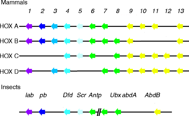