The Interactive Fly
Genes involved in tissue and organ development
Genes of the peripheral nervous system
The Interactive Fly
Genes involved in tissue and organ development
The peripheral nervous system consists of sensory neurons. There are two types. Type I neurons innervate the sensory organs to which they are related by lineage. Each of these sensory organs is thought to be derived from a single ectodermal precursor (sensory organ precursor or SOP) which gives rise to one or several monodendritic neurons and several support cells. Type I sensory organs have been classified into two major groups: first, mechano- or chemosensory organs that have external sensory structures in the cuticle such as bristles, campaniform, and basiconical sensilla (external sensory organs), and second, chordotonal organs that are internally located stretch receptors. In addition the larval PNS also contains numerous type II neurons with multiple dendrites. These neurons, with one exception, do not seem to be associated with support cells. Multiple dendrite neurons are thought to function as stretch or touch receptors. Multiple dendritic neurons are derived from three sources, one group from external sensory organ lineages, a second set from chordotonal neurons and a third set is unrelated to sensory organs (Brewster, 1995).
For information about the development of the antennal olfactory sense organs see Olfactory Receptors.
Although the ability to sense temperature is critical for many organisms, the underlying mechanisms are poorly understood. Using the calcium reporter yellow cameleon 2.1 and electrophysiological recordings, thermosensitive neurons were identified and their physiologic responses were examined in Drosophila larvae. In the head, terminal sensory organ neurons show increased activity in response to cooling by ~1°C, heating reduces their basal activity, and different units show distinct response patterns. Neither cooling nor heating affects dorsal organ neurons. Body wall neurons show a variety of distinct response patterns to both heating and cooling; the diverse thermal responses are strikingly similar to those described in mammals. These data establish a functional map of thermoresponsive neurons in Drosophila larvae and provide a foundation for understanding mechanisms of thermoreception in both insects and mammals (Liu, 2003).
To identify neurons responding to changes in temperature, an optical approach using yellow cameleon 2.1 (YC2.1), an engineered, calmodulin-based, Ca2+-sensitive protein, was used. Its two fluorophores, cyan fluorescent protein (CFP) and yellow fluorescent protein (YFP), comprise a fluorescence resonance energy transfer (FRET)-capable pair; a conformational change in the protein causes FRET to increase when the Ca2+ concentration rises. Cameleon fluorescence has been used to measure intracellular (or cytosolic) Ca2+ concentration, [Ca2+]i, in vitro and in vivo (in C. elegans). Transgenic Drosophila larvae were developed that express cameleon in their neurons, and FRET was assayed to monitor activity in the peripheral neurons as the temperature was changed. The FRET measurements, plus electrophysiologic and behavioral assays, indicate that the terminal organ is a thermosensitive structure that responds to cool temperatures. Some body wall neurons also showed FRET changes with temperature shifts, and in contrast to the terminal organ, they responded to warm temperatures (Liu, 2003).
The pan-neuronal promoter elav was used to drive expression of the YC2.1 variant of cameleon with the Gal4-UAS system. Heterozygous transgenic larvae (elav-Gal4/+;UAS-YC2.1/+) showed fluorescence signal in all neurons. For example, cameleon fluorescence was distributed in neurons of the lateral pentascolopidial chordotonal organs in a diffuse pattern in the cell bodies and neurites, but not in the cell nuclei. Similar results were obtained in two other lines. To increase fluorescence intensity, homozygous lines (elav-Gal4;UAS-YC2.1) were generated. They produced a more intense fluorescence signal than did the heterozygous line; therefore, homozygous larvae were used for optical recording (Liu, 2003).
The dorsal and terminal organs are the major sensory structures of the larval head. Each organ contains more than 30 bipolar neurons with large dendrites extending to the tip of a dome-like structure where pores open to the environment. Cameleon was expressed in the larval head in terminal and dorsal organ neurons. Terminal and dorsal organ YFP/CFP ratio images were measured at 18, 10 and 40°C. When the temperature fell, the terminal organ YFP/CFP ratio and the calculated [Ca2+]i increased, and when the temperature rose, terminal organ [Ca2+]i fell. In contrast to the terminal organ, temperature variations had little effect on [Ca2+]i in the dorsal organ, salivary gland or trachea (Liu, 2003).
At 18°C, the fluorescence ratio from terminal organ neurons fluctuates spontaneously; this was reflected in the variance of the fluorescence ratio (sigma2). These fluctuations are not an artifact, since the fluorescence ratio in the adjacent dorsal organ and in the salivary gland and trachea did not fluctuate, and the sigma2 was much lower. Cooling increases and heating decreases terminal organ sigma2, but has little effect on sigma2 in the other cell types. These findings suggest that terminal organ neurons have substantial spontaneous activity, even at room temperature, and the large [Ca2+]i fluctuations raise the possibility that some neurons might show oscillating activity (Liu, 2003).
Although the data do not allow distinguishing whether the changes in [Ca2+]i are upstream or downstream of action potential firing, these results indicate that the larval terminal organ contains thermosensory neurons that respond to cooling. Their response profile resembles the behavior of the most commonly observed mammalian cold receptors. The lack of thermoreceptor activity in dorsal organ neurons provides an important control showing the specificity of temperature sensing by the terminal organ: both terminal and dorsal organs contain sensory neurons, they lie adjacent to each other in the larval head; they are exposed to the same temperature stimuli, and cameleon fluorescence from both organs can be examined in the same image. These data also indicate that the cameleon protein itself does not respond to temperature changes (Liu, 2003).
To further test the hypothesis that terminal organ neurons detect cool temperatures and to obtain an independent assessment of the response of thermosensitive neurons, a glass electrode was inserted into early third-instar larval terminal or dorsal organs and extracellular recordings were obtained. Dorsal organ recordings showed no activity at room temperature or any response to warming or cooling. Terminal organ neurons, however, showed spontaneous activity at room temperature. This difference between terminal and dorsal organ basal activity is consistent with the difference in sigma2 in the optical measurements. Depending on the electrode position, recordings were obtained with one or two units. Thirty-six such recordings were analyzed (Liu, 2003).
When larvae are cooled by placing a cold metal block in their vicinity, terminal organ neurons respond in one of three ways. A type-I response is an increased firing frequency that adapts during the time course of the stimulus. This response occurred in 20 of 36 recordings. A type-II response, which occurred in 3 of 36 recordings, showed cold-induced oscillatory activity. This oscillating electrical activity may explain, in part, the increase in FRET sigma2 observed on cooling the terminal organ. In contrast to the type-I response, the onset of the type-II response is slow, and after the cold stimulus is removed, oscillations persist for some time before returning to baseline. A type-III response involves a transient cold-induced reduction in activity; it occurred in 13 recordings (Liu, 2003).
When a warm block was substituted for the cold one, spontaneous activity fell with all three types of response. For example, in recordings showing a type-I response, a warm stimulus reduced activity from 16.7 +/- 2.4 spikes/s to 7.4 +/- 2.9 spikes/s. After removing the warm block, neuronal activity recovered to baseline within 5 s (Liu, 2003).
On exposure to the cold stimulus, temperature will fall, and the closer the stimulus to the terminal organ, the more rapid the fall. In type-I cells, faster cooling elicits a greater increase in neuronal activity, the maximal increase in activity occurs 1 s later, at a time when that terminal organ temperature was calculated to have fallen 0.30°C. With the cold block at position 1, the maximal increase in spike rate occurs at 16 s when a 0.43°C decline was calculated to have occured. At the intermediate position 2, a fall of 0.45°C at 7 s was calculated when activity was maximal. Thus, the response to a cold stimulus seems to be a function of the rate of cooling and the change in temperature (Liu, 2003).
To change temperature by another means, a Peltier-based device was used to cool the solution bathing the larval body. Recordings were studied that showed a type-I response and lowered temperature 0.1-1°C from room temperature. This cooling increased spike frequency by 20%. Thus, similar results were obtained with different methods of lowering temperature and with independent means of measuring the response (Liu, 2003).
Because cooling stimulates terminal organ activity, it was hypothesized that disrupting terminal organ function would blunt the behavioral response to a reduced temperature. Earlier work showed that the GH86 promoter drives expression in the terminal organ; it also drives expression in the dorsal organ, epidermis, enocytes and pharyngeal muscle. The GH86-Gal4 promoter was used to drive a UAS-tetanus toxin light chain transgene (UAS-TNT-C), which specifically degrades synaptobrevin and thereby blocks neurotransmitter release. Because both the GH86-Gal4 and UAS-TNT-C transgenes are located on the X chromosome, only female F1 larvae that contained both transgenes were studied. The GH86 x TNT-C cross showed a reduced preference for 18°C compared with 11°C. These data suggest that larvae use terminal organ thermosensors to sense cool temperatures. Larval terminal organs contain more than 30 neurons and also likely respond to other sensory stimuli. For example, some terminal organ neurons may be involved in the response to low concentrations of salt (Liu, 2003).
To test thermosensitivity at another site, neurons in lateral body segments 5-8 were examined. At this location, there was less interference from fluorescence of the central nervous system and salivary glands than in segments 1-4. Depending on the location of the neurons, it was possible to measure fluorescence from some individual neurons. In other cases, clusters of neurons were studied because single neurons could not be reproducibly identified. However, temperature-dependent changes in the clusters likely arise from single neurons because the fluorescence changes often occur at a single spot within the cluster (Liu, 2003).
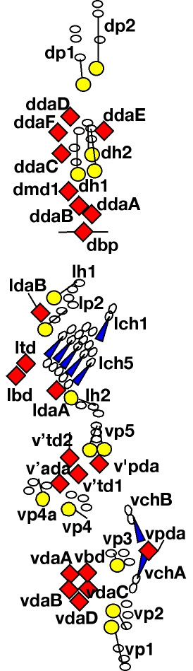
Multidendritic neurons show the greatest response to temperature changes, and the amplitude of [Ca2+]i response varies for different neurons and clusters. Different neurons also show distinct thermosensory responses. For example, for neurons in clusters 2 and 3, and neuron ddaB, cooling reduces and heating elevates [Ca2+]i. This response is the opposite of that in the terminal organ. The neurons in cluster 1 and lch5 increase [Ca2+]i when the temperature is either raised or lowered from the preferred temperature of 18°C. The distinct response patterns observed suggest that different neurons carry specific information to the central nervous system (Liu, 2003).
These data provide a functional map of thermosensitive neurons in Drosophila larvae. Neurons with different temperature responses were to be anatomically segregated. Moreover, within different regions there was a striking diversity in the behavior of thermosensitive neurons (Liu, 2003).
Terminal organ cold sensors show activity at room temperature, and this activity increases with cooling and falls with heating. Thus, these thermosensors are poised to respond whenever temperature changes, even slightly. Moreover, terminal organ function is apparently required for a normal response to cool temperature because expressing the tetanus toxin light chain in the terminal organ blunts the behavioral preference for 18°C versus 11°C. The calculations of the temperature shifts induced by a cold block and the measurements of bath temperature indicate that the terminal organ responds to changes of ~1°C. Thus, these data explain how larvae respond to a change in temperature, but how they respond to absolute temperatures remains unclear. The answer probably lies in central integration of output from the complex mixture of thermosensory neurons. In this regard, thermoreceptors with oscillating discharges may be particularly important to sensing absolute temperatures. It has also been suggested that the substantial complexity of thermoreceptive cell types may increase the sensitivity of the system(Liu, 2003).
The data suggest a striking similarity between thermoreceptor physiology in Drosophila and mammals. In both organisms, different types of neurons encode the response to cold and heat stimuli. It was found that in Drosophila, the most common type of terminal organ cold receptive neurons show a characteristic response to cold and heat; they spontaneously discharge at room temperature, cooling reduces the frequency of nerve impulses, and warming decreases activity. This type of cold receptor neuron is also very common in mammals. Additionally, an oscillatory response to cooling was found in 3 of 36 larval terminal organ neurons. The preliminary observations suggest that oscillatory activity may be more common if temperature is reduced more rapidly and to a greater extent. Interestingly, there are several reports of oscillatory activity in mammalian thermoreceptors. Whether this type of activity results from coordinated effects of temperature on multiple channels or on a single type of channel is unknown. Cluster 1 and Ich5 neurons in Drosophila increase activity on both warming and cooling. Mammals also contain these so-called 'paradoxical' temperature receptors. It will be interesting to learn whether this activity is generated by two different temperature-responsive ion channel receptors that are both expressed in a single neuron. Finally, some Drosophila neurons (multidendritic neurons ddaB and clusters 2 and 3) increase their activity during heating and reduce activity during cooling. This pattern of activity also exists in many mammalian warm receptors (Liu, 2003).
These findings reveal a diverse pattern of thermosensory response in larval neurons and provide new insight into the physiology of temperature sensing in Drosophila. Moreover, the results demonstrate common thermosensory response patterns between distantly related animal species. Given the potential relationship between temperature sensing and pain, this work may provide a basis for additional insight into nociception. Thus, these studies help pave the way toward a better understanding of the molecular mechanisms of thermoreception in both insects and mammals (Liu, 2003).
This work describes unknown aspects of chordotonal organ (ChO) morphogenesis revealed in post-embryonic stages through the use of new fluorescently labeled markers. Towards the end of embryogenesis a hitherto unnoticed phase of cell migration commences in which the cap cells of the ventral ChOs elongate and migrate towards their prospective attachment sites. This migration and consequent cell attachment generates a continuous zigzag line of proprioceptors, stretching from the ventral midline to a dorsolateral position in each abdominal segment. The observation that the cap cell of the ventral-most ChO attaches to a large tendon cell near the midline provides the first evidence for a direct physical connection between the contractile and proprioceptive systems in Drosophila. This analysis has also provided an answer to a longstanding enigma that is what anchors the neurons of the ligamentless ventral ChOs on their axonal side. A new type of ChO attachment cell was identified that binds to the scolopale cells of these organs, thus behaving like a ligament cell, but on the other hand exhibits all the typical features of a ChO attachment cell and is critical for the correct anchoring of these organs (Halachmi, 2016).
In mammals, pain is regulated by the combination of an ascending stimulating and descending inhibitory pain pathway. It remains an intriguing question whether such pain pathways are of ancient origin and conserved in invertebrates. This study reports a new Drosophila pain model and use it to elucidate the pain pathways present in flies. The model employs transgenic flies expressing the human capsaicin receptor TRPV1 in sensory nociceptor neurons, which innervate the whole fly body, including the mouth. Upon capsaicin sipping, the flies abruptly displayed pain-related behaviors such as running away, scurrying around, rubbing vigorously, and pulling at their mouth parts, suggesting that capsaicin stimulated nociceptors in the mouth via activating TRPV1. When reared on capsaicin-containing food, the animals died of starvation, demonstrating the degree of pain experienced. This death rate was reduced by treatment both with NSAIDs and gabapentin, analgesics that inhibit the sensitized ascending pain pathway, and with antidepressants, GABAergic agonists, and morphine, analgesics that strengthen the descending inhibitory pathway. These results suggest Drosophila to possess intricate pain sensitization and modulation mechanisms similar to mammals, and it is proposed that this simple, non-invasive feeding assay has utility for high-throughput evaluation and screening of analgesic compounds (Jang, 2023).
Animals' response to a stimulus in one sensory modality is usually influenced by other modalities. One important type of multisensory integration is the cross-modal modulation, in which one sensory modality modulates (typically inhibits) another. Identification of the mechanisms underlying cross-modal modulations is crucial for understanding how sensory inputs shape animals' perception and for understanding sensory processing disorders. However, the synaptic and circuit mechanisms that underlie cross-modal modulation are poorly understood. This is due to the difficulty of separating cross-modal modulation from multisensory integrations in neurons that receive excitatory inputs from two or more sensory modalities(5)-in which case it is unclear what the modulating or modulated modality is. This study reports a unique system for studying cross-modal modulation by taking advantage of the genetic resources in Drosophila. Gentle mechanical stimuli was shown to inhibit nociceptive responses in Drosophila larvae. Low-threshold mechanosensory neurons inhibit a key second-order neuron in the nociceptive pathway through metabotropic GABA receptors on nociceptor synaptic terminals. Strikingly, this cross-modal inhibition is only effective when nociceptor inputs are weak, thus serving as a gating mechanism for filtering out weak nociceptive inputs. These findings unveil a novel cross-modal gating mechanism for sensory pathways (Pan, 2023).
Mechanical nociception is an evolutionarily conserved sensory process required for the survival of living organisms. Previous studies have revealed much about the neural circuits and sensory molecules in mechanical nociception, but the cellular mechanisms adopted by nociceptors in force detection remain elusive. To address this issue, the mechanosensation of a fly larval nociceptor (class IV da neurons, c4da) was studied using a customized mechanical device. C4da were found to be sensitive to mN-scale forces and make uniform responses to the forces applied at different dendritic regions. Moreover, c4da showed a greater sensitivity to localized forces, consistent with them being able to detect the poking of sharp objects, such as wasp ovipositor. Further analysis reveals that high morphological complexity, mechanosensitivity to lateral tension and possibly also active signal propagation in dendrites contribute to the sensory features of c4da. In particular, it was discovered that Piezo and Ppk1/Ppk26, two key mechanosensory molecules, make differential but additive contributions to the mechanosensitivity of c4da. In all, these results provide updates into understanding how c4da process mechanical signals at the cellular level and reveal the contributions of key molecules (Liu, 2022).
This study has several unexpected findings: (1) velvet ant venom, which is extremely effective at triggering pain in humans, also activates the nociceptive neurons of an insect; (2) a single venom peptide (Do6a) is responsible for potent activation of insect nociceptors and is also the most abundant peptide within the venom; (3) Do6a does not produce signs of nociception in mice, indicating that it is an insect-specific venom mechanism; (4) velvet ant stings are sufficiently aversive to override the prey drive of a predatory insect (Borjon, 2024).
Because the venom can activate neurons of such distant phyla as insects and mammals the venom was expected to find that it acts through an evolutionarily ancient nociceptive mechanism. Instead, the venom cocktail has multiple mechanisms: a general mechanism that is widely effective across the animal kingdom, and a specific mechanism that is tailored to target the nociceptive system of insects. That the most abundant venom peptide acts specifically against insects suggests that interactions with predatory insects is a more important selective pressure for the evolution of velvet ant venom than interactions with vertebrates. Velvet ants are parasitoids of ground dwelling bees and wasps and it is possible that the venom is also used in defensive encounters that may happen when they enter the nests of these hosts (Borjon, 2024).
Despite their ability to avoid potentially dangerous stimuli, whether insects "feel pain" remains a subject of considerable debate and controversy. The current results show that, similar to defensive compounds that evoke noxious sensations by activating pain receptors in mammals (eg. capsaicin of pepper plants), insects can exploit nociceptive pathways of other insects to trigger aversion. Although the mechanism of action for velvet ant venom is distinct in mammals and insects, the fact that the venom converged to target the analogous sensory system in these widely divergent taxa provides further support for the functional similarity between insect nociception and mammalian pain systems (Borjon, 2024).
Axonemal Dynein DNAH5 is Required for Sound Sensation in Drosophila Larvae. Neurosci Bull Somatosensory neurons (SSNs) that detect and transduce mechanical, thermal, and chemical stimuli densely innervate an animal's skin. However, although epidermal cells provide the first point of contact for sensory stimuli, understanding of roles that epidermal cells play in SSN function, particularly nociception, remains limited. This study show that stimulating Drosophila epidermal cells elicits activation of SSNs including nociceptors and triggers a variety of behavior outputs, including avoidance and escape. Further, this study found that epidermal cells are intrinsically mechanosensitive and that epidermal mechanically evoked calcium responses require the store-operated calcium channel Orai. Epidermal cell stimulation augments larval responses to acute nociceptive stimuli and promotes prolonged hypersensitivity to subsequent mechanical stimuli. Hence, epidermal cells are key determinants of nociceptive sensitivity and sensitization, acting as primary sensors of noxious stimuli that tune nociceptor output and drive protective behaviors (Yoshino, 2025).
Spatiotemporal mechanisms generating neural diversity are fundamental for understanding neural processes. This study investigated how neural diversity arises from neurons coming from identical progenitors. In the dorsal thorax of Drosophila, rows of mechanosensory organs originate from the division of sensory organ progenitor (SOPs). In each row of the notum, an anteromedial located central SOP divides first, then neighbouring SOPs divide, and so on. This centrifugal wave of mitoses depends on cell-cell inhibitory interactions mediated by SOP cytoplasmic protrusions and Scabrous, a secreted protein interacting with the Delta/Notch complex. Furthermore, when this mitotic wave was reduced, axonal growth was more synchronous, axonal terminals had a complex branching pattern and fly behaviour was impaired. The temporal order of progenitor divisions influences the birth order of sensory neurons, axon branching and impact on grooming behaviour. These data support the idea that developmental timing controls axon wiring neural diversity (Lacoste, 2022).
To study how functional neuronal diversity can be generated from a homogenous set of neural precursors, advantage was taken of the invariant way in which sensory organs are located on the dorsal epithelium of Drosophila. This spatial configuration greatly facilitated the study of the relative timing of SOP division and the identification of a distinct temporal wave of SOP mitosis. Asynchrony in mitotic reactivation timing has been described in Drosophila larva neuroblasts. This differential timing is related to two cell cycle arrests: one population of neuroblasts is arrested in G2 while another population is arrested in G0. G2-arrested neuroblasts resume mitosis earlier than those in G0-arrest. As in this system, it has been proposed that this particular order of division ensures that neurons form appropriate functional wiring. It is relevant that other temporal processes controlling the wiring of peripheral receptors with the central nervous system have been described in the Drosophila eye, another highly organised structure. It is conceivable that these temporal patterning mechanisms of neurogenesis, to date identified only in organised tissues, could be more widespread (Lacoste, 2022).
A core aspect of this work was to link cellular level of complexity (timing of SOP division) with uppermost level (behaviour). In this context, evidences are presented showing that the cleaning reflex was impaired when the SOP mitotic wave was disrupted. The cleaning reflex has been traditionally analysed after stimulation of macrochaetes rather than microchaetes as in the present work. Macro- and microchaetes have different patterns of terminal axon arborisation. As such, it is remarkable that this fly behaviour was significantly affected by altering the timing of microchaete precursor division in the dorsal thorax. This study showed that the SOP mitotic wave leads to a progressive neurogenesis along each row of microchaetes. This, in turn, would likely induce a particular pattern of microchæte axon arrival in the thoracic ganglion required for the proper organisation of the neuropila in the central nervous system. Although this study has documented this progressive axonogenesis, the strict pattern of axon arrival into the ventral ganglion is not known. It would depend on the order of birth of neurons, and on the geometry of axon projections that fasciculate to form the dorsal mesothoracic nerves in the ganglion. In any case, this study shows that, when sca function was specifically downregulated during the SOP mitotic wave, axonogenesis occurs almost simultaneously in each row of microchaetes. This certainly impairs the pattern of axon arrival into the ganglion leading to ectopic axon branching and changes in fly behaviour. It would be interesting to know whether these impairments are specifically due to neurogenesis occurring simultaneously. To test this, it is necessary to find a way to induce different patterns of SOP mitotic entry, for instance, a centripetal wave or a random order. If the observed effect is specifically due to the simultaneity, normal behaviour would be expected to be associated with other patterns of SOP division (Lacoste, 2022).
We observed that the first SOP to divide (SOP0) was always located in the anteromedial region of each row. This may reflect the existence of a pre-pattern that causes SOPs located in that region to start dividing earlier than the others. Although the anteromedial region corresponds approximately to the posterior limit of expression of the transcription factor BarH1, no factors specifically expressed in this region have yet been identified. Alternatively, as the location of SOP0 is modified when Sca function was impaired, an interesting possibility is that SOP0 is selected by an emergent process related to cell-cell interaction in the epithelium, rather than by a passive pre-pattern that organises the first events in the notum (Lacoste, 2022).
This study presents evidence indicating that the secreted glycoprotein Scabrous, which is known to interact with the N-pathway to promote neural patterning, controls the kinetics of SOP mitosis in the notum. In proneural clusters, cells that express high levels of Dl and Sca become SOPs, while surrounding epithelial cells activate the N-pathway to prevent acquisition of a neural fate. In eye and notum systems, Sca modulates N-activity at a long range. Indeed, during eye development, sca is expressed in intermediate clusters in the morphogenic furrow and transported posteriorly in vesicles through cellular protrusions to negatively control ommatidial cluster rotation. Similarly, in the notum, SOP protrusions extend beyond several adjacent epithelial cells in which Dl and Scabrous are detected. The current data show that shorter protrusions (obtained after rac1N17 overexpression conditions) as well as loss of function of Dl or sca make the mitotic wave more synchronous. Since, no reduction of the global level of sca expression associated with the wave progression was observed, it is plausible that Sca, required to maintain SOPs in G2 arrest, is delivered focally through protrusions that are difficult to follow with in vivo analysis. Although this possibility is favored, it cannot be formally ruled out that Rac1N17 overexpression affects Sca secretion per se without affects sca expression (Lacoste, 2022).
As in neuroblasts, G2 arrest in SOP cells is due to the downregulation of the promitotic factor Cdc25/String. Thus, overexpression of string in SOPs induces a premature entry into mitosis, while overexpression of negative regulators, like Wee1, maintain these cells in arrest. Possibly Sca negatively regulates string expression, perhaps through the N-pathway that it is known to control the level of String. Alternatively, it has been recently shown that the insulin-pathway also regulates String level. Moreover, in muscle precursors, cell proliferation is induced by the insulin-mediated activation of the N-pathway. These observations raise the interesting possibility that, in this system, insulin activates the N-pathway and Sca modulates this activation. Further investigations will be required in order to identify the link between Scabrous, the N/Dl- and insulin-pathways in the resumption of mitosis in SOPs (Lacoste, 2022).
During nervous system development, the complex patterns of neuronal wiring are achieved through the interaction between neuronal cell surface receptors and their chemoattractive or repulsive ligands present in the environment. An essential condition for proper axon guidance is the competence of neurons to respond to these environmental clues. It is generally agreed that neuron competence depends on the specific expression of transcriptional factors regulating their identity. This study shows that the timing of neuron formation is also a factor controlling their terminal morphology. It is proposed that the SOP mitotic wave induces a particular pattern of arrival of microchæte axons in the thoracic ganglion. This pattern establishes a specific framework of guidance cues on which circuits will be built and ultimately influencing an organism’s behaviour. These findings support the idea that, in addition to genetic factors, neurogenic timing is a parameter of development in the mechanisms controlling neural branching (Lacoste, 2022).
Interactions between epithelial cells and neurons influence a range of sensory modalities including taste, touch, and smell. Vertebrate and invertebrate epidermal cells ensheath peripheral arbors of somatosensory neurons, including nociceptors, yet the developmental origins and functional roles of this ensheathment are largely unknown. This study describes an evolutionarily conserved morphogenetic mechanism for epidermal ensheathment of somatosensory neurites. Somatosensory neurons in Drosophila and zebrafish were found to induce formation of epidermal sheaths, which wrap neurites of different types of neurons to different extents. Neurites induce formation of plasma membrane phosphatidylinositol 4,5-bisphosphate microdomains at nascent sheaths, followed by a filamentous actin network, and recruitment of junctional proteins that likely form autotypic junctions to seal sheaths. Finally, blocking epidermal sheath formation destabilized dendrite branches and reduced nociceptive sensitivity in Drosophila. Epidermal somatosensory neurite ensheathment is thus a deeply conserved cellular process that contributes to the morphogenesis and function of nociceptive sensory neurons (Jiang, 2019).
Animals control their developmental schedule in accordance with internal states and external environments. In Drosophila larvae, it is well established that nutrient status is sensed by different internal organs, which in turn regulate production of insulin-like peptides and thereby control growth. In contrast, the impact of the chemosensory system on larval development remains largely unclear. A genetic screen was performed to identify gustatory receptor (Gr) neurons regulating growth and development; Gr28a-expressing neurons were found to be required for proper progression of larval growth. Gr28a is expressed in a subset of peripheral internal sensory neurons, which directly extend their axons to insulin-producing cells (IPCs) in the central nervous system. Silencing of Gr28a-expressing neurons blocked insulin-like peptide release from IPCs and suppressed larval growth during the mid-larval period. These results indicate that Gr28a-expressing neurons promote larval development by directly regulating growth-promoting endocrine signaling in a stage-specific manner (Ohhara, 2022).
Grs are a group of transmembrane chemosensory receptors expressed in external and pharyngeal gustatory neurons as well as in internal sensory neurons. They have a central role in sensation of various environmental cues, including nutrients and noxious compounds, to regulate feeding behavior. However, the importance of Gr-expressing neurons in regulating larval growth remains mostly unclear. This study screened 66 Gr-Gal4 lines using a neural silencer UAS-Kir2.1 to identify GRNs regulating larval growth. Notably, most Gr-Gal4 lines expressed in various populations of GRNs did not induce any major developmental defect. Gr-expressing gustatory neurons thus seem to be mostly dispensable for larval development in standard lab conditions. Alternatively, it is possible that GRNs can compensate for loss of other functionally analogous GRNs during development (Ohhara, 2022).
This study identified Gr28a-expressing internal sensory neurons as a novel regulator of larval development. The results suggest that Gr28a-expressing v'td neurons regulate IPC activity and thereby control growth rate mainly between 24 and 72 hAH. At around 60 hAH, Drosophila larvae attain the minimum body weight required for initiation of metamorphosis in normal schedule, which is known as the 'critical weight checkpoint'. Considering that the attainment of critical weight is promoted by insulin signaling, one possibility is that Gr28a-expressing v'td neurons are involved in the attainment of critical weight through regulation of ILP secretion. Although regulatory mechanisms of ILP secretion by v'td neurons remain unclear, it is speculated that Gr28a-expressing v'td neurons innervate IPCs to regulate their responsiveness to insulinotropic/insulinostatic signaling pathways, including fat body-derived endocrine signals during this growth period (Ohhara, 2022).
An important unanswered question is what types of sensory inputs are received by Gr28a-expressing v'td neurons. Their dendrites are associated with tracheal branches and exposed to the hemolymph, suggesting that these neurons may respond to humoral cues. Considering that a developmental delay observed in Gr28a mutant animals was milder than that of the v'td neuron-silenced animals, it is most likely that v'td neurons sense humoral cues not only via Gr28a but also through other receptors such as Gr28b that are known to be expressed in v'td neurons. It has been reported that Gr28a and Gr28b mediate RNA sensing and that Gr28a is also required for sensing ribonucleosides including uridine and inosine (Mishra, 2018). Thus, one possibility is that Gr28a-expressing v'td neurons regulate larval growth in accordance with the amount of RNA and ribonucleosides diffused in the hemolymph. Interestingly, depletion of extracellular adenosine by administration of extracellular adenosine deaminase, an enzyme converting adenosine to inosine, promotes cell proliferation in vitro, suggesting that extracellular adenosine inhibits proliferation. Furthermore, loss of adenosine deaminase causes a delay in pupariation. It is therefore conceivable that Gr28a-expressing v'td neurons monitor inosine concentration in the hemolymph to control insulin signaling and growth rate in accordance with inosine availability. In addition, a recent study has reported that Gr28b.c, which is co-expressed with Gr28a in v'td neurons (Qian, 2018), mediates sensation of plant-derived saponin, an amphipathic glycoside, in external sensory organs in the adult stage, although whether dietary saponin is incorporated into hemolymph is unknown. Further studies are required to elucidate sensory cues affecting the activity of Gr28a-expressing v'td neurons and their biological significance (Ohhara, 2022).
In summary, this study identified body wall-associated Gr28a neurons as stage-specific insulinotropic sensory neurons. GRNs regulating ILP release and systemic growth have not been reported previously. This study thus provides an important basis to further elucidate neuroendocrine pathways regulating insect growth and development (Ohhara, 2022).
Drosophila dEAAT2, a member of the excitatory amino-acid transporter (EAAT) family, has been described as mediating the high-affinity transport of taurine, which is a free amino-acid abundant in both insects and mammals. However, the role of taurine and its transporter in hearing is not clear. This study reports that dEAAT2 is required for the larval startle response to sound stimuli. dEAAT2 was found to be enriched in the distal region of chordotonal neurons where sound transduction occurs. The Ca(2+) imaging and electrophysiological results showed that disrupted dEAAT2 expression significantly reduced the response of chordotonal neurons to sound. More importantly, expressing dEAAT2 in the chordotonal neurons rescued these mutant phenotypes. Taken together, these findings indicate a critical role for Drosophila dEAAT2 in sound transduction by chordotonal neurons (Sun, 2018).
The sense organs of adult Drosophila, and holometabolous insects in general, derive essentially from imaginal discs and hence are adult specific. Experimental evidence presented in this
study, however, suggests a different developmental design for the three largely gustatory sense organs located along the pharynx. In a comprehensive cellular analysis, it is shown that the
posteriormost of the three organs derives directly from a similar larval organ and that the two other organs arise by splitting of a second larval organ. Interestingly, these two larval organs
persist despite extensive reorganization of the pharynx. Thus, most of the neurons of the three adult organs are surviving larval neurons. However, the anterior organ includes some sensilla that
are generated during pupal stages. Also, apoptosis is observed in a third larval pharyngeal organ. Hence, the experimental data show for the first time the integration of complex, fully
differentiated larval sense organs into the nervous system of the adult fly and demonstrate the embryonic origin of their neurons. Moreover, they identify metamorphosis of this sensory system as
a complex process involving neuronal persistence, generation of additional neurons and neuronal death. The conclusions are based on combined analysis of reporter expression from P[GAL4] driver
lines, horseradish peroxidase injections into blastoderm stage embryos, cell labeling via heat-shock-induced flip-out in the embryo, bromodeoxyuridine birth dating and staining for programmed
cell death. They challenge the general view that sense organs are replaced during metamorphosis (Gendre, 2003).
The external gustatory sensilla of the Drosophila larva appear to follow the general holometabolan fate: they degenerate during metamorphosis and are replaced by adult-specific sensilla that
derive from the labial imaginal disc. This study examines whether this rule also applies to the internal gustatory system that is located along the pharyngeal tube. Interestingly, the adult
pharynx derives essentially from small, densely packed imaginal cells that comprise the clypeolabral bud, which is closely associated with the larval pharyngeal skeleton. Does this imply that
adult pharyngeal sensilla are born during metamorphosis, like their external counterparts, or do the anatomical similarities of certain larval and adult pharyngeal organs rather suggest
persistence of sensilla through metamorphosis (Gendre, 2003)?
The data prove that most of the neurons of the three major adult pharyngeal sense organs are persisting larval neurons that were born in the embryo. This is unlike other adult sensory
neurons, nearly all of which derive from imaginal discs. This interpretation relies on two independent experimental approaches for demonstrating embryonic birth dates (the use of the embryonic
lineage tracer HRP and cell labeling by FLPout at late embryonic stages, a novel use of this technique). The experimental data are supported by anatomical observations showing: (1) an almost
identical organization of the larval posterior pharyngeal sense organ (pps) and the adult dorsal cibarial sense organ (dcso); (2) the presence of the pps and dorsal pharyngeal sense organ (dps)
sensilla continuously through metamorphosis; (3) an uninterrupted expression of the P[GAL4] lines used in these two organs, and (4) the persistence of dendrites and axons in all surviving
neurons (Gendre, 2003).
HRP injected at the syncytial blastoderm stage becomes incorporated into every cell upon cellularization. During subsequent development, the marker remains at high levels in cells that divide
only a few times but becomes diluted in cells that undergo repeated divisions. Consequently, labeling in the adult is expected in many neurons of the central nervous system known to be
persisting larval neurons (e.g. optic lobe pioneers) but should be absent from tissues derived from imaginal discs. This corresponds to what was observed and leads to the postulation of an
embryonic origin for the elements containing high HRP levels in adult pharyngeal sense organs (Gendre, 2003).
This interpretation is supported by the FLPout experiments performed at late embryonic stages with the neuron-specific MJ94 line. In adults deriving from this treatment, exclusively
single labeled neurons were detected in sensillum 7 of the labral sense organ (lso), containing eight neurons, and in the five multiply innervated sensilla of the vcso and dcso. Although the
cell lineage of these sensilla was not studied, they are probably homologous to other multineuronal terminal-pore gustatory sensilla, which derive from a common sensory mother cell. Indeed,
apart from its eight neurons, sensillum 7 of the lso corresponds to a typical insect gustatory sensillum in terms of fine structural and cellular organization, containing no more than three
accessory cells. Hence, the single labeled neurons in this sensillum and in all sensilla of the vcso and dcso must have been postmitotic during FLPout. This agrees with the observation that
formation of head nerves is complete by embryonic stage 15 (Gendre, 2003).
Could these neurons have remained immature during larval life, differentiating only during metamorphosis, similar to subsets of postmitotic cells in the larval central nervous system CNS?
This is thought to be rather unlikely because it would require either the entire sensillum or subsets of neurons in multineuronal sensilla to remain immature. Moreover, there is no indication
for immature neurons from tracing their development with the marker line mCD8-GFP. Thus, it is suggested that all the neurons of the dcso and vcso, and sensillum 7 of the lso derive from
mature, functional larval neurons. Also, continuous reporter expression through metamorphosis suggests that one of the mononeural lso sensilla (perhaps sensillum 3) might be another persisting
larval sensillum (Gendre, 2003).
The fact that the pps and dps persist through metamorphosis is remarkable given the origin of the adult labrum and cibarium from imaginal cells of the clypeolabral bud. Massive labeling of
pharyngeal epithelial cells was observed after early pupal BrdU application. Moreover, the pharyngeal cuticle is shed and regenerates, a process that includes the cuticular part of the sensilla
in question. Perhaps the birth of additional accessory cells during metamorphosis (e.g. in the dcso or vcso, containing exclusively persisting neurons) is related to this modification. Formation
of new cuticular structures is also known from persisting external sensilla during larval molts, but the survival of pharyngeal sensilla during the extensive remodeling of the pharynx remains
stunning. The morphogenetic movements observed in the sensory system certainly reflect these dramatic changes (Gendre, 2003).
Why is the larval pharyngeal sensory apparatus largely conserved through metamorphosis? Small subsets of neurons associated with leg imaginal discs or with abdominal segments persist through
metamorphosis. In the fly Phormia, such leg-disc-associated neurons remain immature, implying that they are non-functional. Laser ablation studies suggest that persisting neurons might
help adult afferents to navigate from the imaginal discs to their central targets. Whether they become truly integrated in the adult nervous system or die after reaching adulthood (having
completed their pathway role) remains to be shown (Gendre, 2003).
The data demonstrate for the first time experimentally the integration of larval sensory neurons into the adult nervous system of Drosophila. Particularly striking and novel is the fact that
entire, fully differentiated larval sense organs become incorporated. Also, this is the first observation of metamorphic survival in the chemosensory system (Gendre, 2003).
Concerning the persisting neurons of the lso, a pathway function for the newly developing afferents toward and inside the central nervous system is certainly possible. However, the
integration of the surviving pharyngeal neurons into the adult sensory system invites other interpretations. For example, these neurons and/or their central projections might be particularly
precious, allowing, for example, the persistence of specific feeding-associated gustatory tasks through metamorphosis. As an alternative explanation, survival might be due to reasons of economy,
a principle that governs the metamorphosis of the nervous system. Although neuronal reorganization is indispensable owing to the changing demands of larval and adult life, it is kept at a
minimum, as shown by the survival of most larval interneurons and motor neurons. Sophisticated adult sense organs, however, might be easier to build de novo than by the transformation of simple
larval organs, explaining the almost complete replacement of the larval sensory system. Why pharyngeal sense organs do not follow this general rule might relate to their largely conserved
function at the two stages of life (analyzing the quality of ingested food of similar composition). The presence of larva-specific and adult-specific sensilla, however, suggests the existence of
stage-specific gustatory tasks (Gendre, 2003).
Neurogenesis depends on a family of proneural transcriptional activator
proteins, but the 'proneural' function of these factors is poorly understood,
in part because the ensemble of genes they activate, directly or indirectly,
has not been identified systematically. A direct approach to this
problem has been undertaken in Drosophila. Fluorescence-activated cell sorting was used to recover
a purified population of the cells that comprise the 'proneural clusters' from
which sensory organ precursors of the peripheral nervous system (PNS) arise.
Whole-genome microarray analysis and in situ hybridization was then used to
identify and verify a set of genes that are preferentially expressed in
proneural cluster cells. Genes in this set encode proteins with a diverse array
of implied functions, and loss-of-function analysis of two candidate genes
shows that they are indeed required for normal PNS development. Bioinformatic
and reporter gene studies further illuminate the cis-regulatory codes that
direct expression in proneural clusters (Reeves, 2005).
The PNC cells that express the proneural genes achaete (ac)
and scute (sc) comprise only a small fraction of the wing
imaginal disc of the late third-instar Drosophila larva. It is
anticipated that this might frustrate attempts to characterize PNC-specific
gene expression in unfractionated wing discs (e.g., by comparison of wild-type
and ac-sc mutant tissue). Accordingly, PNC cells were purified by
using fluorescence-activated cell sorting (FACS). As a PNC-specific marker,
a GFP reporter was chosen representing the Bearded family gene E(spl)m4.
m4 is strongly and specifically expressed
in PNCs, and a cis-regulatory module has been identified sufficient to recapitulate this activity. Wing imaginal discs were dissected
from late third-instar larvae carrying the m4-GFP transgene and
dissociated in trypsin-EDTA; cells with fluorescence greater than that of
w1118 control cells (GFP-positive cells) and cells with
fluorescence comparable to the control (GFP-negative cells) were recovered separately by FACS (Reeves, 2005).
Transcripts from several genes
known to be expressed in domains of the wing disc outside of PNCs (en,
hh, and twi) were found to be greatly depleted in the
GFP-positive cell population. These negative controls provide further evidence of successful separation of PNC cells from other disc cells (Reeves, 2005).
Since the microarray data clearly associates
expression of known genes preferentially with the expected cell populations,
43 candidates not known to be
expressed in wing imaginal discs were chosen for further analysis. Candidate
genes for which cDNA clones were available from the Drosophila Gene
Collection were favored. The selected genes exhibit a wide variety of GFP+/GFP-
expression ratios in the microarray data, and their products have a broad
spectrum of predicted functions (Reeves, 2005).
In situ hybridization was employed as a
secondary screening method, both to verify that candidate genes selected from
these microarray data are expressed in wing imaginal discs, and to determine
their specific patterns of transcript accumulation. The wing disc expression
patterns observed can be sorted into three major classes: PNC patterns, SOP
patterns, and overlapping patterns.
Five of the 43 selected candidate genes exhibit a complete PNC pattern of
expression, while 3 other candidates
are expressed in subsets of PNCs; phyl is expressed in the
SOP and in a subset of non-SOP cells in each PNC. An unexpected 18 candidates are expressed in the
presumptive SOP cells of the wing disc. Fourteen of these SOP genes are
expressed in a complete pattern of SOPs, whereas the remaining four are expressed
either late in SOP development or in subsets of SOPs. The existence of
the latter group suggests that the cell sorting strategy made it possible to
identify genes that are expressed preferentially in just a few cells of the
wing disc. Overall, 27 (63%) of the tested candidates were found to display
PNC- or SOP-specific expression patterns. This is likely to be an underestimate
of the true success rate of the microarray analysis, since 23 genes known to be
expressed in these patterns are not included in the statistic, though they were
reidentified in the screen (Reeves, 2005).
In addition to those expressed specifically
in PNCs and SOPs, a small group of candidate genes was found that is expressed
in patterns that overlap PNCs but appears to be distinct from them.
Detection of this class of genes is an important confirmation
of the efficacy and unbiased nature of the experimental approach (Reeves, 2005).
Patterned expression of the
proneural genes ac and sc defines the PNCs for most external
sensory bristles in adult Drosophila, and ac-sc function is
required for PNC and SOP gene expression, as well as for specification of the
SOP cell fate. Fifteen of the genes identified by the combined cell
sorting/microarray approach also require proneural gene function for their
expression. In an ac− sc− proneural
mutant background, transcript accumulation from members of both the PNC
(CG11798, CG32434/loner, edl, PFE) and SOP
(CG3227, CG30492, CG32150, CG32392, Men,
qua) classes is lost from PNCs that require ac-sc function.
This result is further
evidence that the approach has identified bona fide PNC genes, and it
demonstrates that expression of these ten genes is, directly or indirectly,
downstream of the bHLH activators encoded by ac and sc. The data
further show that the PNC-specific imaginal disc expression of the previously
studied genes mira, phyl, rho, Spn43Aa, and Traf1
is likewise downstream of proneural gene function (Reeves, 2005).
The identification of sets of genes comprising the genetic programs deployed in
PNCs and SOPs by the action of proneural proteins offers a powerful opportunity
to investigate the regulatory organization of these programs. Specifically, it was of interest
to find out (1) which genes are directly activated by proneural
regulators, and which indirectly, and (2) the nature of the
cis-regulatory sequences and their cognate transcription factors that
distinguish PNC- versus SOP-specific target gene expression. This analysis was initiated
by examining potential regulatory sequences of several of the
genes that have been identified for the presence of conserved, high-affinity proneural
protein binding sites of the form RCAGSTG. The initial approach was to ask
whether evolutionarily conserved clusters of these binding sites identify
cis-regulatory modules of the appropriate specificity. To date, this
strategy has proven very successful. Genomic DNA fragments bearing
proneural protein binding site clusters associated with CG11798,
edl, Traf1, CG32434/loner, and rho confer
PNC-specific activity on a heterologous promoter,
while similar modules from CG32150, mira, and PFE drive
SOP-specific expression. In three cases, double
labeling with the SOP marker anti-Hindsight (Hnt) reveals that PNC-specific
expression of the reporter gene includes the SOP as well as the non-SOP cells.
Mutation of the proneural protein binding sites in four of the
enhancer-bearing fragments severely reduces (CG11798) or abolishes (CG32150,
edl, Traf1) reporter gene
expression in PNCs/SOPs. Such results indicate that these genes are indeed
direct targets of activation by proneural proteins in vivo (Reeves, 2005).
Holometabolous insects like Drosophila carry out two major
phases of PNS neurogenesis, one in embryogenesis to form the larval PNS, and a
second in the late larval and early pupal stages to construct the adult PNS.
Many known genes participate in both phases. Accordingly, it was of interest to
determine whether genes identified as being expressed in imaginal disc PNCs
or SOPs are also expressed in the developing larval PNS. In situ hybridization reveals that,
among others, the PNC genes CG11798 and CG32434/loner and the SOP
genes CG3227, CG32150, and CG32392 are indeed expressed in
embryonic PNCs and SOPs, respectively (Reeves, 2005).
To determine whether this combined cell
sorting/microarray/in situ hybridization approach had indeed identified gene
functions required for proper PNS development, loss-of-function
alleles of two loci, CG11798 and CG3227, were generated. These were chosen
because (1) transcript accumulation from both genes was detected in the
primordia of both the larval and adult PNSs; (2) both genes encode proteins
with conserved domains; and (3) mobilizable P element transposon insertions
were available adjacent to these genes (Reeves, 2005).
CG11798 is predicted to encode
a probable transcription factor with four zinc finger domains.
Loss-of-function alleles of the gene were generated
by mobilizing KG03781, a P element located immediately downstream. A
precise excision of the P transposon and two partial deletions of the
CG11798 coding region were recovered and characterized by sequencing.
Deletions 19E and 34E are both homozygous lethal
during early larval stages, and both confer clear defects in the development of
the larval PNS. 19E causes the loss
or misplacement of sensory neurons marked by mAb 22C10 and sensory organ accessory cells marked
by anti-Prospero (αPros). Deletion 34E confers an even more severe PNS phenotype
and removes or misplaces many more 22C10-positive and Pros-positive
sensory organ cells in each hemisegment. The difference in the
severity of the 19E and 34E mutant phenotypes may be due to the fact that the
latter deletes a larger portion of the CG11798 coding region, including
the codons for the four zinc fingers. As a control genotype, use was made of the
precise excision (PE) derivative of the KG03781 transposon insertion. No
PNS mutant phenotype was detected in homozygous PE embryos,
demonstrating that the defects observed in
the 19E and 34E deletion homozygotes do not result from a second-site mutation
on the original KG03781 chromosome. The results of complementation tests
led to the
conclusion that CG11798 corresponds to the previously described
charlatan (chn) locus (Reeves, 2005).
To generate loss-of-function alleles of CG3227,
the P element transposon KG07404, inserted
just upstream of the gene, was mobilized. Imprecise
excision created two deletions, 23B and 23I.
Homozygosity for either results in nearly complete lethality before adulthood.
Mosaic adult flies carrying FLP/FRT-generated mutant clones exhibit a severe
PNS defect in which most mechanosensory bristles within the clonal territory
not only lack shaft structures but also bear multiple socket structures,
suggestive of shaft-to-socket cell fate transformations. The major defects observed in sensory
structures in both the larval and adult PNSs prompted giving CG3227 the new name insensitive (insv) (Reeves, 2005).
insv is predicted to encode a protein containing a conserved C-terminal domain of
unknown function called DUF1172. DUF1172 was
first recognized in the vertebrate NAC1 proteins, transcription factors that
also contain BTB/POZ protein-protein interaction domains. Alignment of
arthropod and vertebrate DUF1172s reveals that the domain is large
(approximately 125 amino acids) and contains a highly conserved central region
of alternating polar/charged residues and nonpolar residues.
This is the first described loss-of-function phenotype for a gene encoding a
DUF1172 domain protein (Reeves, 2005).
Several known or potential components of
signaling pathways were uncovered in this analysis as exhibiting either PNC- or
SOP-specific expression. These include genes encoding a putative G
protein-coupled receptor (CG31660), a receptor tyrosine kinase
(Ror), a regulator of G protein signaling (loco), and a modulator
of Ets protein activity (edl). Earlier studies have linked both G
protein function and Ras/MAPK signaling to the development of Drosophila
sensory bristles, but much remains
to be learned about their roles in this process. These findings suggest functions
in PNS development for both known and previously uncharacterized signaling
pathway components (Reeves, 2005).
Perhaps surprisingly, the data indicate the PNS-specific
expression in imaginal discs of several genes predicted to encode metabolic
enzymes, including a uridine phosphorylase (CG6330), a
maleylacetoacetate isomerase (CG9363), and a malate dehydrogenase
(Men). Exceptional metabolic requirements or signaling activities in
developing sensory organs may underlie these observations (Reeves, 2005).
Loss-of-function analysis of two genes identified by the cell
sorting/microarray/in situ hybridization approach, one expressed in PNCs
(CG11798/chn) and one in SOPs (CG3227), confirms that they are
indeed required for normal PNS development in Drosophila. Deletion
mutations of CG3227 (insensitive)
cause severe defects in the specification and differentiation of
sensory organ cells in the adult PNS, as assayed in mosaic clones. Particularly
prevalent is an apparent transformation of the shaft cell to the fate of its
sister, the socket cell; this is the same phenotype conferred by
loss-of-function mutations in N pathway antagonists such as Hairless and
numb. The definition of a loss-of-function phenotype for a DUF1172 gene
should prove valuable in investigating the in vivo function of this
uncharacterized protein domain (Reeves, 2005).
Certain SOP-specific genes, exemplified by
sens and phyl, are
required for the execution of the SOP fate itself. insv, by contrast,
represents a distinct class of SOP gene, required not for the fate of this
cell, but for the specification and/or differentiation of one or more of its
progeny. Thus, SOP-specific (or, more generally, precursor-specific) gene
expression can serve the same function as maternal gene
expression -- providing gene products essential to the development of
descendants. It is anticipated that a number of the SOP genes identified
will prove to act similarly (Reeves, 2005).
The function of proneural
bHLH proteins in Drosophila PNS development is complex, since they not
only activate in SOPs genes that promote the neural precursor cell fate (e.g.,
ac and sc themselves, sens and
phyl); they also activate in non-SOPs genes involved in inhibiting
this fate (e.g., genes of the Enhancer of split Complex).
The nature of the cis-regulatory
'codes' (combinations of transcription factor binding sites) that
distinguish the PNC versus SOP expression specificities is of particular interest.
One code has been identified for the expression of N-responsive genes in the non-SOP
cells of the PNC that consists of binding sites for the proneural proteins plus
sites for the N-activated transcription factor Suppressor of Hairless (Su(H)). Importantly, none
of the PNC modules identified in this study includes a conserved high-affinity
Su(H) site, yet at least three of them do mediate direct transcriptional
activation by the proneural proteins. Moreover, the expression driven by these
new PNC modules includes the SOP, whereas the 'Su(H) plus
proneural' code directs expression that excludes it.
These findings indicate the existence of at least one novel code
for PNC expression, and of a heretofore hypothetical class of genes -- ones
that are directly regulated by the proneural proteins in PNCs/SOPs but are
evidently not activated in response to N-mediated lateral inhibitory signaling,
perhaps because they are not involved in the inhibitory process (Reeves, 2005).
The proneural genes were first identified by their function
in the ectoderm in specifying neural cell fates, and they have been studied
almost exclusively in that context in both vertebrates and invertebrates.
However, it has become clear that these genes function as well in the other two
germ layers. The Drosophila proneural gene lethal of scute
(l'sc) is required to specify the fates of muscle progenitor
cells in the embryonic mesoderm, and the same gene (and probably also sc) is required
for the adult midgut precursor (AMP) cell fate in the embryonic endoderm. In both
of these nonectodermal settings, a striking parallel with neurogenesis is seen
in the manner in which proneural genes function in close association with the N
pathway to select individual precursor cells. In the mesoderm,
l'sc is deployed in 'pro-muscle clusters' from
which single muscle progenitors emerge by N-mediated 'lateral
inhibition'; in the endoderm, where proneural gene expression is initially
uniform, AMPs are spaced apart from each other by N signaling in a manner very
reminiscent of the spacing of microchaete bristles on the adult thorax.
The mouse proneural protein Atoh1 (Math1) has been shown to be
required for the specification of nonneural secretory cell precursors in the
intestinal epithelium. Thus, proneural transcription factors are not dedicated specifiers
of neural cell fates; rather, they appear to be very effective in first
conferring on a group of cells the potential to adopt a particular cell fate
and then promoting the selection of an individual committed progenitor from
within that group. This suggests the existence of a 'core' set of
genes that function downstream of the proneural proteins in all such contexts,
with other sets of genes contributing to context-dependent (e.g., germ
layer-specific) programs. Further investigation of the genes identified in this
study should permit a test of this intriguing hypothesis (Reeves, 2005).
Neurons establish diverse dendritic morphologies during development, and a major challenge is to understand how these distinct developmental programs might relate to, and influence, neuronal function. Drosophila dendritic arborization (da) sensory neurons display class-specific dendritic morphology with extensive coverage of the body wall. To begin to build a basis for linking dendrite structure and function in this genetic system, da neuron axon projections were analyzed in embryonic and larval stages. It was found that multiple parameters of axon morphology, including dorsoventral position, midline crossing and collateral branching, correlate with dendritic morphological class. A class-specific medial-lateral layering of axons in the central nervous system formed during embryonic development was identified; this layering allows different classes of da neurons to develop differential connectivity to second-order neurons. The effect of Robo family members on class-specific axon lamination was examined, and a forward genetic approach has also been taken to identify new genes involved in axon and dendrite development. For the latter, the third chromosome was screened at high resolution in vivo for mutations that affect class IV da neuron morphology. Several known loci, as well as putative novel mutations, were identified that contribute to sensory dendrite and/or axon patterning. This collection of mutants, together with anatomical data on dendrites and axons, should begin to permit studies of dendrite diversity in a combined developmental and functional context, and also provide a foundation for understanding shared and distinct mechanisms that control axon and dendrite morphology (Grueber, 2007).
Drosophila dendritic arborization (da) neurons have been
segregated into four classes (classes I-IV) that reflect arbor complexity,
arbor size and the length of terminal branches. The
cell bodies are distributed in ventral, ventral', lateral and dorsal
clusters between the epidermis and muscles, spreading dendrites across the
body wall, and axons to the ventral nerve cord. It was reasoned that if
morphological classes correspond to at least partially distinct sensory
systems, then their axons may have divergent morphologies and target
non-overlapping regions of the ventral nerve cord, where information will be
relayed to second-order neurons. Previous studies have found that most da
neurons arborize together in a common fascicle in the ventral CNS, with a
subset, including at least some class I da neurons, projecting to more dorsal
neuropil. In light of studies showing distinct morphological types of da neurons, mosaic analysis with a repressible cell marker (MARCM) was used to examine the morphology of da neuron dendrites and axons in third instar larvae. As a MARCM driver Gal4109(2)80 was used; this labels all multidendritic (md) sensory neurons, including those belonging to the da subgroup. Owing to the sparse labeling of central neurons, the 109(2)80 driver combined with MARCM allowed resolution of axon morphology of individual neurons (Grueber, 2007).
Data was collected from wild-type da neuron clones in segments A2-A7 to
identify their axon projections in the CNS. Different da
classes showed distinctive types of central projections. Class I neurons were
unique in their projection to the dorsal neuropil.
Class II axons showed collateral branches (branches exiting from the main
axonal trunk, although the timing of their emergence has not been determined)
that were not observed in class III and IV neurons. The
class I neuron vpda also showed such a branch. Class IV neurons
projected axon branches across the midline, but these were only rarely
observed for the other classes. Dorsal and ventral' class IV
axons crossed the midline, but axons from the ventral neuron did not. Each
class IV neuron also showed a large accumulation of branches medial to the
commissural/longitudinal branch bifurcation. The class III
terminals extended in an anteroposterior (AP) orientation and were relatively
unbranched, showing neither the collateral branches observed in class II
neurons, nor contralateral projections observed among class IV neurons (Grueber, 2007).
The axons of class I and class IV neurons also showed evidence of
somatotopic arrangements in the CNS. The trajectory of class I neurons in the
CNS mirrored the polarity of their dendrites in the periphery. Dorsal class I
neurons have distinct polarity with respect to the AP body axis: dendrites of
ddaD extend anteriorly and dendrites of ddaE extend posteriorly.
Likewise, it was found that the ddaD axons extended anteriorly in the CNS, whereas
the ddaE axons extended posteriorly. Among the class IV neurons, only neurons positioned in the dorsal and ventral regions of the body wall, but not the lateral region,
extended axons across the midline, fitting with principles of somatotopy established for body wall bristle neurons. These data together demonstrate that da neuron classes have
distinguishing axon terminals, and that neurons in the same class show
evidence of somatotopic organization (Grueber, 2007).
The position of sensory axons defines the population of possible
second-order targets and thus contributes strongly to sensory information
processing in the CNS. Axons of tactile receptors typically project to ventral
areas of the neuropil, whereas strain-sensing or proprioceptive neurons
usually project to more dorsal regions. Fasciclin II-labeled axon tracts provide a frame of reference for assessing dorsoventral (DV) position in the CNS. The DV
positions of axons was studied in 42 ventral nerve cords (VNCs) using MARCM, and 18 VNCs
using the FLP-out system. Both techniques revealed that each class I neuron extended
axons to dorsal regions of the neuropil, terminating just lateral to the
dorsomedial (DM) fascicle. The position of class I axons was therefore indistinguishable
at this level of resolution from the position of the dbd terminal arbor,
implying that information from class I neurons and the putative
stretch-sensing dbd neuron might be processed similarly in the CNS. Class II,
III and IV axons targeted the ventral CNS without obvious class-specific
dorsoventral lamination of terminal position. The positions of
the class II collateral branches were somewhat variable, either terminating on
the ventrolateral (VL) fascicle, or slightly lateral to VL (vdaA often had a more lateral termination). These data together provided anatomical support
for distinct functions among different da neurons, fitting with their distinct
dendritic arbor morphologies. Class II, III and IV axons project similarly to
known tactile afferents, while class I neurons have projections like known
proprioceptive or strain-sensing neurons (Grueber, 2007).
Whether the terminal positions of the ventral-projecting
class II, III and IV neurons could be further distinguished by their position was examined. Short pickpocket (ppk) enhancer sequences can drive gene
expression strongly in all class IV neurons and weakly in class III neurons.
Viewing all class IV neurons together revealed that they crossed the midline
in a single fascicle, that the stereotyped branching at the
commissural-longitudinal junction overlapped for all neurons, and that
longitudinal projections were not always tightly fasciculated. In ppk-eGFP
and ppk-Gal4, UAS-CD8::GFP animals, a strongly labeled
set of medial axons and a weakly labeled, slightly more lateral, layer of
terminals were observed. It is suspected that the weakly labeled axons were class III axons, which may form a layer next to class IV axons. To test this idea,
ppk-Gal4 was introduced into the FLP-out mosaic system. The relative
locations were observed of all class III axons except ddaF (whose axon was labeled too
weakly) and it was found that their major longitudinal projections terminated
immediately lateral to the scaffold of class IV axons (Grueber, 2007).
The ppk reporter lines alone do not label the class II axons, and
thus did not allow determination of whether all da classes form a laminar
organization or only the class III and IV neurons. However, examination of
FLP-out clones produced with Gal4109(2)80, with or without
ppk-eGFP to label class IV neurons, permitted labeling of different
axon groups. It was found that class II neurons with a significant longitudinal
projection formed a third layer of sensory axons that was lateral to both
class III and class IV axons, with class II collateral branches terminating in a distinct, even more lateral, position (Grueber, 2007).
The FLP-out data was confirmed by mapping the relationships of individual
pairs of sensory afferents using the MARCM technique. Within hemisegments, or
in adjacent hemisegments, having two or more da neuron clones
axons were organized (medial>lateral) class IV>class III>class II. Laminar patterning was independent of peripheral cell body position. These data together
indicate the presence of a laminar arrangement of somatosensory axons in the
Drosophila CNS. These data also suggest that somatosensory
information carried by different classes of da neurons might be distinguished
by sensory axon connectivity to second-order targets (Grueber, 2007).
The above FLP-out and MARCM data were collected from third instar larval
stages, so when during development layering of the different classes of axons could be observed was examined. To achieve live two-color discrimination of
different neuronal classes in embryonic and early larval stages
transgenic flies were generated expressing a photoconvertible fluorescent protein, Kaede, and
expression was placed under the control of Gal4109(2)80 in the
presence of ppk-eGFP. The Kaede protein was converted from green to red
fluorescence using a 10-30 second UV pulse and the position of all da
axons was examined relative to ppk-eGFP-labeled class IV axons. As early as the
sensory axon scaffold could be visualized (stage 17), class IV axons occupied
a medialmost layer with respect to other classes. These data
indicate that a laminar pattern develops at least by late embryonic stages and
is maintained without qualitative change in larvae (Grueber, 2007).
Much of the knowledge about somatotopic maps in insect mechanosensory
systems derives from studies of bristle afferents with peripheral receptive
fields that approximate a point source. Drosophila da neurons have
largely overlapping peripheral sensory fields and may, as a group, respond to
several distinct stimuli. How is information from this predominant body wall sensory
system represented in the CNS and what might this organization reveal about
the possible functions of da neurons? Neurons with different
dendritic branching morphologies target distinct regions of the CNS,
supporting the existence of a modality map of da neuron axons. Evidence is provided for nested somatotopic mapping in class I and class IV da
neurons. Individual class I neurons extend their dendritic and axonal arbors
in the same preferred direction along the AP body axis. Class IV axons
project across the midline according to the cell body position along the DV
axis of the body wall, with dorsal and ventral cells, but not more lateral
cells, crossing the midline. It is possible that class II and III neurons also
project in a somatotopic pattern that was not uncovered by this analysis. Thus, da neuron connectivity appears to incorporate both class and position-specific components, with some apparent correlations with dendritic field orientation (Grueber, 2007).
In the context of sensory processing, these data suggest distinct functions
for different morphological classes of da neurons. The class II-IV neurons
target a ventral region of the neuropil; thus information from these neurons
might be processed similarly to ventral-projecting tactile sensory bristle
neurons. Within this ventral region, the class-specific laminar
pattern could allow differential connectivity with second-order interneurons.
Additionally, class II neurons, with their collateral branches, might provide
information to distinct central circuits. The class I neurons targeted a more
dorsal region of the neuropil, which is generally a characteristic of
proprioceptive afferents in insects. Indeed, a class of da neurons in Manduca has been shown to target dorsal neuropil, and to respond to stretch of the cuticle. Many
insect proprioceptors, including chordotonal organs and the bipolar dendrite
neurons, have dendrites oriented in a preferential direction relative to the
body axis. Notably, the primary dendrites of each class I neuron are oriented
dorsally and secondary dendrites are oriented anteriorly and posteriorly. This
arrangement could allow larvae to compare distension along major body axes.
While the anatomical arrangement of their axons suggests distinct functions
for different da neurons, dissecting these different functions will ultimately
require behavioral and physiological studies (Grueber, 2007).
A notable feature of the mapping of da sensory afferents is their
predominant organization into class-correlated mediolateral layers, with class IV neurons in a medial layer, class III neurons intermediate, and class II neurons most
lateral. These layers do not correspond to the medial, intermediate and
lateral fascicles that have their position specified by a Robo combinatorial
code; thus novel molecular cues may contribute to this laminar
organization. Indeed, it has been postulated that the Robo code provides
information about the broad zone that a growth cone targets, while a
complementary fasciculation code fine-tunes pathway choice within that zone. It
is conceivable that Robo proteins could participate in specifying the lateral
position of collateral branches, since Robo3 overexpression in individual neurons
induced ectopic branching from the axon shaft. Although no cell-type-specific expression of Robo3 was detected in neurons that normally show such
branching, it is notable that Robo3 has been implicated in cell-type-specific
patterning decisions of PNS axons, and that Slit2 has been proposed as a positive
regulator of collateral branching of dorsal root ganglion sensory axons (Grueber, 2007).
The mechanisms for targeting of somatosensory afferents should act to
properly position axons of different classes relative to one another. Several
alternative mechanisms could contribute to this positioning. One potentially
important component of axon sorting could involve interactions between
homotypic or heterotypic axons. Heterotypic axons could repel each other to
sort to discrete bundles. Likewise, homotypic axons could adhere to one
another to ensure that they terminate together. Olfactory receptor neuron
axons engage in extensive hierarchical interactions to establish precise
targeting in olfactory glomeruli, and dendrites of da neurons engage in
class-selective interactions during development to ensure their proper spacing. It is
therefore conceivable that intraclass and interclass interactions could
participate in the sorting of somatosensory axons. Axons from different
classes could also project to specific layers that are prepatterned by the
processes of target interneurons or by other axons. Finally, the projection of
da axons to different layers could conceivably reflect a temporal order of
sensory axon arrival in the CNS, similar to the three-way correlation between
mediolateral axon position, physiological function and time of differentiation
among Drosophila wing campaniform sensilla. Among
the ventral cluster da neurons, a group with differentiation that has been
examined in the greatest detail, the class II neurons appear to be the
first-born, followed by class III neurons and class IV neurons. These
data suggest a possible correlation between birth order and axon position in the neuropil, although additional early markers of da neurons and further high-resolution imaging studies are required to further test this scenario (Grueber, 2007).
The molecular basis for insect sensory neuron differentiation, as well as
anatomical studies of somatosensory axon mapping and VNC circuitry, have been
subjects of considerable study, and principles are emerging that link the two
areas into molecular models of connectivity and synaptic specificity. Among
embryonic sensory neurons in Drosophila, there is a three-way
correlation between soma position, proneural transcription factor expression
and axon projection pattern, suggesting that these transcription factors may
endow aspects of modality-specific axonal projections. Such a link was recently established between chordotonal-organ-specific expression of the atonal proneural gene and expression of the Robo3 axon guidance molecule in these same organs.
Misexpression experiments with atonal, robo3 and comm
suggest a model whereby Atonal activates expression of Robo3, which in turn
specifies mediolateral positioning in chordotonal versus bipolar dendrite-type
axon projections. These studies provide an important basis for understanding
the establishment of sensory circuitry in the VNC (Grueber, 2007).
To begin to address the molecular basis of axon and dendrite patterning
using the anatomical framework established for the da neurons, a
forward genetic approach, which has proven a successful means to identify regulators of neuronal morphogenesis, was undertaken. A strength of this screen was the ability to simultaneously assess phenotypes in dendrites and axons at the level of single identifiable neurons. Numerous complementation groups were identified that affect axon patterning, including several mutations with a molecular nature as yet unknown. These mutations should allow identification of new genes involved in axon morphogenesis and place these into the context of their effects on somatosensory axon patterning and circuitry. Given that many mutations affecting dendrite morphogenesis have been identified, it is expected that studies of the mutations identified from the screen will also allow addressing of the similarities and distinctions between axon and dendrite development (Grueber, 2007).
Insects and mammals share similarities of neural organization underlying the perception of odors, taste, vision, sound, and gravity. This study observed that insect somatosensation also corresponds to that of mammals. In Drosophila, the projections of all the somatosensory neuron types to the insect's equivalent of the spinal cord segregated into modality-specific layers comparable to those in mammals. Some sensory neurons innervate the ventral brain directly to form modality-specific and topological somatosensory maps. Ascending interneurons with dendrites in matching layers of the nerve cord send axons that converge to respective brain regions. Pathways arising from leg somatosensory neurons encode distinct qualities of leg movement information and play different roles in ground detection. Establishment of the ground pattern and genetic tools for neuronal manipulation should provide the basis for elucidating the mechanisms underlying somatosensation (Tsubouchi, 2017).
Only three distinct types of sensory information are transmitted directly to the brain by primary neurons [leg gustatory sensilla (gs), chordotonal organs (co), and wing and haltere campaniform sensilla (cs)]. Such connection has also been reported in other insects, suggesting that this might be a general feature across insecta. Whereas only a small portion of leg co neurons project directly to the brain, most wing and haltere cs neurons innervate the brain; these cs neurons are known to detect various aspects of wing-beat force during flight to provide feedback control. Direct projections to the brain would be important for these neurons to enable fast transmission of information about rapidly changing sensory parameters during flight (Tsubouchi, 2017).
It was found that ground detection for wind-induced suppression of locomotion (WISL), which would require slower temporal resolution than flight control, is mediated by both direct and indirect pathways. Primary neurons and secondary interneurons of the same sensory modality tend to converge in specific subregions of the brain, forming modality-specific somatosensory representation. In spite of the similar axon trajectory in the brain, these neurons convey information about leg movement in different ways (Tsubouchi, 2017).
Interneurons associated with the leg co and es terminate in neighboring but different regions of the lateral brain, yet some of them have shared roles in WISL control. Because their signals are transmitted to distinct parts of the brain, yet-unidentified higher-order neurons in the brain should converge those signals to the motor control circuitry (Tsubouchi, 2017).
In this respect, it is important to note that most ascending secondary interneurons identified in this study have presynaptic output sites, not only in the brain but also in the VNC. Local circuitry in the leg neuropil is important for controlling leg movement. Those local neurons are likely candidates that receive output from the ascending interneurons, because axon terminals of sensory neurons hardly have postsynaptic sites. Similar local output has also been found in other sensory modalities; many olfactory and visual projection interneurons have collateral output synapses in the antennal lobe and optic lobes (Tsubouchi, 2017).
There are three pairs of leg neuropils. Among them, the foreleg neuropil has specialized arborization of the gs neurons that exist only in the foreleg. Other than this, no substantial differences of arborization patterns were found between the fore-, mid-, and hindleg neuropils (Tsubouchi, 2017).
The present results provide data for a systematic comparison of the insect somatosensory system with its mammalian counterparts. Insects and mammals share similarities of neural organization underlying the perception of odors, taste, vision, sound, and gravity, and the current data also reveal marked similarity for the mechanosensory system. In insects, some primary neurons project directly to distinct parts of the ventral and lateral brain, whereas others terminate within the VNC. Likewise, in mammals, some neurons project directly to the ventral brain at the medulla oblongata, whereas others terminate within the spinal cord. Modality-specific pathways tend to converge in different subregions of the medulla, as well as in the thalamus of the mammalian brain. Similarly, direct and indirect pathways tend to converge in common subregions of the insect brain, and neurons conveying information about different somatosensory modalities tend to terminate in different subregions. As in mammals, these subregions often lie adjacent to each other in certain parts of the brain; for example, the entire terminal arborizations of the leg co and es secondary interneurons are confined in a 40-μm-wide, 150-μm-tall cylindrical volume in the lateral brain (Tsubouchi, 2017).
Somatosensory signals are sent predominantly to the ipsilateral brain side in insects and contralateral in mammals. Considering that descending neurons tend to project ipsilaterally in insects but contralaterally in mammals, however, somatosensory signals and motor control computation are processed primarily in the same side of the brain in both cases (Tsubouchi, 2017).
Layers of sensory axon terminals in the insect VNC and mammalian spinal cord are also organized in a similar order. Insect multidendritic neurons and mammalian free nerve endings share various characteristics in common: Their dendrites both have free endings without forming particular sense organs to detect pain, temperature, and other submodalities. The md neurons project to the most ventral layer of the VNC, whereas free nerve endings innervate the most dorsal layer of the spinal cord. Axons from the insect external sensilla and mammalian hair receptors, both of which detect haptic contact to the tips of the bristles and hairs, terminate in the second-ventral and second-dorsal layers, respectively. Insect chordotonal organ and mammalian muscle spindle, as well as insect campaniform sensilla and mammalian Golgi tendon organ, also show similarity with respect to their functions in motor control. These receptor systems supply afferents to the most dorsal and most ventral layers in insects and mammals, respectively. A fly's stretch receptors and mammalian Merkel cell neurites-as well as Meissner, Ruffini, and Pacinian corpuscles-terminate in the third-ventral and third-dorsal layer, respectively. Although correspondence between them is less obvious, they similarly detect deformation of the exoskeleton and skin. Thus, functionally comparable somatosensory terminals are layered in reverse order between the two systems. Considering that the dorsoventral axis of the mammalian body is developmentally upside down compared with the insect one, the corresponding order of sensory arrangements is actually conserved exactly between the two systems (Tsubouchi, 2017).
Do corresponding somatosensory cell types express common genes? Modality-dependent molecular specialization is not apparent even within insects or mammals, because the same genes are often expressed in multiple cell types and only a few genes share expression in the corresponding cell types across taxa. This might be a rather general feature; receptor molecules as well as developmental origins of the sensory organs are not identical between insects and mammals also in olfactory and auditory systems, yet sensory centers in the brain share architectural similarities (Tsubouchi, 2017).
With this somatosensory analysis, transphyletic correspondence of neuronal circuitry has been found in all of the sensory modalities. Corresponding organization has been suggested also for associative centers and motor systems. The fact that essentially all important components of the brain system share conserved features across the two evolutionary clades, which have been separated since at least the end of the Ediacaran period more than 550 million years ago, would suggest that basic development programs for the orderly and secrete segregation of those circuits may have evolved before deuterostome-protostome or deuterostomia-ecdysozoa divergence (Tsubouchi, 2017).
Somatic sexual dimorphisms outside of the nervous system in Drosophila melanogaster are largely controlled by the male- and female-specific Doublesex transcription factors (DSXM and DSXF, respectively). The DSX proteins must act at the right times and places in development to regulate the diverse array of genes that sculpt male and female characteristics across a variety of tissues. To explore how cellular and developmental contexts integrate with doublesex (dsx) gene function, this study focused on the sexually dimorphic number of gustatory sense organs (GSOs) in the foreleg. Tha DSXM and DSXF were shown to promote and repress GSO formation, respectively, and their relative contribution to this dimorphism varies along the proximodistal axis of the foreleg. The results suggest that the DSX proteins impact specification of the gustatory sensory organ precursors (SOPs). DSXF then acts later in the foreleg to regulate gustatory receptor neuron axon guidance. These results suggest that the foreleg provides a unique opportunity for examining the context-dependent functions of DSX (Mellert, 2012).
dsx regulates the sexually dimorphic number of GSOs across all tarsal segments of the foreleg: DSXM promotes and DSXF represses the development of certain GSOs. The effects of this regulation are apparent by 8 h APF, when the GSOs are first identified, and the spatiotemporal pattern of DSX implies that dsx determines the number of gustatory SOPs. dsx exhibits a surprising degree of context sensitivity: the relative importance of DSXM and DSXF varies along the proximodistal axis of the foreleg and, during the course of GSO development, DSXF progresses from regulating cell fate to regulating axon guidance (Mellert, 2012).
Given that dsx controls the formation of the other sexually dimorphic cuticular structures of the fly, as well as the number of GSOs in segment T1 of the foreleg, it was anticipated that dsx would regulate the sex-specific GSO numbers in segments T2–T4 of the foreleg. However, the manner in which this regulation is achieved across the tarsal segments was surprising. Although each of the T1–T4 foreleg tarsal segments produces more GSOs in males than in females, in two segments this difference is achieved by promoting formation of several GSOs in males (via the action of DSXM), in one segment it is achieved by repressing the formation of several GSOs in females (via the action of DSXF), and in another segment, both DSXM and DSXF act to regulate GSO number. This is more complicated than the simpler a priori expectation that the function of dsx would be the same across the T1–T4 foreleg segments (Mellert, 2012).
That DSXM and DSXF can be utilized differentially has been previously established. In the fat body, female-specific expression of Yp1 and Yp2 depends on up-regulation by DSXF in females and down-regulation by DSXM in males. Thus, in dsx null flies, both sexes express these genes at equivalent levels. Similarly, DSXF activates and DSXM represses expression at the bric-a-brac locus to generate sex-specific pigmentation in the abdominal epithelium. In these two cases, both DSX proteins contribute to regulation of a single trait, similar to the regulation of GSO number in T2. In contrast, desatF is activated by DSXF in oenocytes to produce female-specific pheromones without influence from DSXM. This single isoform-mediated regulation bears similarity to the regulation of foreleg GSOs in T1, T3 and T4. Whereas the previous studies found that DSXM or DSXF were differentially utilized to sculpt sexually dimorphic traits arising from developmentally distinct tissues, this study found that these transcription factors can be differentially utilized across a single developmental field–the epithelium of the foreleg disc. Moreover, the differential roles of DSXM and DSXF in different tarsal segments suggest that each segment may have independently evolved a molecular mechanism for integrating sexual and proximodistal axis information within the foreleg disc to produce more GSOs in the male (Mellert, 2012).
Attempts were made to determine when the function of dsx impacts neurogenesis to generate the numbers of GSOs. Although the details of foreleg GSO development have not been specifically reported, studies of the mechanosensory macrochaete lineages of the notum provide a basic framework for the multi-step process of sensory organ neurogenesis. The initiating event is patterned expression of the proneural genes ac and sc, which imparts the potential to produce SOPs to specific clusters of epithelial cells across the disc epithelium. Subsequent cell-cell interactions within the cluster typically specify a single SOP. The nascent SOP must then sustain its fate and undergo a series of stereotyped cell divisions to produce all of the cells of the sensory organ. Any of the molecular processes that underlie these stages could be influenced by the functions of dsx (Mellert, 2012).
Of interest was the broad distribution of the DSX proteins across the T1–T4 foreleg disc epithelium before and at 0 h APF, a time when the gustatory SOPs are specified. Because the number of DSX-positive cells far exceeded the number of gustatory SOPs necessary to give rise to the GSOs, it was inferred that dsx is acting prior to or during SOP formation. This is consistent with the frequent colocalization of DSX with AC in proneural clusters, which suggests that dsx might act within these cells to determine whether the SOP fate is promoted in males or repressed in females. The broad distribution of the DSX proteins could ensure that sexual information is available for integration with positional information across the foreleg disc epithelium to guide sexually dimorphic development (Mellert, 2012).
In contrast to T2–T4, DSX was not apparent in the foreleg epithelium of T5, which produces a sexually monomorphic GSO number. Thus, the presence of DSX in the epithelium correlates with the adult sexual dimorphism in GSO number, consistent with the notion that dsx is expressed at the right time and place to impact SOP selection in the foreleg. However, at 0 h APF DSX was observed in two nascent sensory organs expressing ase-lacZ. It is speculated that these sensory organs correspond to the GSOs containing GRNs that express pickpocket 25 (ppk25), which is enriched in males and required for their normal response to female pheromones. Thus, the presence of DSX in the nascent GSOs may forecast sexually dimorphic gene expression in the adult GSO (Mellert, 2012).
In addition to specifying foreleg GSO numbers, a temporally distinct function was observed for dsx in the GRNs. During pupal development, GRN axons project proximally along the leg nerves and into the VNC, and here the behavior of the axon depends on the activities of FRUM or DSXF. In males, FRUM promotes crossing of the VNC midline by the axons, but in females, DSXF represses this behavior. This dual regulation causes GRN axons to project across the VNC midline only in males. Two competing hypotheses have been proposed to explain the action of DSXF: 1) DSXF directly affects axon guidance in differentiating GRNs; or 2) only male-specific GRNs are competent to cross the VNC midline and DSXF indirectly affects midline crossing by repressing formation of the male-specific GSOs. Having shown that post-mitotic expression of DSXF in FruM-expressing GRNs subsequent to the establishment of GSO number prevents midline crossing, the second hypothesis is now rejected. Moreover, the early sexual information that impacts GSO number does not irreversibly determine sex-specific development of the GRNs as they continue to be sensitive to the action of DSXF (and presumably FRUM). Because dsx and fru are classically thought of as acting in parallel, it was intriguing to find both genes regulating the same phenotype in a common set of GRNs. Determining whether they coregulate a common set of target genes or independently regulate distinct targets will be of great interest (Mellert, 2012).
Because DSXM and DSXF differentially impact GSO numbers in different tarsal segments, and DSXF regulates the later process of axon guidance, the identity of the genes directly regulated by dsx during foreleg development likely changes with spatiotemporal context. Although it is currently unknown which genes are directly regulated by dsx in the foreleg epithelium or the GSO lineage, the available data on in vivo DSX binding sites may reveal genes that are known to be involved in peripheral neurogenesis or axon guidance. The challenge will then be to determine if such candidates exhibit sexually dimorphic expression in the different tarsal segments at different developmental time points. In this way, development of the foreleg GSOs presents a unique opportunity for investigating how dsx function is integrated with spatiotemporal context across a changing developmental landscape (Mellert, 2012).
Metazoans detect and differentiate between innocuous (non-painful) and/or noxious (harmful) environmental cues using primary sensory neurons, which serve as the first node in a neural network that computes stimulus specific behaviors to either navigate away from injury-causing conditions or to perform protective behaviors that mitigate extensive injury. The ability of an animal to detect and respond to various sensory stimuli depends upon molecular diversity in the primary sensors and the underlying neural circuitry responsible for the relevant behavioral action selection. Recent studies in Drosophila larvae have revealed that somatosensory class III multidendritic (CIII md) neurons function as multimodal sensors regulating distinct behavioral responses to innocuous mechanical and nociceptive thermal stimuli. Recent advances in circuit bases of behavior have identified and functionally validated Drosophila larval somatosensory circuitry involved in innocuous (mechanical) and noxious (heat and mechanical) cues. However, central processing of cold nociceptive cues remained unexplored. This study implicates multisensory integrators (Basins), premotor (Down-and-Back) and projection (A09e and TePns) neurons as neural substrates required for cold-evoked behavioral and calcium responses. Neural silencing of cell types downstream of CIII md neurons led to significant reductions in cold-evoked behaviors and neural co-activation of CIII md neurons plus additional cell types facilitated larval contraction (CT) responses. It was further demonstrated that optogenetic activation of CIII md neurons evokes calcium increases in these neurons. Collectively, this study demonstrates how Drosophila larvae process cold stimuli through functionally diverse somatosensory circuitry responsible for generating stimulus specific behaviors (Patel, 2023).
During asymmetric division, fate assignation in daughter cells is mediated by the partition of determinants from the mother. In the fly sensory organ precursor cell, Notch signalling partitions into the pIIa daughter. Notch and its ligand Delta are endocytosed into Sara endosomes in the mother cell and they are first targeted to the central spindle, where they get distributed asymmetrically to finally be dispatched to pIIa. While the processes of endosomal targeting and asymmetry are starting to be understood, the machineries implicated in the final dispatch to pIIa are unknown. This study shows that Sara binds the PP1c phosphatase and its regulator Sds22. Sara phosphorylation on three specific sites functions as a switch for the dispatch: if not phosphorylated, endosomes are targeted to the spindle and upon phosphorylation of Sara, endosomes detach from the spindle during pIIa targeting (Loubery, 2017).
Asymmetric cell division plays many roles in development. In particular, stem cells divide asymmetrically to self-renew while also forming differentiated cells. Asymmetric cell division involves the specific partitioning of cell fate determinants (RNA, proteins or organelles) in one of the two sibling daughter cells. The Sensory Organ Precursor cells (SOPs) of the Drosophila notum are a model system of choice to unravel the molecular mechanisms of asymmetric cell division (Loubery, 2017).
The division of each SOP gives rise to a pIIa and a pIIb daughter cell and, after two more rounds of asymmetric cell divisions, to the four cells of the sensory organ: the outer cells (shaft and socket) are progeny of the pIIa, while the pIIb forms the inner cells (sheath and neuron) and a glial cell that rapidly undergoes apoptosis. The Notch signalling pathway controls cell fate determination in this system: a signalling bias between the pIIa-pIIb sibling cells is essential to obtain a correct lineage (Loubery, 2017).
The asymmetric dispatch of cell fate determinants during SOP division is governed by the polarity of the dividing cell. The Par complex (composed by the aPKC, Par-3 and Par-6 proteins) is the master regulator of the establishment of this polarity. Downstream the Par complex, Notch signalling is regulated by endocytosis and endosomal trafficking through four independent mechanisms: (1) The E3 Ubiquitin ligase Neuralized is segregated to the pIIb cell, where it induces the endocytosis and thereby the activation of the Notch ligand Delta; (2) Recycling endosomes accumulate in the perinuclear region of the pIIb cell, in which they enhance the recycling and activation of Delta; (3) The endocytic proteins α-adaptin and Numb are segregated to the pIIb cell, where they inhibit the Notch activator Sanpodo; (4) During SOP mitosis, Sara endosomes transport a signalling pool of Notch and Delta to the pIIa cell, where Notch can be activated. Asymmetric Sara endosomes have also been shown to operate in the larval neural stem cells (Coumailleau, 2009) as well as in the adult intestinal stem cells in flies, where they also play a role during asymmetric Notch signalling. In fish, Sara endosomes mediate asymmetric cell fate assignation mediated by Notch during the mitosis of neural precursor of the spinal cord (Loubery, 2017).
Sara endosomes are a subpopulation of Rab5-positive early endosomes characterised by the presence of the endocytic protein Sara. Sara directly binds the lipid phosphatidyl-inositol-3-phosphate and both molecules are found at the surface of these endosomes. A pulse-chase antibody uptake assay has been established to monitor the trafficking of endogenous internalised Notch and Delta and showed that both Notch and Delta traffic through Sara endosomes. Furthermore, it was shown that Sara endosomes are specifically targeted to the pIIa cell during SOP division, mediating thus the transport of a pool of Notch and Delta that contribute to the activation of Notch in the pIIa. The Notch cargo and its Uninflatable binding partner are required for this asymmetric dispatch. Targeting of Sara endosomes to the central spindle is mediated by a plus-end-directed kinesin, Klp98A. The asymmetric distribution of endosomes at the central spindle results from a higher density of microtubules in pIIb with their plus ends pointed towards pIIa15 (Loubery, 2017).
This study shows that the Sara protein itself controls both the targeting and the final dispatch of Sara endosomes to the pIIa daughter cell. Sara binds and is a target of the PP1 phosphatase complex. The phosphorylation state of Sara functions as a switch that enables the targeting of Sara endosomes to the central spindle of the dividing SOP, and their subsequent detachment from the central spindle, which is necessary to allow their movement to the pIIa daughter cell (Loubery, 2017).
Previous work has shown that a subpopulation of Rab5 early endosomes positive for Sara are asymmetrically dispatched into the pIIa daughter cell during cytokinesis of the SOP. This was monitored by following in vivo either GFP-Sara or internalized Delta or Notch, which reach the Sara endosomes 20 min after their endocytosis in the mother cell. These vesicles were termed iDelta20' endosomes. In contrast, the pools of Notch in endosomal populations upstream or downstream of the Sara endosomes (that is, the Rab5 early endosomes with low Sara levels and the Rab7 late endosomes, respectively) were segregated symmetrically. Rab5 endosomes show different levels of Sara signal: by a progressive targeting of Sara to the Rab5 endosomes, Rab5 early endosomes mature into Sara endosomes. This prompts the question whether the levels of Sara in endosomes correlate indeed with their asymmetric behaviour (Loubery, 2017).
To study the relationship between the levels of Sara in endosomes and their targeting to the spindle, Matlab codes were written to perform automatic 3D-tracking of the Sara endosomes. Sara endosomes were detected by monitoring a GFP-Sara fusion, which was overexpressed through the UAS/Gal4 system. This way, the position of the endosomes, their displacement towards and away from the central spindle was monitored as well as the levels of Sara. In addition, the position was detected automatically of the Pon cortical crescent, which forecasts the side of the cell that will become the pIIb cell (Loubery, 2017).
The localization of endosomes was studied with respect to a 2 μm-wide box centered in the central spindle during SOP mitosis. The enrichment was measured of endosomes in this central spindle as a function of time. Two phases were observed in the movement of the endosomes during mitosis: (1) targeting to the central spindle and (2) departure into the pIIa cell. The endosomes are progressively accumulating in the central spindle area from the end of metaphase (~450 s before abscission) through anaphase and during cytokinesis until they are enriched at the central spindle by about 10-fold at 250 s before abscission (Loubery, 2017).
Subsequently, the endosomes depart from the central spindle area into the pIIa cell. By fitting an exponential decay to the profile of abundance of the endosomes at the central spindle, the characteristic residence time of the endosomes at the central spindle was measured after the recruitment phase: after recruitment, endosomes remain at the central spindle 98±9.8 s before they depart into one of the daughter cells, preferentially the pIIa cell (Loubery, 2017).
To address a potential role of Sara on central spindle targeting and asymmetric segregation, the behaviour was tracked and quantified of the endosomes in a Sara loss of function mutant (Sara12) and in conditions of Sara overexpression in the SOP (Neur-Gal4; UAS-GFP-Sara). In Sara12 SOPs, targeting of iDelta20' endosomes to the cleavage plane is severely impaired. Consistent with the fact that the asymmetric dispatch of endosomes to pIIa requires first their targeting to the central spindle as previously shown, in Sara12 SOPs the dispatch to the pIIa daughter is strongly affected. A slight bias (60% pIIa targeting) is, however, retained in the mutant, consistent with a previous report (Loubery, 2017).
Conversely, overexpression of Sara increases targeting to the central spindle. In these conditions, Sara is found not only in Rab5 endosomes, but also in Rab7 late endosomes as well as in the Rab4 recycling endosomes. Correlating with this, Rab4, Rab5 and Rab7 endosomes, which are not all recruited to the central spindle in wild-type conditions, are now targeted to the central spindle upon Sara overexpression and are asymmetrically targeted (Loubery, 2017).
Furthermore, consistent with the correlation that is observed between the levels of Sara at the endosomes and their displacement towards the cleavage plane, quantification of central spindle targeting of the Sara endosomes upon its overexpression shows that targeting of the endosomes to the cleavage plane is increased by a factor of 2.5 in these conditions. These observations indicate that Sara plays a crucial role on the targeting of the endosomes to the spindle and the subsequent dispatch of the Notch/Delta containing endosomes to pIIa. Does this play a role during Notch-dependent asymmetric cell fate assignation? (Loubery, 2017).
Sara function contributes to cell fate assignation through asymmetric Notch signalling, but this activity is redundantly covered by Neuralized. Neuralized E3 Ubiquitin ligase does play an essential role during the endocytosis and activation of the Notch ligand Delta. Therefore, during larval development, Neuralized is essential for Notch-mediated lateral inhibition in the proneural clusters, which leads to the singling-out of SOP cells from the proneural clusters. Later, during pupal development, Neuralized appears as a cortical crescent in the pIIb side of the dividing SOPs, thereby biasing Delta activation in the pIIb cell and asymmetric activation of Notch in pIIa6 (Loubery, 2017).
Consistently, a partial loss of function of Neuralized by RNAi interference in the centre of the notum (Pnr>NeurRNAi Control) showed lateral inhibition defects in the proneural clusters, causing the appearance of supernumerary SOPs as well as asymmetric Notch signalling defects in the SOP lineage, leading to supernumerary neurons and loss of the external shaft/socket cells in the lineage. The remaining Neuralized activity in this partial loss of function condition allows many sensory organs (more than forty in the centre of the notum) to perform asymmetric cell fate assignation and to develop, as in wild type, into structures containing at least the two external cells (Loubery, 2017).
In Pnr>NeurRNAi, Sara12/Df(2R)48 transheterozygote mutants, the number of supernumerary SOPs is increased by 35% with respect to the Pnr>NeurRNAi controls (668±38 versus 498±52). This indicates that during lateral inhibition, Sara endosomes contributes to Notch signalling. This general role of Sara is uncovered when the Neuralized activity during Notch signalling is compromised (Loubery, 2017).
In the case of Neuralized, its localization to the anterior cortex biases Notch signalling to be elicited in the pIIa cell. This is the same in the case of Sara endosomes: asymmetric dispatch of Sara endosomes also biases Notch signalling to pIIa10. Indeed, in Pnr>NeurRNAi, Sara12/Df(2R)48 transheterozygote mutants, the number of bristles (external shaft/socket cells) in the notum is strongly reduced at the expense of supernumerary neurons compared to the Pnr>NeurRNAi controls. This indicates that Notch-dependent asymmetric cell fate assignation in the SOP lineage is synergistically affected in the Sara/Neuralized mutant. This implies that the SOP lineages which still could generate bristles with lower levels of Neuralized function in Pnr>NeurRNAi need Sara function to perform asymmetric cell fate assignation: in Pnr>NeurRNAi, Sara12/Df(2R)48 and Pnr>NeurRNAi, Sara12/Sara1 transheterozygote mutants, these lineages failed to perform asymmetric signalling, causing the notum to be largely bald. Therefore, Sara contributes to Notch signalling and asymmetric cell fate assignation, as observed in conditions in which other redundant systems for asymmetric Notch signalling are compromised (Loubery, 2017).
Both Neuralized and Sara play general roles in Notch signalling: they are both involved in lateral inhibition at early stages and, at later stages, in asymmetric cell fate assignation. Indeed, both Neuralized and Sara mutants show early defects in lateral inhibition and, accordingly, they show supernumerary SOPs. In addition, Neuralized and Sara mutant conditions also show defective Notch signalling during cell fate assignation in the SOP lineage and therefore cause the transformation of the cells in the lineage into neurons. In this later step, Notch signalling is asymmetric. The possibility that both Sara and Neuralized play key roles in ensuring the asymmetric nature of this signalling event is only correlative: in the case of Neuralized, it is enriched in the anterior cortex of the cell, which will give rise to pIIb; in the case of Sara, (1) both Delta and Notch are cargo of these endosomes, (2) cleaved Notch is seen in the pIIa endosomes and (3) Sara endosomes are dispatched asymmetrically to pIIa10. It is tantalizing to conclude that the asymmetric localization of these two proteins mediate the asymmetric nature of Notch signalling in the SOP lineage, but further assays will be necessary to unambiguously address this issue. Clonal analysis is unfortunately a too slow assay to sort out the specific requirement of these cytosolic factors (Sara and Neuralized) in the pIIa versus the pIIb cell (Loubery, 2017).
Sara mediates the targeting of Notch/Delta containing endosomes to the central spindle and could contributes to Notch-mediated asymmetric signalling in the SOP lineage. What machinery controls in turn the Sara-dependent targeting of endosomes to the central spindle? Previous proteomic studies uncovered bona fide Sara-binding factors, including the Activin pathway R-Smad, Smox17 and the beta subunit of the PP1c serine-threonine phosphatase (PP1β(9C)). In an IP/Mass Spectrometry approach, those interactions were confirmed and in addition to PP1β(9C), two of the other three Drosophila isoforms of PP1c: PP1α(87B) and PP1α(96A) were found. Furthermore, the PP1c regulatory subunit Sds22 was found, suggesting that Sara binds the full serine-threonine PP1 phosphatase complex. The interaction with Sds22 was confirmed by immunoprecipitation of overexpressed Sds22-GFP and western blot detection of endogenous Sara in the immunoprecipitate (Loubery, 2017).
Prompted by these results, whether the PP1 complex plays a role in the asymmetric targeting of the Sara endosomes was explored by manipulating the activity of Sds22, the common regulatory unit in all the complexes containing the different PP1 isoforms. Sds22 was overexpressed specifically during SOP mitosis, by driving Sds22-GFP under the Neur-Gal4 driver with temporal control by the Gal80ts system. In SOPs where PP1-dependent dephosphorylation is enhanced by overexpressing Sds22, the Sara endosomes fail to be dispatched asymmetrically toward the pIIa daughter cell (Loubery, 2017).
The role of PP1-dependent dephosphorylation in the SOP was examined by knocking down Sds22 (through a validated Sds22-RNAi). Loss of function Sds22 did also affect the asymmetric targeting of endosomes. These data uncover a key role for phosphorylation and PP1-dependent dephosphorylation as a switch that contributes to the asymmetric targeting of Sara during asymmetric cell division (Loubery, 2017).
The observations raise the question of which is the step in the asymmetric dispatch of the endosomes that is controlled by the levels of phosphorylation: central spindle targeting, central spindle detachment or targeting to the pIIa cell? PP1/Sds22-dependent dephosphorylation controls a plethora of mitotic events, including mitotic spindle morphogenesis, cortical relaxation in anaphase, epithelial polarity and cell shape, Aurora B activity and kinetochore-microtubule interactions as well as metabolism, protein synthesis, ion pumps and channels. Therefore, to establish the specific event during the asymmetric dispatch of Sara endosomes that is controlled by PP1/Sds22 dephosphorylation, focus was placed on the phosphorylation state of Sara itself and its previously identified phosphorylation sites. This allowed specific interference with this phosphorylation event and thereby untangle it from other cellular events also affected by dephosphorylation (Loubery, 2017).
PP1/Sds22 was shown to bind Sara. It has previously been shown that mammalian Sara itself is phosphorylated at multiple sites and that the level of this Sara phosphorylation is independent on the level of TGF-beta signalling. Three phosphorylation sites have been identified at position S636, at position S709, and at position S774 in Sara protein and these sites were confirmed by Mass Spectrometry of larval tissue expressing GFP-Sara. Phosphorylation of Sara had been previously reported to be implicated in BMP signalling during wing development. However, the role of these three phosphorylation sites during asymmetric division are to date unknown (Loubery, 2017).
ProQ-Diamond phospho-staining of immunoprecipitated GFP-Sara confirmed that Sara is phosphorylated. To test whether PP1/Sds22 controls the phosphorylation state of Sara, ProQ-Diamond stainings of GFP-Sara were performed with and without down-regulation of Sds22. Downregulating Sds22 induced a 40%-increase in the normalized quantity of phosphorylated Sara, showing that PP1/Sds22 does control the phosphorylation state of Sara (Loubery, 2017).
To study the role of Sara phosphorylation during asymmetric targeting of the endosomes, the mitotic behaviour of the endosomes was analyzed in conditions of overexpression of mutant versions of Sara where (1) the three phosphorylated Serines (at position S636, S709, and S774) were substituted by Alanine (phosphorylation defective: GFP-Sara3A) or (2) the PP1 interaction was abolished by an F678A missense mutation in the PP1 binding domain (hyper-phosphorylated: GFP-SaraF678A). Neither mutation affects the general levels of abundance of the Sara protein in SOPs, the targeting of Sara itself to the endosomes, nor the residence time of Sara in endosomes as determined by FRAP experiments. Also, the targeting dynamics of internalized Delta to endosomes are not affected in these mutants (Loubery, 2017).
Upon overexpression of GFP-Sara3A in SOPs, the rate of targeting of the endosomes to the central spindle is greatly increased. In addition, GFP-Sara3A shows impaired departure from the spindle: while the residence time of Sara endosomes at the central spindle after their recruitment is around 100 s in wild type, GFP-Sara3A endosomes stay at the spindle significantly longer (151±21 s). In GFP-Sara3A endosomes, impaired departure leads to defective asymmetric targeting to the pIIa cell while, in wild type, departure from the central spindle occurs well before abscission, in the GFP-Sara3A condition, endosomes that did not depart are caught at the spindle while abscission occurs. These data indicate that the endosomal targeting to the central spindle is greatly favoured when these three sites in Sara are dephosphorylated and suggest that the departure from the microtubules of the central spindle requires that the endosomes are disengaged by phosphorylation of Sara (Loubery, 2017).
Loss of Sara phosphorylation in these sites impairs disengagement from the central spindle. Conversely, impairing Sara binding to the PP1 phosphatase results in defective targeting to the central spindle. Indeed, when binding of Sara to the PP1/Sds22 phosphatase is impaired in the GFP-SaraF678A overexpressing SOP mutants, Sara endosomes fail to be targeted to the spindle. Mistargeted away from the central spindle, the GFP-SaraF678A endosomes fail thereby to be asymmetrically targeted to the pIIa cell. Loss and gain of function phenotypes of the Phosphatase regulator Sds22 during endosomal spindle targeting support the role of Sara phosphorylation during targeting to the central spindle microtubules suggested by the GFP-Sara3A and GFP-SaraF678A experiments (Loubery, 2017).
What are the functional consequences on signalling of impaired phosphorylation/dephosphorylation in Sara mutants? The presence of Sara in endosomes is itself essential for Notch signalling. Sara loss of function mutants show a phenotype in SOP specification (supernumerary SOPs) as well as during fate determination within the SOP lineage (all cells in the lineage acquire a neural fate). In addition, this study showed that Sara is also essential for the targeting of endosomes to the spindle: in the absence of Sara, endosomes fail to move to the spindle in the SOP. They are therefore dispatched symmetrically, but those endosomes do not mediate Notch signalling. As a consequence, both daughters fail to perform Notch signalling in sensitized conditions in which Neuralized is compromised. The result is a Notch loss of function phenotype: the whole lineage differentiates into neurons (Loubery, 2017).
In both Sara3A and SaraF678A mutants, because of reasons that are different in the two cases (either they do not go to the spindle or their departure from the spindle is impaired), functional Sara endosomes are dispatched symmetrically (Fig. 6a,b,e). In contrast to the situation in the Sara loss of function mutant, those endosomes are functional Sara signalling endosomes, which can mediate Notch signalling in both cells. Therefore, these mutations are consistently shown to cause a gain of function Sara signalling phenotype: supernumerary sockets are seen in the lineages (88% of the lineages for Sara3A and 82% of the lineages for SaraF678A). A milder version of this phenotype can be also seen by overexpressing wild-type Sara (34% of the lineages) consistent again with some gain of function Notch signalling phenotype when Sara concentrations are elevated. In summary, this implies that the 3A and F678A mutations impair the phosphorylation state of Sara (with consequences in targeting), but not its function in Notch signalling (Loubery, 2017).
These results indicate that Sara itself plays a key, rate limiting role on the asymmetric targeting of the endosomes by controlling the targeting to the spindle and its departure. Maturation of the early endosomes by accumulating PI(3)P leads to accumulation of the PI(3)P-binding protein Sara to this vesicular compartment. At the endosome, the phosphorylation state of Sara indeed determines central spindle targeting and departure: in its default, dephosphorylated state, Sara is essential to engage the endosomes with the mitotic spindle. Phosphorylation of Sara disengages the endosomes from the central spindle allowing the asymmetric departure into the pIIa cell (Loubery, 2017).
The control of cell shape during cytokinesis requires a precise regulation of mechanical properties of the cell cortex. Only few studies have addressed the mechanisms underlying the robust production of unequal-sized daughters during asymmetric cell division. This study reports that unequal daughter-cell sizes resulting from asymmetric sensory organ precursor divisions in Drosophila are controlled by the relative amount of cortical branched Actin between the two cell poles. This was demonstrated by mistargeting the machinery for branched Actin dynamics using nanobodies and optogenetics. It is thereby possible to engineer the cell shape with temporal precision and thus the daughter-cell size at different stages of cytokinesis. Most strikingly, inverting cortical Actin asymmetry causes an inversion of daughter-cell sizes. These findings uncover the physical mechanism by which the sensory organ precursor mother cell controls relative daughter-cell size: polarized cortical Actin modulates the cortical bending rigidity to set the cell surface curvature, stabilize the division and ultimately lead to unequal daughter-cell size (Daeden, 2023).
This study shows that developmental cell death plays a critical role in the morphogenesis of multicellular proprioceptors in Drosophila. The most prominent multicellular proprioceptive organ in the fly larva, the pentascolopidial (LCh5) organ, consists of a cluster of five stretch-responsive sensory organs that are anchored to the cuticle via specialized attachment cells. Stable attachment of the organ to the cuticle is critical for its ability to perceive mechanical stimuli arising from muscle contractions and the resulting displacement of its attachment sites. This study shows that five attachment cells are born within the LCh5 lineage, but three of them are rapidly eliminated, normally, by apoptosis. Strong genetic evidence attests to the existence of an autophagic gene-dependent safeguard mechanism that guarantees elimination of the unwanted cells upon perturbation of the apoptotic pathway prior to caspase liberation. The removal of the three superfluous cells guarantees the right ratio between the number of sensory organs and the number of attachment cells that anchor them to the cuticle. This accurate matching seems imperative for the attachment of cell growth and functionality and is thus vital for normal morphogenesis and functionality of the sensory organ (Avetisyan, 2019).
Insect load sensors, called campaniform sensilla (CS), measure strain changes within the cuticle of appendages. This mechanotransduction provides the neuromuscular system with feedback for posture and locomotion. Owing to their diverse morphology and arrangement, CS can encode different strain directions. Nano-computed tomography and finite-element analysis were used to investigate how different CS morphologies within one location-the femoral CS field of the leg in the fruit fly Drosophila-interact under load. By investigating the influence of CS substructures' material properties during simulated limb displacement with naturalistic forces, it was shown that CS substructures (i.e. socket and collar) influence strain distribution throughout the whole CS field. Altered socket and collar elastic moduli resulted in 5% relative differences in displacement, and the artificial removal of all sockets caused differences greater than 20% in cap displacement. Apparently, CS sockets support the distribution of distal strain to more proximal CS, while collars alter CS displacement more locally. Harder sockets can increase or decrease CS displacement depending on sensor location. Furthermore, high-resolution imaging revealed that sockets are interconnected in subcuticular rows. In summary, the sensitivity of individual CS is dependent on the configuration of other CS and their substructures (Dinges, 2022).
Flexible mapping between activity in sensory systems and movement parameters is a hallmark of motor control. This flexibility depends on the continuous comparison of short-term postural dynamics and the longer-term goals of an animal, thereby necessitating neural mechanisms that can operate across multiple timescales. To understand how such body-brain interactions emerge across timescales to control movement, whole-cell patch recordings were performed from visual neurons involved in course control in Drosophila. The activity of leg mechanosensory cells, propagating via specific ascending neurons, is critical for stride-by-stride steering adjustments driven by the visual circuit, and, at longer timescales, it provides information about the moving body's state to flexibly recruit the visual circuit for course control. Thus, these findings demonstrate the presence of an elegant stride-based mechanism operating at multiple timescales for context-dependent course control. It is proposed that this mechanism functions as a general basis for the adaptive control of locomotion (Fujiwara, 2022).
Individual sensory neurons can be tuned to many stimuli, each driving unique, stimulus-relevant behaviors, and the ability of multimodal nociceptor neurons to discriminate between potentially harmful and innocuous stimuli is broadly important for organismal survival. Moreover, disruptions in the capacity to differentiate between noxious and innocuous stimuli can result in neuropathic pain. Drosophila larval Class III (CIII) neurons are peripheral noxious cold nociceptors and innocuous touch mechanosensors; high levels of activation drive cold-evoked contraction (CT) behavior, while low levels of activation result in a suite of touch-associated behaviors. However, it is unknown what molecular factors underlie CIII multimodality. This study showed that the TMEM16/anoctamins subdued and This study has shown that CIII cold nociceptors make use of excitatory Cl- currents to selectively encode cold. A current working hypothesis in light of these findings is that cold-evoked, TRP-channel mediated Ca2+ currents activate Cl- channel (CaCCs), which due to differential expression of ncc69 and kcc, result in depolarizing Cl- currents, enhancing neural activation in response to cold. These results support a role for subdued, white walker, and ncc69 in selectively facilitating CIII-dependent cold nociception and not mechanosensation, thereby participating in mechanisms that allow CIII neurons to differentiate between sensory modalities. While these results provide strong evidence for Ca2+-dependent mechanisms in the rapid response of CIII neurons to cooling, these studies also suggest that additional Ca2+-independent mechanisms may also contribute to complex processes in these neurons that function in driving spiking activity, sensitization, and/or cold acclimation at colder temperaturespain (Himmel, 2023).
As subdued has been previously characterized as a CaCC, its role is consistent with the hypothesis outlined above. However, the evolution of subdued has been implicitly debated in the literature, with suggestion that it may be more closely related to ANO6. Phylogenetic analysis carried out for this study strongly evidences that subdued is part of the bilaterian ANO1/ANO2 subfamily of CaCCs. Moreover, the phylogeny suggests that insects have no direct ANO6 homologue, as the diversification of ANO3, ANO4, ANO5, ANO6, and ANO9 occurred after the protostome-deuterostome split. The role of subdued in cold nociception therefore may constitute functional homology in the bilaterian ANO1/ANO2 subfamily, as mammalian ANO1 has been shown to participate in nociception alongside mammalian TRP channels. However, the possibility of convergent evolution cannot yet be ruled due to the absence of evidence of function in other taxa (Himmel, 2023).
In contrast, white walker has not been demonstrated to function as, or be closely related to, CaCCs. Phylogeny evidences that White walker is part of the metazoan ANO8 subfamily; one important function of mammalian ANO8 is to tether the endoplasmic reticulum (ER) and plasma membrane (PM), thereby facilitating inter-membrane Ca2+ signaling. Therefore, a speculative hypothesis is that White walker likewise serves to couple the ER and PM, and that subsequently, ER-dependent Ca2+ signaling might promote the CIII cold response. In fact, a recent study (published contemporaneously with this study) has shown that ER-related Ca2+-induced Ca2+ release mechanisms are required for cold nociception (Patel et al., 2022). However, ANO8 has been shown to conduct Cl- heterologously (Tian, 2012), so Cl- channel function in Drosophila cannot be ruled out a priori. As white walker appears to be broadly expressed in neural tissues, white walker may function as a fundamental component of insect neural machinery and is therefore likely to be a gene of interest in future studies (Himmel, 2023).
In addition to the functions outlined above, anoctamins-including Subdued (Le, 2019)-are known to function as lipid scramblases. A plausible alternative hypothesis is therefore that Subdued and/or White walker function as lipid scramblases as part of unidentified signaling cascades critical to noxious cold transduction (Himmel, 2023).
The results of ncc69 knockdown behavior, Cl--channel optogenetics, and Cl- electrophysiology experiments are consistent with the hypothesis that CIII neurons make use of atypical excitatory Cl- currents. However, no effect on cold-evoked CIII activity was observed. in response to ncc69 knockdown. It may be the case that this knockdown only affects electrical activity at very noxious temperatures; the inability to detect deficiencies in cold-evoked neural activity may therefore be due to limitations in the electrophysiology prep, which limit the ability to cool below 10°C. These results are still curious; however, as subdued and white walker knockdowns result in electrophysiological defects at less-noxious (10°C) and innocuous (15°C) temperature drops; despite this discrepancy, these electrophysiological differences do not correlate with behavioral differences at 10°C and decreased activity thus may not be behaviorally relevant at this temperature. Moreover, although there is substantial Bayesian evidence of an effect on % strong CT under kcc overexpression, this difference was not evidenced by traditional frequentist statistics, the phenotype did not clearly mimic ncc69 knockdown, nor was a difference seen in the mean peak magnitude of CT response. In totality, these results may suggest that CIII Cl- homeostasis involves other cotransporters which can adapt in either function or expression in response to loss or gain of function. Given the importance of this system to behavior selection, CIII Cl- homeostasis will make an interesting target for future experimentation (Himmel, 2023).
Importantly, this study has shown that overexpression of ncc69-a fly orthologue of NKCC1-is sufficient for driving a sensitization of cold nociception in larvae. As dysregulation of NKCC1 and kcc is associated with neuropathic pain in mammals, it is posited that altered larval cold nociception constitutes a new system in which to study neuropathic pain. Importantly, this system is wholly genetic and does not require injury or other methods of invoking nociceptive sensitization, making it a high throughput and easily accessible tool. Interestingly, RNAi knockdown of kcc did not mirror the ncc69 overexpression phenotype. It is speculated that this is because native kcc expression levels are low enough that knockdown does not sufficiently disrupt Cl- homeostasis. This might also be because of hypothetical unknown mechanisms of compensation, as discussed above (Himmel, 2023).
While it has been often stated that neuropathic pain is maladaptive, there is growing support for the hypothesis that neuropathic pain has its mechanistic bases in adaptive nociceptive sensitization-a mechanism by which organisms are more readily able to respond to danger following insult. Nerve injury has been previously shown to cause nociceptive sensitization in adult Drosophila and has been hypothesized to be protective. Moreover, it has been recently shown that hyperexcitability of CIII neurons is coincident with cold acclimation, the mechanism by which insects adapt to dips in temperature. One speculative hypothesis is that changes in expression levels of SLC12 transporters underlie these shifts in cold acclimation-induced cold sensitivity. This would be consistent with a study demonstrating that a number of genes in Drosophila involved in ion homeostasis are differentially regulated following cold acclimation. If this speculation is veridical, insect thermal acclimation may serve as an example of how ‘maladaptive’ injury and neuropathic sensitization can confer an adaptive advantage. It is therefore possible that these findings, and continued study, will lead to not only advances relevant to human health but also better understanding of nervous system evolution and the evolution of mechanisms underlying neuropathic sensitization and pain (Himmel, 2023).
Proprioceptive feedback is critically needed for locomotor control, but how this information is incorporated into central proprioceptive processing circuits remains poorly understood. Circuit organization emerges from the spatial distribution of synaptic connections between neurons. This distribution is difficult to discern in model systems where only a few cells can be probed simultaneously. Therefore, this study turned to a relatively simple and accessible nervous system to ask: how are proprioceptors' input and output synapses organized in space, and what principles underlie this organization? Using the Drosophila larval connectome, a map was generated of the input and output synapses of 34 proprioceptors in several adjacent body segments (5-6 left-right pairs per segment). The spatial organization of these synapses was characterized, and this organization was compared to that of other somatosensory neurons' synapses. Three distinguishing features of larval proprioceptor synapses were found: (1) Generally, individual proprioceptor types display segmental somatotopy. (2) Proprioceptor output synapses both converge and diverge in space; they are organized into six spatial domains, each containing a unique set of one or more proprioceptors. Proprioceptors form output synapses along the proximal axonal entry pathway into the neuropil. (3) Proprioceptors receive few inhibitory input synapses. Further, it was found that these three features do not apply to other larval somatosensory neurons. Thus, this study has generated the most comprehensive map to date of how proprioceptor synapses are centrally organized. This map documents previously undescribed features of proprioceptors, raises questions about underlying developmental mechanisms, and has implications for downstream proprioceptive processing circuits (Greaney, 2023).
Circuits that process proprioceptive information are essential to locomotor control. This study describes the anatomical organization of the first stage of proprioceptive processing circuits: the input and output synapses of proprioceptors. Four anatomical features were identified that differentiate Drosophila larval proprioceptors from other somatosensory neurons. (1) All Drosophila larval proprioceptors project to a region of the CNS that is dorsal to other somatosensory projections, in agreement with previous reports. All but vbd project to a common region, the 'central domain'. (2) Nearly all proprioceptor types display hemisegmental somatotopy, meaning their own outputs do not cross the midline and tend to repeat, but do not overlap, across adjacent segments. (3) Drosophila larval proprioceptors make proximal and distal output synapses along the axon, leading to the complex mapping of proprioceptive outputs into multiple spatial domains. The presence of proximal output synapses in proprioceptors cannot be explained by these neurons' connections to motor neurons. (4) Drosophila larval proprioceptors receive few presynaptic inputs, in agreement with previous reports, and this study newly concludes that few inputs are inhibitory. In summary, this study has described the spatial logic and distinctive features that characterize the organization of Drosophila larval proprioceptive synapses (Greaney, 2023).
This study focus on six proprioceptive neurons: dbd, vbd, ddaD, ddaE, vpda, and dmd1. It is not argued that these are the only neurons that sense proprioceptive information in the Drosophila larva. Focus was placed on this set because they are widely agreed to be proprioceptive in nature. However, in adult insects, chordotonal neurons are proprioceptive, and in larvae, chordotonal and md cIV neurons may respond to self-movement. Significant anatomical differences exist between the six 'proprioceptive neurons,' chordotonal neurons, and md cIV neurons. These differences do not rule out that chordotonal or md cIV neurons encode proprioceptive information, but they do raise questions about how different anatomies arise during development and how they contribute to different circuit-level properties (Greaney, 2023).
This study takes advantage of an EM dataset of the larval CNS to gain nanometer-resolution insight into the cell biological, morphological, and spatial features of proprioceptive neurons. EM data was used to generate a detailed map of larval proprioceptor inputs and outputs across three body segments, the most complete map of its kind. However, EM datasets come with their own set of limitations: first, it cannot be assumed synapses are functional or their strengths determined, which might lead to overlooked patterns related to functional connectivity. Second, reconstructions are subject to occasional annotation errors. Lastly, the EM dataset is from one animal at one developmental stage and may reflect idiosyncrasies in this animal's development or transient patterns (e.g., synapses that will be pruned over time (Greaney, 2023).
Finally, this study has not described proprioceptors' postsynaptic partners in this study, leaving open many important lines of follow-up inquiry. The number of postsynaptic partners is likely to be in the hundreds, as in Drosophila, each presynaptic site can contact multiple postsynaptic sites, each potentially belonging to a unique neuron (Greaney, 2023).
Proprioceptive processing circuits have long been thought to be characterized by the properties of divergence and convergence. At the anatomical level, divergence and convergence have largely been understood by examining the projection patterns of individual proprioceptor afferents, leading to identification of regions of the CNS innervated by different sensory types. In this study, thanks to the larval EM dataset, the existence of both convergence and divergence can be demonstrated at the level of proprioceptive outputs in space, and patterns are described in how specific proprioceptor types converge and diverge at this level. These descriptions are a first step in unraveling the spatial complexity of the larval proprioceptive system and extracting organizational principles, discussed below Anatomical divergence of larval proprioceptive output synapses is underpinned by two main strategies (Greaney, 2023).
First, hemisegmental somatotopy segregates the outputs of (most) proprioceptive types across adjacent segments. Hemisegmental somatotopy may be important from the point of view of downstream partners. A limited number of downstream partners have been identified in other studies, including both local interneurons (e.g., Jaam neurons) and intersegmental interneurons (e.g., late-born Even-skipped Lateral interneurons). The presence of both local and intersegmental downstream partners implies that proprioceptive information is likely both to be used within a segment and to be simultaneously distributed to other segments. In the case of local downstream neurons, whose dendrites are largely restricted to one segment of neuropil, a matching restriction of partner proprioceptor synapses could help establish segmentally repeated circuits that are responsible for the local implementation of a proprioceptive processing computation. Such local computations have been suggested for, e.g., computing a local bending angle or correcting left-right asymmetries. Other somatosensory neurons (e.g., nociceptors) do not respect the principle of hemisegmental somatotopy, implying that such local output restrictions may be more important for proprioceptive processing than for other somatosensory processing circuits (Greaney, 2023).
Second, individual proprioceptors distribute output synapses along their axons at both proximal and distal locations. In the case of dbd, only the proximal synapses contact motor neurons, suggesting a potential separation of downstream partners by synapse location. However, it cannot be currently determined whether this finding is the exception or the rule. Furthermore, most proprioceptor types distribute synapses into multiple domains, one of which is typically the central domain. The exception is vbd, whose outputs are not distributed to the central domain but instead contribute to the midline domain. This raises the additional question of why vbd alone locates its outputs in this region: does it participate in distinct proprioceptive processing circuits from other proprioceptors? Future analysis of downstream partners should help resolve these questions (Greaney, 2023).
Anatomical convergence of output synapses reveals interesting patterns regarding what proprioceptive information could be combined in space. There are six proprioceptor types in Drosophila larvae, whose outputs could be spatially combined in many patterns. However, given the total number of possibilities, evidence is found for a rather limited number of combinations. Five domains consist of interdigitated outputs from multiple proprioceptor types. Only in the central and midline domains are outputs combined from multiple segments. Furthermore, in these domains, indiscriminate mixing of all proprioceptors is not seen; rather, some proprioceptors appear to 'skip' the domains in their own segment of neuropil and form outputs only in adjacent segments. The limited number of output combinations and specificity in how neurons converge across segments may indicate combinations of feedback signals that are important to integrate into downstream circuits. For instance, in the case of the central domain, convergence may create an area where locomotor-related proprioceptive feedback is integrated. Outputs in this domain likely encode information related to contraction of the current segment (dmd1), as well as information about the movement of either of the adjacent boundaries: movement forward of the anterior boundary (ddaE, and plausibly vpda, from the anterior segment), or movement backward of the posterior boundary (ddaD from the posterior segment) . This information could signal that a locomotor wave is progressing from the segment in question into the next (Greaney, 2023).
Using the current dataset, in which a large fraction of proprioceptors' downstream targets have yet to be identified and reconstructed, it is not possible to currently confirm whether spatial convergence corresponds to shared downstream target neurons or, conversely, whether spatial divergence leads to unique downstream neurons. This lacuna leaves many interesting questions to be answered. For instance, how unique are the downstream target neurons that receive input from each distinct spatial domain? Is spatially divergent proprioceptive information kept separated at the level of these second-order interneurons, or instead combined by these neurons across spatial domains and/or hemisegments? A few examples have already been described of downstream interneurons that make their own outputs on both sides of the midline and/or across segment boundaries, including some of the Even-skipped Lateral neurons and the A27h neuron, both of which contribute to coordinating (hemi-)segmental contractions during locomotion. Examples are also known of downstream interneurons, such as Jaam interneurons, that receive proprioceptive inputs from both the left and right side of a segment. Either type of second-order interneuron connectivity could quickly combine proprioceptive information that was spatially segregated at the level of sensory outputs. It is noted that, even in cases where downstream target neurons combine information across multiple spatial domains (such as the Jaam neurons), the local divergence/convergence of proprioceptive subtypes this study describea may still be important for local dendritic computations (Greaney, 2023).
Thus, from the functional perspective, an important next step will be to identify and reconstruct all downstream targets of proprioceptive neurons. This will help elucidate the extent to which spatial convergence and divergence of proprioceptive synapses actually represent integration or distribution of proprioceptive signals, respectively. From the developmental perspective, an important next step will be to understand the genetic control of synapse placement, in cases both of convergence and divergence (Greaney, 2023).
Presynaptic inhibition has been repeatedly described across proprioceptive systems and is considered a near-universal feature of proprioceptive sensory processing. Presynaptic inhibition of proprioceptive feedback is thought to play many roles in motor control, including reducing the gain of proprioceptive signals, stabilizing reflexes, and preventing oscillations that could otherwise be caused by delayed feedback. This study found few inputs to proprioceptors alongside evidence that many inputs are excitatory rather than inhibitory. This suggests that larval proprioceptors are distinctive, likely being subject to little to no presynaptic inhibitory control. What aspects of Drosophila larval body or behavior could explain this? In contrast to most proprioceptive systems that have been studied in depth (e.g., adult fly), larvae lack limbs; they move primarily using peristaltic contractions of consecutive body segments. This difference in body form and locomotor strategy could lead to differences in the organization of locomotor circuits that obviate the need for reflex stabilization or phase-dependent gating. Indeed, the 'mission accomplished' model proposed for the role of proprioceptive feedback in larval locomotion does not apparently depend on presynaptic inhibition. Alternatively, gain control may be important for some aspects of proprioceptive processing but implemented without presynaptic inhibition: via properties of the sensory neurons themselves or of their interaction with the body that reduce synaptic transmission depending on the animal's behavioral state or the neuron's firing history. Future modeling and experiments are needed to determine what properties of proprioceptors, or properties of proprioceptive processing, differentiate this sensory modality in larvae from other somatosensory modalities (Greaney, 2023).
Altogether, these results provide the most comprehensive and detailed map to date of a proprioceptive system's earliest stage of central organization. This map opens the door to developmental and functional studies that will ultimately elucidate the relationship between the anatomical and functional organization of proprioceptive networks, a relationship that is fundamental for understanding how proprioception contributes to larval motor control (Greaney, 2023).
Drosophila Transmembrane channel-like (Tmc) is a protein that functions in larval proprioception. The closely related TMC1 protein is required for mammalian hearing and is a pore-forming subunit of the hair cell mechanotransduction channel. In hair cells, TMC1 is gated by small deflections of microvilli that produce tension on extracellular tip-links that connect adjacent villi. How Tmc might be gated in larval proprioceptors, which are neurons having a morphology that is completely distinct from hair cells, is unknown. This study used high-speed confocal microscopy both to measure displacements of proprioceptive sensory dendrites during larval movement and to optically measure neural activity of the moving proprioceptors. Unexpectedly, the pattern of dendrite deformation for distinct neurons was unique and differed depending on the direction of locomotion: ddaE neuron dendrites were strongly curved by forward locomotion, while the dendrites of ddaD were more strongly deformed by backward locomotion. Furthermore, GCaMP6f calcium signals recorded in the proprioceptive neurons during locomotion indicated tuning to the direction of movement. ddaE showed strong activation during forward locomotion, while ddaD showed responses that were strongest during backward locomotion. Peripheral proprioceptive neurons in animals mutant for Tmc showed a near-complete loss of movement related calcium signals. As the strength of the responses of wild-type animals was correlated with dendrite curvature, it is proposed that Tmc channels may be activated by membrane curvature in dendrites that are exposed to strain. These findings begin to explain how distinct cellular systems rely on a common molecular pathway for mechanosensory responses (He, 2019).
For stimuli in motion, sensory systems must encode the direction of movement. This is perhaps best studied in the visual system, where neurons in the vertebrate and invertebrate retina are activated by moving edges in a visual scene. In the retina, specific neurons are tuned to be activated by stimuli moving in a preferred direction but are inhibited by stimuli with non-preferred motion. More poorly understood is how mechanosensory systems might encode the direction of movement. Nevertheless, direction selectivity has been observed in several mechanosensory systems. In the best-understood example, the hair cells of the inner ear show a preferred mechanosensory response when the actin-rich bundles of stereocilia are displaced toward the microvilli on the taller side of the bundle. Another example is found in the neurons that innervate the mechanosensory bristles of adult Drosophila. These neurons are activated by forces that displace the bristle toward the body, but not by displacements away from the body. Similarly, texture sensing in the adult fly proboscis involves a directional deflection of taste bristles that depends on Drosophila Tmc (Zhang, 2016). Low-threshold mechanoreceptors with lanceolate endings that innervate hair follicles in the mouse respond preferentially to deflection of hairs in the caudal to rostral direction (He, 2019).
This study has also discovered another example of preferred directional mechanosensory responses in identified non-ciliated sensory neurons of the Drosophila larva. The ddaE neuron shows preferential responses to forward locomotion, while the ddaD neuron responds preferentially to backward locomotion. Interestingly, the molecular basis of these mechanosensory responses depends on the Drosophila Tmc gene, which encodes
a putative ion channel gene that is homologous to a pore forming subunit of the mechanotransduction channel of mammalian hearing (TMC1) (He, 2019).
In the hair cell, direction selectivity is an emergent property of the actin-rich bundle of stereovilli. The villi possess extracellular tip-links that transmit tugging forces to the mechanosensory channels localized near the tips of the actin bundles. The tip-link tension that is needed for mechanosensory channel gating is generated when the bundle is deflected toward the tallest side, but not when deflected toward the shortest side. A dimeric TMC1 protein complex comprises an ion channel that may be activated by the tugging forces of the tip-link. It is remarkable that Drosophila proprioceptive neurons, which bear no apparent structural resemblance to the inner ear hair cell, rely on a homologous gene (Tmc) for mechanosensory responses that are direction sensitive. These observations raise interesting questions for future study. How can Tmc family channel members function for mechanosensation in such structurally distinct cells as class I neurons and hair cells? Do class I neurons possess extracellular or intracellular links that are involved in activating the Tmc channels? If not, it may be that membrane curvature or tension alone is an important feature for the activation Tmc channels. The latter idea is consistent with proposed models for activation of mechanosensory transduction channels via the forces imposed on them by the plasma membrane (He, 2019).
An additional question that comes from these studies underlies finding the mechanism that generates the preferred direction responses of the class I neurons. Several potential possibilities are envisioned that are not mutually exclusive. The first possibility is that the direction preference is entirely explained by the magnitude of dendrite curvature that occurs in the different neurons during forward and backward movement. Estimates of dendrite curvature were found to be higher in ddaE relative to ddaD during forward locomotion and higher in ddaD than in ddaE during backward locomotion. Thus, in the current experiments, the degree of curvature was correlated with the strength of the calcium signals that was observed in the different neurons during movement. Although the total curvature, and the peak GCaMP signals, were higher for the cells in the preferred direction, these findings may not provide a complete explanation for the direction-selective responses. For instance, evidence for possible differences in adaptation mechanisms is found in the sustained recordings on the tracking microscope, which revealed a higher baseline calcium level in neurons that were responding to prolonged bouts of movement in the preferred direction (He, 2019).
A second possibility would invoke a circuit mechanism that involves inhibition. The current results have shown that the dendrite deformations observed in ddaE and ddaD occur at distinct phases of the segmental contraction cycle. During forward locomotion, ddaE dendrites deform earlier than those of ddaD, and the dendrites of ddaD deform earlier during reverse locomotion. Thus, the more strongly activated neuron is the first to experience deformation, and it is possible that inhibition of the less strongly activated cell occurs during the delay. This model has similarities to the mechanisms that allow starburst amacrine cells to shape responses of direction-selective ganglion cells of the vertebrate retina (He, 2019).
A third possibility is that dendrite deformations that progress in a distal to proximal direction are more strongly activating than
those that progress in proximo-distal direction. Ionic currents that progress from distal to proximal might summate at a spike initiation zone reflected by calcium signals at the cell soma. In contrast, proximal-to-distal dendrite deformations would show reduced summation since the currents would progress in a direction that is moving away from the cell body. This model predicts passive dendrites in class I neurons that lack strongly voltage-gated currents.
Fourth, as with other mechanosensory systems the cellular transduction machinery of the class I neurons may be constructed with an inherent asymmetry that causes it to be more sensitive to the forces that are generated in the preferred direction of movement. This model is appealing due to the involvement of the Tmc family of ion channels in the mechanically driven responses of both the class I neurons and hair cells of the inner ear. Thus, the cellular ultrastructure of the Tmc-dependent transduction machinery of class I neurons will be a fascinating subject for future study (He, 2019).
Finally, the results indicate that the responses of the class I neurons are consistent with the previously proposed mission-accomplished model, but this study adds into this model the feature of direction selectivity. The highest responses of the neurons coincide with the phase of the segmental contraction cycle in which the muscles of the segment are most fully contracted (i.e., mission accomplished). The timing of this peak class I response may facilitate the progression of the wave of neural activity in the larval ganglion to initiate contraction of the next segment, and the signals may also help to terminate the contraction of the preceding segment and within the contracting segment of the traveling wave. It is noteworthy that neurons of the larval ganglion have been identified that show specific activity during bouts of forward locomotion and backward locomotion, respectively. In addition, the larva has a suite of neurons beyond ddaE and ddaD that are thought to participate in proprioception. These neurons include the chordotonal neurons, the bipolar dendritic neurons, and possibly the dmd1 neuron. The activities of many of these neurons (such as the bipolar dendritic neurons, dmd1, and the class I cells ddaD, ddaE, and vpda) have been recently investigated using SCAPE microscopy of moving larvae (see the accompanying paper by Vaadia (2019), and the results indicate that each cell shows a relatively unique response that is timed to various phases of the forward locomotion contraction cycle (as was also seen with ddaE and ddaD). As the larval connectome mapping proceeds, it will be interesting to determine how sensory input from each of these neurons impacts CNS circuits that are specifically engaged during forward and backward locomotion, respectively (He, 2019).
Proprioceptors provide feedback about body position that is essential for coordinated movement. Proprioceptive sensing of the position of rigid joints has been described in detail in several systems; however, it is not known how animals with a flexible skeleton encode their body positions. Understanding how diverse larval body positions are dynamically encoded requires knowledge of proprioceptor activity patterns in vivo during natural movement. This study used high-speed volumetric swept confocally aligned planar excitation (SCAPE) microscopy in crawling Drosophila larvae to simultaneously track the position, deformation, and intracellular calcium activity of their multidendritic proprioceptors. Most proprioceptive neurons were found to activate during segment contraction, although one subtype was activated by extension. During cycles of segment contraction and extension, different proprioceptor types exhibited sequential activity, providing a continuum of position encoding during all phases of crawling. This sequential activity was related to the dynamics of each neuron's terminal processes, and could endow each proprioceptor with a specific role in monitoring different aspects of body-wall deformation. This study demonstrates this deformation encoding both during progression of contraction waves during locomotion as well as during less stereotyped, asymmetric exploration behavior. The results provide powerful new insights into the body-wide neuronal dynamics of the proprioceptive system in crawling Drosophila, and demonstrate the utility of the SCAPE microscopy approach for characterization of neural encoding throughout the nervous system of a freely behaving animal (Vaadia, 2019).
This study demonstrates a new approach for live volumetric imaging of sensory activity in behaving animals, leveraging an optimized form of high-speed SCAPE microscopy. This methodology was used to examine the activity patterns of a heterogeneous collection of proprioceptive neurons during crawling, as well as during more complex movements such as head turning and retraction, to determine how larvae sense body-shape dynamics. Imaging revealed 3D distortion of proprioceptive dendrites during movement and GCaMP activity that occurred coincident with dendritic deformations. It is noted that the results are consistent with a complementary study (He, 2019), which examined ddaD and ddaE dorsal proprioceptors and also demonstrated increased activity during dendrite folding. The He study elucidated that this deformation-dependent signaling is reliant on the mechanosensory channel TMC (Vaadia, 2019).
This survey of the full set of hypothesized multidendritic proprioceptors in behaving larvae revealed that most neurons (all class I neurons, dmd1, and vbd) increase activity during segment contraction. By contrast, dbd neurons showed increased activity during segment stretch, which is consistent with previous electrophysiological recordings of dbd in a dissected preparation. The temporal precision afforded by high-speed SCAPE microscopy further revealed that different proprioceptors exhibit sequential onset of activity during forward crawling. Timing of activity was associated with distinct dendrite morphologies and movement dynamics, suggesting that proprioceptors monitor different features of segment deformation. The complementary sensing of segment contraction versus stretch in class I, dmd1, and vbd versus dbd neurons provides an additional measure of movement that is conceptually similar to the responses of Golgi tendon organs versus muscle spindles in mammals. Combined, these results indicate that this set of proprioceptors function together to provide a continuum of sensory feedback describing the diverse 3D dynamics of the larval body (Vaadia, 2019).
Prior work suggested that the proprioceptors analyzed in this study have partially redundant functions during forward crawling because silencing different subsets caused similar behavioral deficits, namely slower crawling, whereas silencing both subsets had a more severe effect. Slow locomotion may be a common outcome in a larva that is lacking in part of its sensory feedback circuit, yet the results suggest that each cell type has a unique role. The demonstration of the varying activity dynamics of proprioceptors during crawling and more complex movements indicates that diverse sensory information is available to the larva, and suggests that feedback from a combination of these sensors could be used to infer aspects of speed, angle, restraint, and overall body deformation. This feedback system is likely to be important for a wide range of complex behaviors, such as body bending and nociceptive escape (Vaadia, 2019).
How can an understanding of proprioceptor activity patterns inform models of sensory feedback during locomotion? Electron microscopic reconstruction has shown that ddaD, vbd, and dmd1 proprioceptors synapse onto inhibitory premotor neurons (period-positive median segmental interneurons, A02b) (Schneider-Mizell, 2016), which promote segment relaxation and anterior wave propagation (Kohsaka, 2014). Thus, activity of these sensory neurons may signal successful segment contraction and promote forward locomotion, in part by promoting segment relaxation. Furthermore, vpda neurons provide input onto excitatory premotor neurons A27h, which acts through GABAergic dorsolateral (GDL) interneurons to inhibit contraction in neighboring anterior segments, thereby preventing premature wave propagation (Fushiki, 2016). In this way, vpda feedback could contribute to proper timing of contraction in anterior segments during forward crawling. In contrast to other proprioceptors, dbd neurons are active during segment stretch. Their connectivity also tends to segregate from contraction-sensing neurons, and understanding how the timing of this input promotes wave propagation is an important future question. This study's dynamic recordings of the function of these neurons during not just crawling but also exploration behavior provide essential new boundary data for testing putative network models derived from this anatomical roadmap (Vaadia, 2019).
SCAPE's high-speed 3D imaging capabilities enabled 10 VPS imaging of larvae during rapid locomotion. Fast volumetric imaging not only prevented motion artifacts but also revealed both the 3D motion dynamics and cellular activity associated with crawling behavior. SCAPE's large, 1-mm-wide field of view allowed multiple cells along the larva to be monitored at once, while providing sufficient resolution to identify individual dendrite branches. Because SCAPE data are truly 3D, dynamics could be examined in any section or view. Additionally, fast two-color imaging enabled simultaneous 3D tracking of cells, monitoring of GCaMP activity, and correction for motion-related intensity effects. The demonstration that larvae that are compressed during crawling exhibit altered dendrite deformation, and thus altered proprioceptive signaling, underscores the benefit of being able to image unconstrained larvae, volumetrically, in real time. Furthermore, rapid volumetric imaging allowed for the analysis of sensory responses during non-stereotyped, exploratory head movements in 3 dimensions, revealing activity patterns that could be utilized for encoding of complex, simultaneous movements. This finding also demonstrates the quantitative nature of SCAPE data and its high signal to noise, which enabled real-time imaging of neural responses without averaging from multiple neurons (Vaadia, 2019).
This study provides an example of how high-resolution, high-speed volumetric imaging enabled investigation of the previously intractable question of how different types of proprioceptive neurons encode forward locomotion and exploration behavior during naturalistic movement. Imaging could readily be extended to explore a wider range of locomotor behaviors such as escape behavior, in addition to other sensory modalities such as gustation and olfaction. Detectable signals reveal rich details including the firing dynamics of dendrites and axonal projections during crawling. Waves of activity in central neurons within the ventral nerve cord can also be observed. It is expected that the in vivo SCAPE microscopy platform utilized in this study could ultimately allow complete activity mapping of sensory activity during naturalistic behaviors throughout the larval CNS. Using SCAPE, it is conceivable to assess how activity from proprioceptive neurons modulates central circuits that execute motor outputs, which will provide critical information for a dissection of the neural control of behavior with whole-animal resolution (Vaadia, 2019).
Coordinated animal locomotion depends on the development of functional proprioceptors. While early cell-fate determination processes are well characterized, little is known about the terminal differentiation of cells within the proprioceptive lineage and the genetic networks that control them. This work describes a gene regulatory network consisting of three transcription factors-Prospero (Pros), D-Pax2, and Delilah (Dei)-that dictates two alternative differentiation programs within the proprioceptive lineage in Drosophila. D-Pax2 and Pros control the differentiation of cap versus scolopale cells in the chordotonal organ lineage by, respectively, activating and repressing the transcription of dei. Normally, D-Pax2 activates the expression of dei in the cap cell but is unable to do so in the scolopale cell where Pros is co-expressed. It was further shown that D-Pax2 and Pros exert their effects on dei transcription via a 262 bp chordotonal-specific enhancer in which two D-Pax2- and three Pros-binding sites were identified experimentally. When this enhancer was removed from the fly genome, the cap- and ligament-specific expression of dei was lost, resulting in loss of chordotonal organ functionality and defective larval locomotion. Thus, coordinated larval locomotion depends on the activity of a dei enhancer that integrates both activating and repressive inputs for the generation of a functional proprioceptive organ (Avetisyan, 2021).
A central question in developmental biology is how different cells that originate in the same lineage and develop within the same organ, acquire unique identities, properties and specialized morphologies. One of the common mechanisms involved in cell fate diversification within a cell lineage is asymmetric cell division in which cytoplasmic determinants of the mother cell differentially segregate into one of the two daughter cells. This asymmetry is then translated into differential gene expression and the activation of cell-type-specific gene regulatory networks (GRN) that dictate the differentiation programs of cells with unique properties. The transition from a primary cell fate to the characteristic phenotype of a fully differentiated cell involves complex GRNs in which numerous genes regulate each other's expression. Despite this complexity, genetic analyses in well-characterized developmental systems can often reveal elementary interactions in small GRNs which dictate a specific cell fate, or a specific feature of the differentiating cell (Avetisyan, 2021).
Many of the core components and the central processes underlying asymmetric cell divisions and primary cell fate decisions have been uncovered in studies performed on the central and peripheral nervous system (PNS) of Drosophila. The PNS of Drosophila contains two classes of multicellular sensory organs, external sensory organs and chordotonal organs (ChOs), whose lineages share a similar pattern of asymmetric cell divisions. In both types of organs, the neuron and support cells, which collectively comprise the sensory organ, arise from a single sensory organ precursor cell (SOP) through a sequence of precisely choreographed asymmetric cell divisions. Antagonistic interactions involving Notch and Numb are key regulators of the asymmetry generated between each two sibling cells within these lineages. Unlike the primary cell-fate specification, which has been extensively investigated, the process of terminal differentiation of the post-mitotic progeny is poorly understood. This study has used the larval lateral pentascolopidial ChO (LCh5) as a model system to study cell fate diversification within a sensory lineage (Avetisyan, 2021).
The LCh5 organ is composed of five mechano-sensory units (scolopidia) that are attached to the cuticle via specialized epidermal attachment cells. Each of the five scolopidia originates in a single precursor cell that divides asymmetrically to generate five of the six cell types that construct the mature organ: the neuron, scolopale, ligament, cap, and cap-attachment cell. Three of the five cap-attachment cells are rapidly removed by apoptosis, leaving two cap-attachment cells that anchor the five cap cells to the epidermis. Later in development, following the migration of the LCh5 organ from the dorsal to the lateral region of the segment, a single ligament-attachment cell is recruited from the epidermis to anchor the five ligament cells to the cuticle. The mature LCh5 organ responds to mechanical stimuli generated by muscle contractions that lead to relative displacement of the attachment cells and the consequent shortening of the organ (Avetisyan, 2021).
Very little is known about the unique cell-type-specific differentiation programs that characterize each of the ChO cells, whose morphologies and mechanical properties differ dramatically from each other. To address this issue, focus was placed on the transcription factor Taxi wings/Delilah (Dei), an important regulator of cell adhesion (Egoz-Matia, 2011), which is expressed in the four accessory cell types (cap, ligament, cap-attachment and ligament-attachment) but is excluded from the neuron and the scolopale cell. Even though dei is expressed in all four accessory cells, its expression in these cells is differentially regulated. The transcription of dei in the ChO is controlled by two cis-regulatory modules (CRMs): The dei attachment enhancer, located ~2.5 Kb upstream of the dei transcription start site, drives expression in the cap-attachment and ligament-attachment cells (as well as tendon cells), whereas the deiChO-1353 enhancer, an intronic 1353 bp DNA fragment, drives dei expression specifically in the cap and ligament cells. The deiattachment enhancer was shown to be activated by the transcription factor Stripe, which is considered a key regulator of tendon cell development and a known determinant of attachment cell identity. This study provides a high-resolution dissection of the deiChO-1353 enhancer and shows that it integrates both activating and repressive cues to drive dei expression in cap and ligament cells while suppressing it in scolopale cells. D-Pax2/Shaven (Sv), which is expressed in both branches of the cell lineage, is a positive regulator of dei, and Prospero (Pros) inhibits dei expression specifically in the scolopale cell. This small GRN is required for the realization of differentiation programs characterizing cap versus scolopale cell fates and is therefore essential for ChO functionality and coordinated larval locomotion (Avetisyan, 2021).
This work has identified a small GRN that governs the alternative differentiation programs of two cousins once removed cells within the ChO lineage - the cap cell and the scolopale cell. Pros and D-Pax2/Sv are direct regulators of dei that together dictate its expression in the cap cell and its repression in the scolopale cell. Both D-Pax2/Sv and Pros exert their effects on dei transcription via a 262 bp chordotonal-specific enhancer (deiChO-262) in which two D-Pax2/Sv and three Pros binding sites were identified (Avetisyan, 2021).
Following primary cell fate decisions within the ChO lineage, Pros expression becomes restricted to the scolopale cell, whereas D-Pax2/Sv expression becomes restricted to the scolopale and cap cells, similar to its behavior in the external sensory lineages. D-Pax2/Sv activates the expression of dei in the cap cell but is unable to do so in the scolopale cell where Pros is co-expressed. If D-Pax2/Sv activity is compromised, the cap cell fails to express dei and loses some of its differentiation markers, such as the expression of αTub85E. In contrast, if Pros activity is lost, dei is ectopically expressed in the scolopale cell that, as a consequence, acquires some cap cell features including the expression of αTub85E. The observed D-Pax2/Sv- and Pros-associated phenotypes do not reflect genuine cell fate transformations, suggesting that D-Pax2/Sv and Pros do not affect primary cell fate decisions within the ChO lineage. Rather, the observed phenotypes reflect a failure of the cap and scolopale cells to follow the cell type-specific differentiation programs responsible for bringing about their characteristic cellular phenotypes. The D-Pax2/Sv-deficient cap cells fail to express unique differentiation markers (such as αTub85E) and are therefore hardly detectable. It is also possible that the Sv/Pax2-deficient cap cells fail to survive. Thus, the possibility that some of the findings reflect more upstream roles of Sv/D-Pax2 in the specification of cap-cell identity cannot be excluded (Avetisyan, 2021).
The switch between the differentiation programs of cap and scolopale identities cannot be simply explained by the nature of asymmetric cell divisions within the ChO lineage. The effects on the ChO lineage of major regulators of asymmetric cell division, such as Notch and Numb, and the expression pattern of cell differentiation determinants such as Pros and D-Pax2/Sv, were mainly postulated based on knowledge gained by analyzing external sensory lineages. According to the similarity between the lineages, the cap cell parallels the Notch-non-responsive hair (trichogen) cell, whereas the scolopale parallels the Notch- responder sheath (thecogen) cell. Thus, D-Pax2/Sv is expressed in one Notch responder and one non-responder cells in the lineage. The presence of Pros in the Notch-responder cell represses the cap-promoting activity of D-Pax2/Sv. Somewhat similar cousin-cousin cell transformation was found in external sensory organs in the adult where mutations in hamlet transform the sheath cell into a hair cell (parallel to scolopale-to-cap transformation). Ectopic expression of hamlet induced pros expression and repressed the hair shaft-promoting activity of D-Pax2/Sv (Avetisyan, 2021).
In the adult external sensory lineage, Pros was shown to be important for the specification of the pIIb precursor, which gives rise to the neuron and sheath cell (the scolopale counterpart). However, the absence of Pros from the pIIa precursor, which gives rise to the hair and socket cells (the cap and cap-attachment cells counterparts) was even more critical for proper development of this branch of the lineage. This phenomenon is somewhat conserved in the larval ChO. While Pros is required for proper differentiation of the scolopale cell, its absence from the cap cell is critical for adopting the correct differentiation programs within the lineage (Avetisyan, 2021).
Opposing effects of D-Pax2/Sv and Pros activities on cell differentiation have been also identified in the regulation of neuronal versus non-neuronal cell fate decisions in the developing eye, where they play a role in modulating the Notch and Ras signaling pathway. Interestingly, in the R7 equivalence group Pros and D-Pax2/Sv can only alter the cell-type-specific differentiation program of cells that already express the other gene. Similarly, in the ChO lineage, ectopic expression of Pros in the cap and ligament cells transforms the D-Pax2/Sv-positive cap cell toward a scolopale cell identity but does not affect the D-Pax2/Sv-negative ligament cell in a similar fashion, even though the ectopic expression of Pros does repress the transcription of dei in both cell types. Additionally, a loss of Pros activity in the scolopale cell can transform the identity of this cell toward a cap cell identity only in the presence of D-Pax2/Sv (Avetisyan, 2021).
This study has shown that the opposing influences of Pros and D-Pax2/Sv on dei expression is integrated by the deiChO-262 enhancer in both larval and adult ChO lineages. This is the first example of an enhancer that responds to these opposing signals to dictate cell-specific differentiation programs in a sensory lineage. While the identified enhancer is ChO-specific, it is plausible that other enhancers of sensory organ lineage-specific genes encode coupled Pros and D-Pax2/Sv binding sites. The expression of the dei gene in other (non-ChO) organs is regulated via different enhancers. Some of these enhancers are responsible for regulating dei expression in tissues where Pros and Pax2 play opposing roles, such as the eye and wing margin ES organs (the deiwing+eye enhancer; Nachman , 2015). It is beyond the scope of this work, but in the future, it will be interesting to decipher whether these enhancers also serve as molecular platforms for integrating opposing effects of Pax2 and Pros (Avetisyan, 2021).
This study has identified two D-Pax2/Sv and three Pros binding sites in the deiChO-262 enhancer. Apart from D-Pax2/Sv site 2, none of these sites match the published binding motifs for D-Pax2/Sv or Pros. These results agree with recent studies that showed that many transcription factors function in vivo through low-affinity or suboptimal binding sites that differ from their predicted binding motifs. It was suggested that low-affinity binding sites provide specificity for individual transcription factors belonging to large paralogous families, such as the homeodomain family of transcription factors, that share similar DNA-binding preferences. To compensate for their weak binding capabilities, low-affinity binding sites are often organized in homotypic clusters that can increase the cumulative binding affinity of an enhancer. The findings that the homeodomain transcription factor Pros functions through a cluster of low-affinity binding sites in deiChO-262, may represent another example for the suggested tradeoff between transcription factor binding affinity and specificity (Avetisyan, 2021).
It is not known how Pros opposes the effect of D-Pax2/Sv in the context of deiChO-262 to inhibit the expression of dei in scolopale cells. The results suggest that the inhibitory effect of Pros is not mediated through binding competition with D-Pax2/Sv at the D-Pax2/Sv high-affinity site (site 2), since this site does not overlap with a Pros-binding site. The D-Pax2/Sv low-affinity site does overlap with a Pros binding site and mutations in the Pros binding site affect D-Pax2/Sv binding in vitro, however, while being important for robust dei expression, this site is dispensable in the presence of the high-affinity site. It is possible that binding of Pros to the deiChO-262 enhancer targets this sequence to a repressed heterochromatin domain as was recently shown for other Pros target genes in differentiating neurons (Avetisyan, 2021).
How is dei regulated in other ChO cell types? dei is expressed in four out of six cell types comprising the ChO: the cap-attachment and ligament-attachment cells, in which dei transcription is activated by Sr via the deiattachment regulatory module, and the cap and ligament cells in which the expression of dei is regulated via the deiChO-262 enhancer. This study now shows that D-Pax2/Sv activates dei transcription in the cap cell, and that Pros inhibits its expression in the scolopale cell. The identity of the positive regulator/s of dei in the ligament cell, whose cell-fate is determined by the glial identity genes gcm and repo, and the identity of the negative regulator/s of dei in the neuron remains unknown. Interestingly, the expression of dei was found to be altered in response to ectopic expression of gcm in the embryonic nervous system; its expression was upregulated at embryonic stage 11, but was repressed in embryonic stages 15-16. This observation points to GCM as a potential regulator of dei expression in the ligament cells. Another interesting candidate for repressing dei in the sensory neuron is the transcriptional repressor Lola. Lola has been identified as a putative direct regulator of dei in the Y1H screen and was shown to be required in post-mitotic neurons in the brain for preserving a fully differentiated state of the cells. The possible involvement of Gcm and Lola in the regulation of dei awaits further studies. The observed upregulation of the deiChO-262 reporter in the ligament cells of embryos with mutated Pros-binding sites may reflect an early role of Pros in the pIIb precursor before its restriction to the scolopale cell, which prevents dei expression in the ligament cell (Avetisyan, 2021).
Although the loss of dei in the genetic/cellular milieu of the ligament cell (unlike the cap cell), even when accompanied by ectopic expression of Pros, is not sufficient for transforming ligament cell properties towards those of scolopale cells, it is known that the expression of dei in the ligament cell is critical for its proper development. Ligament-specific knockdown of dei leads to failure of the ligament cells to acquire the right mechanical properties and leads to their dramatic over-elongation. By analysing the locomotion phenotypes of larvae homozygous for a dei null allele and the newly generated cap and ligament-specific dei∆ChO allele, it was possible to show that the expression of dei in the cap and ligament cells is crucial for normal locomotion. Thus, it is concluded that the correct expression of dei within the ChO is critical for organ functionality. Surprisingly, the gross morphology of LCh5 of deiΔChO larvae appears normal. Yet, in a way that remains to be elucidated, the Dei-deficient cap and ligament cells fail to correctly transmit the cuticle deformations to the sensory neuron, most likely due to changes in their mechanical properties (Avetisyan, 2021).
Nervous systems have the ability to select appropriate actions and action sequences in response to sensory cues. The circuit mechanisms by which nervous systems achieve choice, stability and transitions between behaviors are still incompletely understood. To identify neurons and brain areas involved in controlling these processes, a large-scale neuronal inactivation screen was combined with automated action detection in response to a mechanosensory cue in Drosophila larva. Behaviors were analyzed from 2.9x10⁵ larvae, and 66 candidate lines were identified for mechanosensory responses out of which 25 for competitive interactions between actions. The neurons in these lines were further characterized in detail and their connectivity was analyzed using electron microscopy. The neurons in the mechanosensory network were found to be located in different regions of the nervous system consistent with a distributed model of sensorimotor decision-making. These findings provide the basis for understanding how selection and transition between behaviors are controlled by the nervous system (Masson, 2020).
In order to identify neurons and brain regions underlying competitive interactions and transitions between actions during mechanosensory responses, a high-throughput inactivation screen was performed where individual neurons and groups of neurons were silenced (using tetanus-toxin) in 567 genetic GAL4 lines in Drosophila larva, and the effects of these manipulations on larval behavioral responses to a mechanosensory cue were examined (Masson, 2020).
The behavioral response of wild-type larvae to the stimulus (air-puff) were characterized and larvae were found to perform a probabilistic sequence of five different actions. An automated approach was developed and used that detects and distinguishes five different discrete behaviors that larvae perform in response to the air-puff. Evidences suggest that the discrete action description is relevant when compared to a continuum approach as parameters associated to larva dynamics tend to naturally cluster. The representation is found to be stable even for large number of larvae while their characteristics (amplitude of actions, duration, size of the larva, shape etc.) can vary significantly. Yet it is pointed out that it does not mean that all behaviors and actions that larvae exhibit are necessarily described as only discrete actions (Masson, 2020).
This analysis was used to describe phenotypes that result from manipulation of different populations of neurons or single neuron types. Phenotypes were found that are consistent with a specific role of neurons in sensory processing or motor control, competitive interactions, and sequence transitions. Neuronal expression data for all of the GAL4 lines used in this screen have been previously published (Li, 2014). The number of neurons that were targeted in the tested lines varies from 1 to 7 pairs on average and smaller number of the GAL4 lines the driver is restricted to a single neuron type. The morphology of top hits were analyzed in more detail using single-cell FLP-out and their connectivity was analyzed using electron-microscopy reconstruction (see Putative pathways in the mechanosensory network)(Masson, 2020).
A framework was developed for selectively identifying circuit elements underlying competitive interactions and sequence transitions. Sensory-processing, sensorimotor decisions, and sequence generation are intertwined processes as the latter two will depend on how the sensory information is processed, and the sequence production mechanistically might depend on competitive interaction between distinct actions as suggested by models of sequence generation like competitive queuing or chains of disinhibitory loops. Nevertheless, the reasoning described below was used to identify neurons selectively involved in competitive interactions that underlie sensorimotor decisions and sequence generation (Masson, 2020).
It was reasoned that, if the stimulus cannot be processed and thus perceived accurately the animals might respond less, by performing less of all or some of the actions. If the sensory processing is affected in the opposite way (hypersensitivity), animals might respond more, and perform more of all or some of the actions normally observed. Thus, the neurons that gave such inactivation phenotypes (of less of one or more actions; or more of one or more actions) could be involved in any aspect of sensory processing or motor control. It cannot be excluded that these larvae responded less because the inactivation of the neurons modulates the overall animal state (Masson, 2020).
However, inactivation of neurons involved in mediating competition between actions is expected to result in increased probability of one action and a decreased probability of one or more other actions (or the converse) as the neuron implementing the competitions will promote one action while inhibiting competing options. Based on this logic, 25 hits (GAL4 lines) were identified that were top candidates for selectively mediating affected competitive interactions. Morphologically the neurons in these lines were characterized using light microscopy of multicolor flip-out and for some of the neurons determined their connectivity by identifying them in the electron microscopy volume. It was found that some of these neurons received input from chordotonal sensory neurons, chordotonal related interneurons or multidendritic class III sensory neurons while others were pre-motor neurons. In addition, other neurons were found that project to or are located in the brain. Taken altogether, the GAL4 lines that were identified as hits drive in neurons that are located in the ventral nerve cord (both abdomen and thorax region), suboesophageal ganglion and brain. This suggests that the networks for competitive interactions between actions that occur in response to air-puff are distributed across the nervous system (Masson, 2020).
The idea that sensorimotor decisions are made 'through a distributed consensus that emerges in competitive populations' and that interactive behaviors require sensorimotor and selection system to function in parallel have emerged in various fields, but it has been challenging to elucidate the neuronal architecture that would implement such sensorimotor decisions. The Drosophila larva, because of its numerical simplicity, small size and the existence of multiple experimental approaches for structural and functional connectivity studies, behavioral genetics, optogenetics etc. is an ideal system for investigating how the outcomes of these competitive interactions at the different sites are integrated across the nervous system to give rise to coherent sensorimotor behaviors (Masson, 2020).
The neural architecture that controls the productions of probabilistic action sequences and establishes the order of the individual elements in the sequence is also poorly understood. This study identified a number of hit line phenotypes that were consistent with an implication of the neurons in ensuring proper ordering of individual elements in the sequence. For example, the neurons in the R45D11 line could be inhibiting reversals from Bend to Crawl and promoting transitions from Bend to Back, while neurons in the R69E06 line could be promoting transitions from Bend to Back-up while preventing reversals from Bend to Hunch. In previous work on a two- element Hunch-Bend sequence in response to an air-puff, it was proposed that transitions to the next element in the sequence and reversal to the previous element are controlled through two different motifs: lateral disinhibition from the neuron driving one behavior onto the neuron driving the following behavior and feedback disinhibition that provides a positive feedback that stabilizes the second behavior and prevents reversals back onto the previous actions (Masson, 2020).
It was speculated that chains of such disinhibitory loops could be a general mechanism for generating longer behavioral sequences. In the case of longer sequences (more than two elements) the maintenance of a selected action (through a positive feedback) after the transition from the previous action has occurred would need to be balanced with promoting the transition from the current onto the following action in the sequence. The candidate neurons in the R45D11 and the R69E06 could represent a starting point for investigating these mechanisms as their phenotype are consistent with preventing reversals from Bend to Hunch and Crawl and promoting transitions from Bend to Back-up (that represent nearly 80% of transitions from Bend). Another category of phenotypes, increase in transitions from Hunch to Back-up and decreased from Hunch to Bend, suggests that asymmetric competitive interactions exist between transitions to Bend and Back-up (from Hunches) where the transitions from Hunch to Bend inhibit transitions from Hunch to Back-up but not the other way around. Such a mechanism would allow a progression of a sequence in a probabilistic way where the transitions from Hunch to Bend are more likely (50%) than to Back-up (less than 15%) (Masson, 2020).
In summary, this screen provides a roadmap for investigating the neural circuit mechanisms underlying the different computations during mechanosensory responses. It also offers a starting point for identifying the mechanisms underlying the competitive interactions between behaviors as well as the transition between individual actions in probabilistic sequences across the nervous system. While the number of neurons that were targeted in the tested lines varies from one to seven pairs on average, and sometimes more, in the case when the lines label multiple neuron types, intersectional strategies can be used to further refine the expression patterns. In the larva, a volume of electron microscope data has been acquired and more than 60% of the nervous system has been reconstructed through collaborative efforts. The synaptic partners of the identified candidate neurons can therefore be further reconstructed in the electron microscopy volume. Combined with EM reconstruction, physiology, and modeling the candidate lists of neurons can be used to relate circuit structure and function across the nervous system and unravel the principles of how the nervous system selects actions and produces action sequences in response to external stimuli (Masson, 2020).
Adhesion-type GPCRs (aGPCRs) participate in a vast range of physiological processes. Their frequent association with mechanosensitive functions suggests that processing of mechanical stimuli may be a common feature of this receptor family. Previous studies reported that the Drosophila aGPCR CIRL sensitizes sensory responses to gentle touch and sound by amplifying signal transduction in low-threshold mechanoreceptors. This study shows that Cirl is also expressed in high-threshold mechanical nociceptors where it adjusts nocifensive behaviour under physiological and pathological conditions. Optogenetic in vivo experiments indicate that CIRL lowers cAMP levels in both mechanosensory submodalities. However, contrasting its role in touch-sensitive neurons, CIRL dampens the response of nociceptors to mechanical stimulation. Consistent with this finding, rat nociceptors display decreased Cirl1 expression during allodynia. Thus, cAMP-downregulation by CIRL exerts opposing effects on low-threshold mechanosensors and high-threshold nociceptors. This intriguing bipolar action facilitates the separation of mechanosensory signals carrying different physiological information (Dannhauser, 2020).
Mechanical forces are detected and processed by the somatosensory system. Mechanosensation encompasses the distinct submodalities of touch, proprioception, and mechanical nociception. Touch plays an important discriminative role and contributes to social interactions. Nociception reports incipient or potential tissue damage. It triggers protective behaviours and can give rise to pain sensations. Thus, physically similar signals can carry fundamentally different physiological information, depending on stimulus intensity. Whereas innocuous touch sensations rely on low-threshold mechanosensory neurons, noxious mechanical stimuli activate high-threshold mechanosensory neurons, i.e. nociceptors. While mechanisms to differentiate these mechanosensory submodalities are essential for survival, little is known how this is achieved at cellular and molecular levels (Dannhauser, 2020).
The activity of nociceptors can be increased through sensitization, e.g. upon inflammation, and decreased through antinociceptive processes, leading to pain relief. In both cases, G protein-coupled receptors (GPCRs) play an important modulatory role. Receptors that couple to heterotrimeric Gq/11 or Gs proteins, like the prostaglandin EP2 receptor, increase the excitability of nociceptors by activating phospholipase C and adenylyl cyclase pathways, respectively. In contrast, Gi/o-coupled receptors, which are gated by soluble ligands like morphine and endogenous opioid neuropeptides generally inhibit nociceptor signalling. In mammalian nociceptors, cell surface receptors that couple to Gi/o proteins are located at presynaptic sites in the dorsal horn of the spinal cord, where they reduce glutamate release, at somata in dorsal root ganglia (DRG), and at peripheral processes, where they suppress receptor potential generation (Dannhauser, 2020).
Research on mechanosensation has focussed mainly on receptors that transduce mechanical force into electrical current, and the function of such mechanosensing ion channels is currently the subject of detailed investigations. In contrast, evidence for mechano-metabotropic signal transfer and compelling models of force conversion into an intracellular second messenger response are limited, despite the vital role of metabotropic modulation in all corners of physiology. Adhesion-type GPCRs (aGPCRs), a large molecule family with over 30 members in humans, operate in diverse physiological settings. Correspondingly, these receptors are associated with diverse human diseases, such as developmental disorders, defects of the nervous system, allergies and cancer. In contrast to most members of the GPCR phylum, aGPCRs are not activated by soluble ligands. Instead, aGPCRs interact with partner molecules tethered to membranes or fixed to the extracellular matrix via their long, adhesive N-terminal domain. This arrangement positions aGPCRs as metabotropic mechanosensors, which translate a relative displacement of the receptor-bearing cell into an intracellular second messenger signal (Dannhauser, 2020).
CIRL (ADGRL/Latrophilin, Lphn), one of the oldest members of the aGPCR family, is expressed in the neuronal dendrites and cilia of Drosophila larval chordotonal organs (ChOs), mechanosensory structures that respond to gentle touch, sound, and proprioceptive input. Here, mechanical stimulation of CIRL triggers a conformational change of the protein and activation via its tethered agonist (Stachel). Signalling by the activated receptor reduces intracellular cAMP levels, which in turn sensitizes ChO neurons and potentiates the mechanically-evoked receptor potential (Scholz, 2017). The current study shows that CIRL also adjusts the activity of nociceptors, which respond to strong mechanical stimuli. Here, too, its function is consistent with Gi/o coupling. However, in contrast to touch-sensitive ChO neurons, nociceptors are sensitized by elevated cAMP concentrations and toned down by an antinociceptive and Stachel-independent action of CIRL. As a result of curtailing cAMP production, CIRL modulates neural processing of noxious harsh and innocuous gentle touch bidirectionally. This elegant signalling logic serves signal discrimination by helping to separate mechanosensory submodalities (Dannhauser, 2020).
This study provides evidence that CIRL, an evolutionarily conserved aGPCR, reduces nociceptor responses to mechanical insult in Drosophila larvae. This modulation operates in the opposite direction to the sensitization of touch sensitive neurons by CIRL. In both types of mechanosensors, these effects are connected to CIRL-dependent decreases of cAMP levels. The opposing cell physiological outcomes, in turn, likely arise from specific adjustments of different effector proteins through cAMP-signalling. Candidate effectors are mechanotransduction channels and ion channels, which are mechanically-insensitive but influence the rheobase, i.e. the threshold current of the sensory neuron (Dannhauser, 2020).
The transient receptor potential (TRP) channel subunits NOMPC (no receptor potential, TRPN), NAN (nanchung, TRPV), and IAV (inactive, TRPV) govern mechanosensation by larval ChO neurons. The mechanically gated ion channel Piezo, the DEG/ENaC subunit Pickpocket, and the TRPN channel Painless, on the other hand, are required for mechanical nociception in Drosophila. It is therefore conceivable that the receptor potential generated by these different mechanotransducers may be modulated in opposite directions, i.e., decreased in ChO neurons and increased in nociceptors, by cAMP/PKA (protein kinase A)-dependent channel phosphorylation. Matching the results in Drosophila, enhanced nociceptor activity in mammals has been linked to elevated cAMP levels. For example, mechanical hyperalgesia during inflammation involves cAMP-modulated HCN channels and sensitization of mammalian Piezo2 via PKA and protein kinase C (PKC)-based signalling. Conversely, Gi/o-coupled receptors, such as opioid, somatostatin, and GABAB receptors, counteract cAMP-dependent nociceptor sensitization. In addition to this second messenger pathway, Gβγ subunits of Gi/o-coupled GPCRs can directly interact with ion channels. Thereby nociceptor signalling can be suppressed via activation of G protein regulated inwardly rectifying K+ channels (GIRK) or by inhibition of voltage-gated Ca2+ channels. Recent work has identified that CIRL2 and CIRL3 promote synapse formation in the mouse hippocampus (Sando, 2019). While Drosophila CIRL may also shape synaptic connectivity, the current results indicate that CIRL modulates the mechanically-evoked activity of nociceptors independently of such an additional function (Dannhauser, 2020).
The present findings show that CIRL decreases the activity of C4da neurons independently of mechanotransduction and that the aGPCR feeds into the same pathway as the adenylyl cyclase. Taken together, this strongly suggests that Gi/o coupling by CIRL regulates cAMP-dependent modulation of ion channels, which control nociceptor excitability. Work in cell culture has put forward a model in which Stachel-dependent and -independent aGPCR activation triggers different downstream signalling pathways. The current study provides evidence in support of such a dual activation model in a physiological setting. The dispensability of an intact Stachel sequence in mechanical nociceptors and its necessity in touch-sensitive neurons argues for alternative activation modes of CIRL in these two types of mechanosensory neurons. This raises the intriguing possibility that such functional differentiation may be connected to specific downstream effects, e.g. Stachel-dependent, phasic modulation of mechanotransduction in ChO neurons versus Stachel-independent, tonic modulation of nociceptor excitability (Dannhauser, 2020).
Many genes display altered expression in DRG neurons in neuropathy. For example, receptors and ion channels involved in sensitization are upregulated, whereas endogenous antinociceptive mechanisms, including opioid receptors and their peptides, are downregulated in certain neuropathy models. Thus, neuropathy not only enhances pro-nociceptive mechanisms but also decreases endogenous antinociceptive pathways. This analysis of rodent DRGs indicates that neuropathy-induced allodynia correlates with reduced Cirl1 expression in IB4-positive non-peptidergic nociceptors, a class of neurons, which have been linked to mechanical inflammatory hypersensitivity. It is therefore tempting to speculate that CIRL operates via a conserved antinociceptive mechanism in both invertebrate and mammalian nociceptors to reduce cAMP concentrations. Future work will have to test this hypothesis by examining a direct causal relation between CIRL activation and nociceptor attenuation in the mammalian peripheral nervous system and to explore whether metabotropic mechanosensing by CIRL is a possible target for analgesic therapy. Limited options for treating chronic pain have contributed to the current opioid epidemic. Opioids are powerful analgesics but have severe side effects and lead to addiction mainly through activation of receptors in the central nervous system. There is thus a strong incentive to develop novel peripherally acting pain therapeutics (Dannhauser, 2020).
The specificity theory, put forward by Sherrington more than 100 years ago, defines nociceptors as a functionally distinct subtype of nerve endings, which are specifically tuned to detect harmful, high-intensity stimuli. The results reported in the present study are consistent with this validated concept and identify a physiological mechanism, which contributes to the functional specialization. On the one hand, CIRL helps set the high activation threshold of mechanical nociceptors, while on the other hand, CIRL lowers the activation threshold of touch sensitive neurons. This bidirectional adjustment, attributable to cell-specific effects of cAMP, moves both submodalities further apart and sharpens the contrast of mechanosensory signals carrying different information (Dannhauser, 2020).
The Drosophila giant fiber (GF) escape circuit is an extensively studied model for neuron connectivity and function. Researchers have long taken advantage of the simple linear neuronal pathway, which begins at peripheral sensory modalities, travels through the central GF interneuron (GFI) to motor neurons, and terminates on wing/leg muscles. This circuit is more complex than it seems, however, as there exists a complex web of coupled neurons connected to the GFI that widely innervates the thoracic ganglion. This study defines four new neuron clusters dye coupled to the central GFI, which were named GF coupled (GFC) 1-4. New transgenic Gal4 drivers were identified that express specifically in these neurons, and both neuronal architecture and synaptic polarity were mapped. GFC1-4 share a central site of GFI connectivity, the inframedial bridge, where the neurons each form electrical synapses. Targeted apoptotic ablation of GFC1 reveals a key role for the proper development of the GF circuit, including the maintenance of GFI connectivity with upstream and downstream synaptic partners. GFC1 ablation frequently results in the loss of one GFI, which is always compensated for by contralateral innervation from a branch of the persisting GFI axon. Overall, this work reveals extensively coupled interconnectivity within the GF circuit, and the requirement of coupled neurons for circuit development. Identification of this large population of electrically coupled neurons in this classic model, and the ability to genetically manipulate these electrically synapsed neurons, expands the GF system capabilities for the nuanced, sophisticated circuit dissection necessary for deeper investigations into brain formation (Kennedy, 2018).
Nociceptive neurons of Drosophila melanogaster larvae are characterized by highly branched dendritic processes whose proper morphogenesis relies on a large number of RNA-binding proteins. Post-transcriptional regulation of RNA in these dendrites has been found to play an important role in their function. This study investigated the neuronal functions of two putative RNA modification genes, RluA-1 and RluA-2, which are predicted to encode pseudouridine synthases. RluA-1 is specifically expressed in larval sensory neurons while RluA-2 expression is ubiquitous. Nociceptor-specific RNAi knockdown of RluA-1 caused hypersensitive nociception phenotypes, which were recapitulated with genetic null alleles. These were rescued with genomic duplication and nociceptor-specific expression of UAS-RluA-1-cDNA As with RluA-1, RluA-2 loss of function mutants also displayed hyperalgesia. Interestingly, nociceptor neuron dendrites showed a hyperbranched morphology in the RluA-1 mutants. The latter may be a cause or a consequence of heightened sensitivity in mutant nociception behaviors (Song, 2020).
Mechanoreceptor cells start the neural pathway of mechanosensation by converting physical stimuli (e.g., force or deformation) into cellular signals. To accomplish this task, they develop specialized mechanosensory organelles (MOs), which are structural–mechanical entities that consist of force-sensitive channels and supporting components, such as the cytoskeleton and extracellular matrix. While force-sensitive ion channels are key signal transducers, supporting components were thought to refine the sensory features of mechanoreceptor cells (e.g., sensitivity and dynamic range). For example, vertebrate inner ear hair cells grow stereocilia that contain intracellular actin bundles and extracellular tip-links, which serve to match the mechanical impedance when stereocilia deflection is converted into a conformational change of the mechanotransduction channels. The touch receptors of C. elegans form a specialized sensory complex containing the extracellular matrix (e.g., MEC-5), membrane channels (MEC-4 and MEC-10), and 15-protofilament microtubules (MEC-7 and MEC-12) to sense tactile signals. Recent studies suggest that Piezo, a force-sensitive channel that contributes to the perception of various mechanical stimuli, may be tethered to and regulated by F-actin in cells (Wang, 2022). Therefore, MOs are structurally specialized to match the sensory modality of the mechanoreceptors. This raises the question of how the MOs are formed (Song, 2023).
The MOs of Drosophila ciliated mechanoreceptors (i.e., type I mechanoreceptors) have been extensively studied to understand the structural basis of mechanotransduction. Early ultrastructural analysis showed that the main intracellular structure of the MOs is a compound cytoskeletal structure consisting of arrayed microtubules and electron-dense materials (EDMs). Later, it was shown that NompC force-sensitive channels are structurally linked to the microtubules and also formed into arrays in the MO membrane. These observations suggest that the entire MO acts as an integrated mechanosensor with a nanoscopic size. In recent work, it was revealed that short microtubules are required for the formation of the MOs and that the 'kat-60L1/Patronin' module is required to generate the short microtubules (Sun, 2021). An ensuing question is how these short microtubules are stabilized and organized in the MOs (Song, 2023).
Previous work showed that knocking down the expression level of Patronin, a microtubule minus-end–stabilizing protein, reduces the amount of the MO microtubules (Sun, 2021), suggesting that Patronin stabilizes the MO microtubules. However, this study also found that in addition to Patronin, there is a MO-specific microtubule-stabilizing mechanism (Sun, 2021). A previous study showed that DCX-EMAP, a doublecortin (DCX) domain-containing protein, is highly enriched in fly haltere tissue based on the DNA array analysis and that a piggyBac insertion mutant of DCX-EMAP, which may express a truncated protein (Liang, 2014), contains a disrupted microtubule array in the outer segment of campaniform mechanoreceptors. This implies that DCX-EMAP may be key for the assembly or stability of microtubules inside the mechanosensory cilia. However, due to the lack of further genetic and cell biological analysis, the cellular functions and biochemical mechanism of DCX-EMAP remain elusive, especially in the context of the recently resolved structure of the mechanosensory cilia (Song, 2023).
DCX-EMAP belongs to the EMAP (echinoderm-microtubule-associated proteins) family, the members of which are regulators for microtubule dynamics. All EMAP proteins share a conserved TAPE (tandem atypical propeller in EMLs) domain at the carboxyl-terminal, including the HELP (hydrophobic echinoderm-microtubule-associated-like protein) domain and multiple WD40 repeats. The amino-terminal part of EMAP proteins often contains a coiled-coil domain that binds to microtubules through trimerization. Among all EMAP family members, DCX-EMAP is unique because the coiled-coil domain is replaced by a tandem of two DCX domains (Bechstedt, 2010), which has a microtubule-binding/stabilizing activity. It was found that the DCX-domain-containing (DCDC) proteins express in a wide range of cells and show diverse cellular functions. Therefore, it is intriguing to understand how DCX-EMAP couples the functions of the DCX and EMAP families in a specific cellular process, such as the formation of a modified cilium (Song, 2023).
This work studied the formation of fly MOs by analyzing the cellular functions of DCX-EMAP, an essential molecule for fly mechanosensation. The results show that DCX-EMAP specifically expresses in fly mechanosensory cilia and acts as a core organizer for the ultrastructure of the MOs, thereby having a direct contribution to fly mechanotransduction. These findings help take an important step forward in understanding how fly MOs are formed. Additionally, this study provides implications to understand the cellular roles of the DCDC and EMAP family members in ciliary assembly and maintenance (Song, 2023).
This study resolved the 3D ultrastructural organization of the compound cytoskeleton in fly MOs. By studying the function and working mechanism of DCX-EMAP, novel insights were provided into understanding how fly MOs are formed. The key finding is that DCX-EMAP, an essential molecule for fly mechanotransduction, serves as the core ultrastructural organizer of the MOs by locally stabilizing and organizing the microtubule-EDM complex (Song, 2023).
The results demonstrate that the DCX tandem and the TAPE domain (i.e., the HELP + WD40 domains) are all required for the in vivo function of DCX-EMAP. First, in vitro analysis shows that the DCX tandem of DCX-EMAP has a microtubule-binding/stabilizing activity, in which both DCX domains and the structural linkage in between are required. In vivo experiments show that the mutant DCX-EMAP with no or only one DCX domain cannot rescue the cellular and functional phenotypes of DCX-EMAPKO, suggesting that the intact microtubule-binding/stabilizing activity is required for the formation of the MOs. Second, the HELP domain is conserved among all EMAP family members. The HELP domain of DCX-EMAP was shown to be key for the MO-specific localization. However, it is not yet clear how the HELP works at the molecular level. It might interact with other components of the MOs (such as the EDMs) or ciliary motors that are responsible for the directional transport to the MOs. Third, the WD40 domain, a known molecular platform to mediate protein–protein interactions, is also conserved in the EMAP family. It is noted that ΔWD40 could still localize to the MO but the 3D porous structure of the EDMs is absent, suggesting that the WD40 domain of DCX-EMAP is key for the local organization of the EDMs. To refine the in vivo working mechanism of the HELP and WD40 domains in this model, it would be essential to identify more components of the EDMs in future studies (Song, 2023).
Based on these findings, it is proposed that DCX-EMAP serves as a component in the structural link between the microtubules and EDMs in the MOs. More specifically, the DCX tandem promotes the assembly and stabilization of short microtubules in the MOs. The HELP and WD40 domains mediate the localization signal and organize the EDMs. In this model, DCX-EMAP promotes the assembly and stability of the MO microtubules, which would facilitate local accumulation of the EDMs and DCX-EMAP by providing more landing or binding sites. Then, an increase in the amount of DCX-EMAP would in turn promote the assembly or stability of more MO microtubules, thereby mediating a positive feedback loop (Song, 2023).
This study showed that short microtubules, generated by the 'kat-60L1-patronin' module, provide constructional flexibility in the formation of the compound cytoskeleton within the nanoscopic space of the MOs. As a concurrent mechanism, the positive feedback loop mediated by DCX-EMAP would facilitate the full assembly of all other components into a compound cytoskeleton in the MOs. The sensillar structures, in particular the MO membrane and extracellular sheath, could serve as a physical boundary to constrain this positive feedback and control the overall shape/size of the MOs. This point is supported by the observation that in the nompA mutants, where the extracellular contact of the MOs is lost, the morphology of the MOs is largely altered. Based on these considerations, it is concluded that DCX-EMAP acts as a core ultrastructural organizer for the MOs of fly ciliated mechanoreceptors (Song, 2023).
The functions of other DCDCs have also been implicated in cilia assembly and ciliopathy. For example, a missense mutation in dcdc2, which encodes DCDC2, causes human recessive deafness, likely by interfering with the structures of sensory hair cells and the supporting cells. Similar to DCX-EMAP, DCDC2 has a tandem pair of DCX domains at the amino terminus and an unstructured tail of over 200 residues at the carboxyl terminus, where the pathogenetic mutation is. Moreover, it also tends to localize to the distal end of cilia, similar to DCX-EMAP. Although the cellular functions and working mechanism of DCDC2 still await further studies, the expression of the deaf mutant of DCDC2 leads to disrupted ciliary structure, such as cilium branching and dysregulation of ciliary length, suggesting that DCDC2 has an essential role in organizing ciliary structures. This is to some extent similar to the function of DCX-EMAP in fly mechanosensory cilia. The resemblance in the cell biological features of DCDC2 and DCX-EMAP may suggest a common way of how DCDCs work in regulating ciliary structure, e.g., the ciliary tip compartment (Song, 2023).
To respond to the world around them, animals rely on the input of a network of sensory organs distributed throughout the body. Distinct classes of sensory organs are specialized for the detection of specific stimuli such as strain, pressure, or taste. The features that underlie this specialization relate both to the neurons that innervate sensory organs and the accessory cells they comprise. To understand the genetic basis of this diversity of cell types, both within and between sensory organs, single-cell RNA sequencing was performed on the first tarsal segment of the male Drosophila melanogaster foreleg during pupal development. This tissue displays a wide variety of functionally and structurally distinct sensory organs, including campaniform sensilla, mechanosensory bristles, and chemosensory taste bristles, as well as the sex comb, a recently evolved male-specific structure. This study characterized the cellular landscape in which the sensory organs reside, identify a novel cell type that contributes to the construction of the neural lamella and resolved the transcriptomic differences among support cells within and between sensory organs. The genes that distinguish between mechanosensory and chemosensory neurons were identified, a combinatorial transcription factor code was resolved that defines 4 distinct classes of gustatory neurons and several types of mechanosensory neurons, and the expression of sensory receptor genes were matched to specific neuron classes. Collectively, this work identifies core genetic features of a variety of sensory organs and provides a rich, annotated resource for studying their development and function (Hopkins, 2023).
Axon regeneration helps maintain lifelong function of neurons in many animals. Depending on the site of injury, new axons can grow either from the axon stump (after distal injury) or from the tip of a dendrite (after proximal injury). However, some neuron types do not have dendrites to be converted to a regenerating axon after proximal injury. For example, many sensory neurons receive information from a specialized sensory cilium rather than a branched dendrite arbor. It was hypothesized that the lack of traditional dendrites would limit the ability of ciliated sensory neurons to respond to proximal axon injury. This hypothesis was tested by performing laser microsurgery on ciliated lch1 neurons in Drosophila larvae and tracking cells over time. These cells survived proximal axon injury as well as distal axon injury, and, like many other neurons, initiated growth from the axon stump after distal injury. After proximal injury, neurites regrew in a surprisingly flexible manner. Most cells initiated outgrowth directly from the cell body, but neurite growth could also emerge from the short axon stump or base of the cilium. New neurites were often branched. Although outgrowth after proximal axotomy was variable, it depended on the core DLK axon injury signaling pathway. Moreover, each cell had at least one new neurite specified as an axon based on microtubule polarity and accumulation of the endoplasmic reticulum. It is concluded that ciliated sensory neurons are not intrinsically limited in their ability to grow a new axon after proximal axon removal (Stone, 2023).
Unlike many cell types, neurons are not typically replaced if damaged. Therefore, regeneration of damaged cellular domains is critical for maintenance of neuronal function. While axon regeneration has been documented for several hundred years, it has only recently become possible to determine whether neurons respond to dendrite removal with regeneration. Regrowth of dendrite arbors has been documented in invertebrate and vertebrate model systems, but whether it leads to functional restoration of a circuit remains unknown. To test whether dendrite regeneration restores function, larval Drosophila nociceptive neurons were used. Their dendrites detect noxious stimuli to initiate escape behavior. Previous studies of Drosophila sensory neurons have shown that dendrites of single neurons regrow after laser severing. This studybremoved dendrites from 16 neurons per animal to clear most of the dorsal surface of nociceptive innervation. As expected, this reduced aversive responses to noxious touch. Surprisingly, behavior was completely restored 24 h after injury, at the stage when dendrite regeneration has begun, but the new arbor has only covered a small portion of its former territory. This behavioral recovery required regenerative outgrowth as it was eliminated in a genetic background in which new growth is blocked. It is concluded that dendrite regeneration can restore behavior (Hertzler, 2023).
Posttranslational modification of tubulin increases the dynamic complexity and functional diversity of microtubules. Acetylation of α-tubulin at Lys-40 is a highly conserved posttranslational modification that has been shown to improve the flexibility and resilience of microtubules. The in vivo functions of α-tubulin acetylation was studied by knocking-out Atat, the Drosophila α-tubulin acetyltransferase, and by mutating Lys-40 to Arg in α1-tubulin. A reduction in the dendritic arborization of larval class I dendritic arborization (da) neurons was found in both mutants. The dendritic developmental defects in atat mutants could be reversed by enhancing the stability of microtubules either through knocking down the microtubule severing protein Katanin 60 or through overexpressing tubulin-specific chaperone E, suggesting that α-tubulin deacetylation impairsed dendritic morphology by decreasing the stability of microtubules. Using time-lapse recordings, this study found that atat and α1-tubulinK40R mutations dramatically increased the number of dendritic protrusions that were likely to be immature dendritic precursors. Finally, it was shown that both Atat and α-tubulin acetylation were required in class I da neurons to control larval locomotion. These findings add novel insight into the current knowledge of the role of α-tubulin acetylation in regulating neuronal development and functions (Niu, 2023).
Although the molecular mechanisms governing abscission of isolated cells have largely been elucidated, those underlying the abscission of epithelial progenitors surrounded by epidermal cells (ECs), connected via cellular junctions, remain largely unexplored. This study investigated the remodeling of the paracellular diffusion barrier ensured by septate junctions (SJs) during cytokinesis of Drosophila sensory organ precursors (SOPs). SOP cytokinesis involves the coordinated, polarized assembly and remodeling of SJs in the dividing cell and its neighbors, which remain connected to the former via membrane protrusions pointing towards the SOP midbody. SJ assembly and midbody basal displacement occur faster in SOPs than in ECs, leading to quicker disentanglement of neighboring cell membrane protrusions prior to midbody release. As reported in isolated cells, the endosomal sorting complex required for the transport-III component Shrub/CHMP4B is recruited at the midbody and cell-autonomously regulates abscission. In addition, Shrub is recruited to membrane protrusions and is required for SJ integrity, and alteration of SJ integrity leads to premature abscission. This study uncovers cell-intrinsic and -extrinsic functions of Shrub in coordinating remodeling of the SJs and SOP abscission (Bruelle, 2023).
GM130 is a matrix protein that is conserved in metazoans and involved in the architecture of the Golgi apparatus. In neurons, Golgi apparatus and dendritic Golgi outposts (GOs) have different compartmental organizations, and GM130 localization is present in both, indicating that GM130 has a unique Golgi-targeting mechanism. This study investigated the Golgi-targeting mechanism of the GM130 homologue, dGM130, using in vivo imaging of Drosophila dendritic arborization (da) neurons. The results showed that two independent Golgi-targeting domains (GTDs) with different Golgi localization characteristics in dGM130, together determined the precise localization of dGM130 in both the soma and dendrites. GTD1, covering the first coiled-coil region, preferentially targeted to somal Golgi rather than GOs; whereas GTD2, containing the second coiled-coil region and C-terminus, dynamically targeted to Golgi in both soma and dendrites. These findings suggest that there are two distinct mechanisms by which dGM130 targets to the Golgi apparatus and GOs, underlying the structural differences between them, and further provides new insights into the formation of neuronal polarity (Cheng, 2023).
The limited axon regeneration capacity of mature neurons often leads to insufficient functional recovery after damage to the central nervous system (CNS). To promote CNS nerve repair, there is an urgent need to understand the regeneration machinery in order to develop effective clinical therapies. To this aim, this study developed a Drosophila sensory neuron injury model and the accompanying behavioral assay to examine axon regeneration competence and functional recovery after injury in the peripheral and central nervous systems. Specifically, a two-photon laser was used to induce axotomy and performed live imaging to assess axon regeneration, combined with the analysis of the thermonociceptive behavior as a readout of functional recovery. Using this model, it was found that the RNA 3'-terminal phosphate cyclase (Rtca), which acts as a regulator for RNA repair and splicing, responds to injury-induced cellular stress and impedes axon regeneration after axon breakage. How the Drosophila model is used to assess the role of Rtca during neuroregeneration is described (Wang, 2023).
This study dissected the functional roles of a previously uncharacterized gene, CG3995, in cell type-specific Dendritic development in Drosophila melanogaster. CG3995, which was named bedwarfed (bdwf), encodes a zinc-finger BED-type protein that is required for proportional growth and branching of dendritic arbors. It also exhibits nucleocytoplasmic expression and functions in both transcriptional and translational cellular pathways. At the transcriptional level, a reciprocal regulatory relationship was demonstrated between Bdwf and the homeodomain transcription factor (TF) Cut. Cut positively regulates Bdwf expression and that Bdwf acts as a downstream effector of Cut-mediated dendritic development, whereas overexpression of Bdwf negatively regulates Cut expression in multidendritic sensory neurons. Proteomic analyses revealed that Bdwf interacts with ribosomal proteins and disruption of these proteins resulted in phenotypically similar dendritic hypotrophy defects as observed in bdwf mutant neurons. It was further demonstrated that Bdwf and its ribosomal protein interactors are required for normal microtubule and F-actin cytoskeletal architecture. Finally, these findings reveal that Bdwf is required to promote protein translation and ribosome trafficking along the dendritic arbor. These findings shed light on the complex, combinatorial, and multi-functional roles of transcription factors (TFs) in directing the diversification of cell type-specific dendritic development (Bhattacharjee, 2023).
Pruning that selectively eliminates unnecessary or incorrect neurites is required for proper wiring of the mature nervous system. During Drosophila metamorphosis, dendritic arbourization sensory neurons (ddaCs) and mushroom body (MB) γ neurons can selectively prune their larval dendrites and/or axons in response to the steroid hormone ecdysone. An ecdysone-induced transcriptional cascade plays a key role in initiating neuronal pruning. However, how downstream components of ecdysone signalling are induced remains not entirely understood. This study identified that Scm, a component of Polycomb group (PcG) complexes, is required for dendrite pruning of ddaC neurons. Two PcG complexes, PRC1 and PRC2, are important for dendrite pruning. Interestingly, depletion of PRC1 strongly enhances ectopic expression of Abdominal B (Abd-B) and Sex combs reduced, whereas loss of PRC2 causes mild upregulation of Ultrabithorax and Abdominal A in ddaC neurons. Among these Hox genes, overexpression of Abd-B causes the most severe pruning defects, suggesting its dominant effect. Knockdown of the core PRC1 component Polyhomeotic (Ph) or Abd-B overexpression selectively downregulates Mical expression, thereby inhibiting ecdysone signalling. Finally, Ph is also required for axon pruning and Abd-B silencing in MB γ neurons, indicating a conserved function of PRC1 in two types of pruning. This study demonstrates important roles of PcG and Hox genes in regulating ecdysone signalling and neuronal pruning in Drosophila. Moreover, these findings suggest a non-canonical and PRC2-independent role of PRC1 in Hox gene silencing during neuronal pruning (Bu, 2023).
The development of neuronal connectivity requires stabilization of dynamic axonal branches at sites of synapse formation. Models that explain how axonal branching is coupled to synaptogenesis postulate molecular regulators acting in a spatiotemporally restricted fashion to ensure branching toward future synaptic partners while also stabilizing the emerging synaptic contacts between such partners. This study investigated this question using neuronal circuit development in the Drosophila brain as a model system. Epidermal growth factor receptor (EGFR) activity is required in presynaptic axonal branches during two distinct temporal intervals to regulate circuit wiring in the developing Drosophila visual system. EGFR is required early to regulate primary axonal branching. EGFR activity is then independently required at a later stage to prevent degradation of the synaptic active zone protein Bruchpilot (Brp). Inactivation of EGFR results in a local increase of autophagy in presynaptic branches and the translocation of active zone proteins into autophagic vesicles. The protection of synaptic material during this later interval of wiring ensures the stabilization of terminal branches, circuit connectivity, and appropriate visual behavior. Phenotypes of EGFR inactivation can be rescued by increasing Brp levels or downregulating autophagy. In summary, this study identified a temporally restricted molecular mechanism required for coupling axonal branching and synaptic stabilization that contributes to the emergence of neuronal wiring specificity (Dutta, 2023).
The complexity of neuronal circuit wiring patterns, driven to a significant extent by the degree of axonal branching during development, is thought to be key to the emergence of behavior. In the mammalian motor cortex for example, axon branch complexity can allow a single neuron to innervate very distant ipsi- and contra-lateral cortical and sub-cortical targets. The unique branching pattern of L-fibers supports the nocturnal lifestyle of the sweat bee M. genalis, while intrinsic variation in medulla dorsal cluster neurons
(M-DCN) connectivity underlies individual variation in Drosophila visual response behavior. Understanding the spatial, temporal, and mechanistic coordination of axonal branching and synaptogenesis during development is critical for a more complete description of the emergence of both conserved patterns and individual variation of neuronal circuit diagrams (Dutta, 2023).
This study asked if, when, and how molecular factors that play key roles in regulating axon branching, synapse formation, and membrane degradation interact to produce specific presynaptic patterns, neuronal circuit connectivity and behavior using the developing Drosophila visual system as a model. This diverse list of cellular processes is tied together by their collaborative effects during the development of synaptic connectivity. Axo-dendritic branching and synapses formation have long been known to depend on each other during synaptotropic growth of branches based on synapse stabilization. For example, branch stabilization during synaptotropic growth has also been shown to depend on synaptic activity and locally restricted calcium signals in developing Drosophila dendrites. Developmental interactions of axonal and dendritic processes are a major contributor, and can sometimes predict, the adult synaptic connectivity. By contrast, cell biological processes like cytoskeletal dynamics or membrane degradation have long been described as a "permissive" basis for more "instructive" molecular mechanisms of synaptic specification. However, to the extent that such cell biological mechanisms directly contribute to branching and synapse formation, they become parts of a composite instruction that cannot be pinned on a single molecular mechanism but require the consideration of several collaborating factors to understand a neuron’s choice to branch and form a synapse. Hence, an integrative analysis of molecular recognition, signaling, and cell biological machinery is necessary to mechanistically understand how branching contributes to adult synaptic connectivity (Dutta, 2023).
Numerous molecular and cellular mechanisms have been implicated in the spatiotemporal control of how synaptic partners are brought together for synapse formation. Over the last 25 years investigating the role of EGFR in nervous systems has demonstrated its roles in neural stem cell maintenance, astrocyte and oligodendrocyte maturation, axon regeneration, and more recently in neurite outgrowth and branching. In addition, roles have been demonstrated for the cell biological regulation of filopodial dynamics and autophagic degradation in establishing specific synaptic connectivity. How such basic cellular processes are coupled to the process of patterning axonal branches and how the three processes—branching, synaptogenesis, and autophagy—are coordinated in time to establish circuit-specific wiring diagrams has remained unclear. These processes are indeed coupled but only during a very specific temporal interval through EGFR activity preventing the autophagic degradation of the synaptic AZ protein Brp. Inactivation of EGFR during this specific critical interval in late development, but not before or after, causes Brp degradation, circuit wiring changes, and altered visually driven behaviors (Dutta, 2023).
An interesting observation is the difference in dynamics upon EGFR inactivation and knockdown of Brp. While both result in increased secondary branches, loss of Brp leads to a more dynamic state of these branches even in the adult brain. In contrast, suppression of EGFR function causes these secondary branches to be largely static, even though EGFR inactivation also causes Brp loss. This is likely because suppression of EGFR function also impairs actin dynamics which are required for filopodial dynamics, while Brp loss does not affect EGFR activity and thus does not impair actin dynamics. These observations underline the critical importance of live imaging for discovering and interpreting otherwise seemingly similar phenotypes. These observations are also consistent with the genetic hierarchy observed whereby EGFR regulates Fluorescently tagged BrpD3 (or Brpshort) levels, but Brp does not appear to regulate EGFR levels. Again, live imaging combined with temporally restricted inactivation of EGFR allowed dissection of the temporal logic of this genetic hierarchy and discover the role of EGFR activity as coupling mechanism between branching dynamics, local degradation, and synapse formation (Dutta, 2023).
Observations show that the coupling between branching, autophagy, and synaptogenesis is only required during a very specific temporal interval which, perhaps not surprisingly, coincides with active synaptogenesis in the developing fly brain. Analysis of the temporal sequence of trafficking of various molecules indicates that this developmental critical interval is opened by the spatial and temporal coincidence of EGFR and BrpD3 in exploratory branches. Similarly, it is likely that the critical interval ends once synaptic contacts between DCNs and their postsynaptic cells have been established. What remains to be explored is how exactly synapse formation ends the interactive feedback between branch dynamics and synapse degradation. One possibility is that postsynaptic dendrites provide molecular signals to presynaptic axonal branches to limit AZ degradation. Another possibility is that the initiation of spontaneous activity alters local endolysosomal recycling to reduce degradation and favor maintenance, for example, through the activity-dependent regulation of local translation of resident mRNAs. A third possibility is the late arrival of presynaptic proteins that protect the AZ, reduce autophagy, or alter EGFR function. Clearly, none of these mechanisms are mutually exclusive, and it is speculated that a combination of such feedback mechanisms would be the best way to ensure a robust closing of the developmental critical interval discovered in this study. Flies, similar to vertebrates, undergo a critical period of post-natal experience-dependent synaptic pruning with unknown molecular mechanism. It would be interesting to explore whether the mechanism uncovered in this study also plays a role in synaptic regulation and neural circuit plasticity during that critical period. It is possible that similar molecular modules are used re-iteratively to regulate synaptic homeostasis and presynaptic branching in experience-independent and -dependent brain development (Dutta, 2023).
Previous work has shown that an increase in local autophagy also led to a decrease of adult synapses in R7 photoreceptor neurons, albeit by a different mechanism. In R7 photoreceptors, local autophagosome formation at the tips of synaptogenic filopodia is accompanied by engulfment of synaptic seeding factors but not Brp, followed by filopodial collapse and thus less filopodial availability to form synapses. In DCNs, axonally localized autophagosome formation is accompanied by engulfment of Brp but not synaptic seeding factors, leading to a destabilization and reduction of mature synapses. Hence, the spatiotemporal-specific roles of autophagy in different types of axon terminals during synapse formation are highly context specific, involve different substrates, and yet lead to similar outcomes. An obvious contextual difference between R7s and DCNs is that only the latter form branched axons and employ synaptotropic-like branch stabilization through mature synapses (Dutta, 2023).
Increasing evidence supports the important roles that stochastic and/or noisy molecular processes play in the early phases of establishing wiring specificity prior to the final step of spatially and temporally restricted local synapse formation and stabilization. While neuronal circuit wiring patterns across the animal kingdom are highly robust, they, almost without exception, show at least some degree of intrinsic variation within and between individuals. Noise in genetically encoded program can ensure robustness of highly reproducible wiring patterns, for example, when stochastic exploration ensures partner finding. Correspondingly, axon branch initiation in DCNs is noisy and exploratory, while the final axonal pattern is robustly stereotypic. The evidence presented in this work demonstrates the importance of intrinsic local control of the feedback between axonal branching and synapse formation to ensure the robust outcome. Such local feedback regulation ensures that genetically encoded noisy molecular and cellular processes such as filopodial growth and retraction and synaptic seeding are coordinated in time and space to produce conserved, robust, yet individually variable, non-random neuronal circuit diagrams (Dutta, 2023).
Drosophila class IV neurons are polymodal nociceptors that detect noxious mechanical, thermal, optical, and chemical stimuli. Escape behaviors in response to attacks by parasitoid wasps are dependent on class IV cells, whose highly branched dendritic arbors form a fine meshwork that is thought to enable detection of the wasp's needle-like ovipositor barb. To understand how mechanical stimuli trigger cellular responses, a focused 405-nm laser was used to create highly localized lesions to probe the precise position needed to evoke responses. By imaging calcium signals in dendrites, axons, and soma in response to stimuli of varying positions, intensities, and spatial profiles, it was discovered that there are two distinct nociceptive pathways. Direct stimulation to dendrites (the contact pathway) produces calcium responses in axons, dendrites, and the cell body, whereas stimulation adjacent to the dendrite (the noncontact pathway) produces calcium responses in the axons only. The noncontact pathway is interpreted as damage to adjacent cells releasing diffusible molecules that act on the dendrites. Axonal responses have higher sensitivities and shorter latencies. In contrast, dendritic responses have lower sensitivities and longer latencies. Stimulation of finer, distal dendrites leads to smaller responses than stimulation of coarser, proximal dendrites, as expected if the contact response depends on the geometric overlap of the laser profile and the dendrite diameter. Because the axon signals to the central nervous system to trigger escape behaviors, it is proposed that the density of the dendritic meshwork is high not only to enable direct contact with the ovipositor but also to enable neuronal activation via diffusing signals from damaged surrounding cells. Dendritic contact evokes responses throughout the dendritic arbor, even to regions distant and distal from the stimulus. These dendrite-wide calcium signals may facilitate hyperalgesia or cellular morphological changes after dendritic damage (Basak, 2021).
Chronic pain is a debilitating condition affecting millions of people worldwide, and an improved understanding of the pathophysiology of chronic pain is urgently needed. Nociceptors are the sensory neurons that alert the nervous system to potentially harmful stimuli such as mechanical pressure or noxious thermal temperature. When an injury occurs, the nociceptive threshold for pain is reduced and an increased pain signal is produced. This process is called nociceptive sensitization. This sensitization normally subsides after the injury is healed. However, dysregulation can occur which results in sensitization that persists after the injury has healed. This process is thought to perpetuate chronic pain. The Hedgehog (Hh) signaling pathway has been previously implicated in nociceptive sensitization in response to injury in Drosophila melanogaster. Downstream of Hh signaling, the Bone Morphogenetic Protein (BMP) pathway has also been shown to be necessary for this process. This study describes a role for nuclear components of BMP's signaling pathway in the formation of injury-induced nociceptive sensitization. Brinker (Brk), and Schnurri (Shn) were suppressed in nociceptors using an RNA-interference (RNAi) "knockdown" approach. Knockdown of Brk resulted in hypersensitivity in the absence of injury, indicating that it normally acts to suppress nociceptive sensitivity. Animals in which transcriptional activator Shn was knocked down in nociceptors failed to develop normal allodynia after ultraviolet irradiation injury, indicating that Shn normally acts to promote hypersensitivity after injury. These results indicate that Brk-related transcription regulators play a crucial role in the formation of nociceptive sensitization and may therefore represent valuable new targets for pain-relieving medications (McParland, 2021).
Mechanical nociception is essential for animal survival. However, the forces involved in nociceptor activation and the underlying mechanotransduction mechanisms remain elusive. This study addressed these problems by investigating nocifensive behavior in Drosophila larvae. Strong poking was shown to stimulate nociceptors with a mixture of forces including shear stress and stretch. Unexpectedly, nociceptors are selectively activated by shear stress, but not stretch. Both the shear stress responses of nociceptors and nocifensive behavior require transient receptor potential A1 (TrpA1), which is specifically expressed in nociceptors. It was further demonstrated that expression of mammalian or Drosophila TrpA1 in heterologous cells confers responses to shear stress but not stretch. Finally, shear stress activates TrpA1 in a membrane-delimited manner, through modulation of membrane fluidity. Together, this study reveals TrpA1 as an evolutionarily conserved mechanosensitive channel specifically activated by shear stress and suggests a critical role of shear stress in activating nociceptors to drive mechanical nociception (Gong, 2022).
Cilia biogenesis relies on intraflagellar transport (IFT), a conserved transport mechanism which functions bi-directionally to bring protein complexes to the growing ciliary tip and recycle signaling and transport proteins between the cilium and cell body. In Drosophila, anterograde IFT is critical for assembly of sensory cilia in the neurons of both chordotonal (ch) organs, which have relatively long ciliary axonemes, and external sensory (es) organs, which have short axonemal segments with microtubules in distal sensory segments forming non-axonemal bundles. Previous work has isolated the beethoven (btv) mutant in a mutagenesis screen for auditory mutants. Although many btv mutant flies are deaf, some retain a small residual auditory function as determined both by behavior and by auditory electrophysiology. This study molecularly characterized the btv gene and demonstrated that it encodes the IFT-associated dynein-2 heavy chain Dync2h1. Morphological changes in Johnston's organ are described as flies age to 30 days, and it was found that morphological and electrophysiological phenotypes in this ch organ of btv mutants become more severe with age. NompB protein, encoding the conserved IFT88 protein, an IFT complex B component, fails to be cleared from chordotonal cilia in btv mutants, instead accumulating in the distorted cilia. In macrochaete bristles, a class of es organ, btv mutants show a 50% reduction in mechanoreceptor potentials. Thus, the btv-encoded Dync2h1 functions as the retrograde IFT motor in the assembly of long ciliary axonemes in ch organs and is also important for normal function of the short ciliary axonemes in es organs (Sharma, 2023).
Although painful stimuli elicit defensive responses including escape behavior for survival, starved animals often prioritize feeding over escape even in a noxious environment. This behavioral priority is typically mediated by suppression of noxious inputs through descending control in the brain, yet underlying molecular and cellular mechanisms are incompletely understood. This study identified a cluster of GABAergic neurons in Drosophila larval brain, designated as SEZ-localized Descending GABAergic neurons (SDGs), that project descending axons onto the axon terminals of the peripheral nociceptive neurons and prevent presynaptic activity through GABA(B) receptors. Remarkably, glucose feeding to starved larvae causes sustained activation of SDGs through glucose-sensing neurons and subsequent insulin signaling in SDGs, which attenuates nociception and thereby suppresses escape behavior in response to multiple noxious stimuli. These findings illustrate a neural mechanism by which sugar sensing neurons in the brain engages descending GABAergic neurons in nociceptive gating to achieve hierarchical interaction between feeding and escape behavior (Nakamizo-Dojo, 2023).
The fruit fly Drosophila melanogaster has provided important insights into how sensory information is transduced by transient receptor potential (TRP) channels in the peripheral nervous system (PNS). However, TRP channels alone have not been able to completely model mechanosensitive transduction in mechanoreceptive chordotonal neurons (CNs). This study shows that, in addition to TRP channels, the sole voltage-gated sodium channel (Na(V)) in Drosophila, Para, is localized to the dendrites of CNs. Para is localized to the distal tip of the dendrites in all CNs, from embryos to adults, and is colocalized with the mechanosensitive TRP channels No mechanoreceptor potential C (NompC) and Inactive/Nanchung (Iav/Nan). Para localization also demarcates spike initiation zones (SIZs) in axons and the dendritic localization of Para is indicative of a likely dendritic SIZ in fly CNs. Para is not present in the dendrites of other peripheral sensory neurons. In both multipolar and bipolar neurons in the PNS, Para is present in a proximal region of the axon, comparable to the axonal initial segment (AIS) in vertebrates, 40-60 μm from the soma in multipolar neurons and 20-40 μm in bipolar neurons. Whole-cell reduction of para expression using RNAi in CNs of the adult Johnston's organ (JO) severely affects sound-evoked potentials (SEPs). However, the duality of Para localization in the CN dendrites and axons identifies a need to develop resources to study compartment-specific roles of proteins that will enable us to better understand Para's role in mechanosensitive transduction (Ravenscroft, 2023).
In the early 20th century, Calvin Bridges and Thomas Morgan identified a number of spontaneous mutations that displayed visible phenotypes in adult flies and subsequent analysis of these mutations over the past century have provided fundamental insights into subdisciplines of biology such as genetics, developmental, and cell biology. One of the mutations they identified in 1915 was named tilt (tt) and was described by Bridges and Morgan as having two visible phenotype characteristics in the wing. The wings were "held out at a wider angle from the body" and had a break in wing vein L3. Subsequent analysis of the tilt phenotype identified another phenotype: the wings were missing a varying number of campaniform sensilla on L3. Though Bridges and Morgan provided an ink drawing of the wing posture phenotype, only the vein and campaniform sensilla loss images have been published. This study confirmed and documented the tilt phenotypes that have been previously described. The penetrance of these phenotypes was also shown: the vein break and the distinct outward wing posture have decreased since its discovery (Houtman, 2023).
Campaniform sensilla (CS) are mechanosensors embedded in the cuticle of insects. They are often found at locations near the joints of leg segments. On legs, CS are generally considered to respond directionally to cuticle bending during legged locomotion. It is currently unclear how CS locations affect strain levels at the CS, but this information is crucial for understanding how CS respond to stimuli. This study presents a parametric finite element model of the femoral CS field for Drosophila hind legs with 12 general and seven CS-specific parameters each. This model allows testing how changes in CS location, orientation and material property affect strain levels at each CS. Experimentally acquired kinematic data was used, and ground reaction forces were computed to simulate in vivo-like forward stepping. The displacements found in this study at the physiological CS field location near the trochanter-femur joint are smaller than those necessary for conformation changes of ion channels involved in signal elicitation. Also, variation of material properties of the CS had little influence on displacement magnitudes at the CS cap where the sensory neuron attaches. Thus, these results indicate that ground reaction forces alone are unlikely to serve CS field activation during forward walking (Saltin, 2025).
Legged locomotion in terrestrial animals is often essential for mating and survival, and locomotor behavior must be robust and adaptable to be successful. This adaptability is largely provided by proprioceptors monitoring positions and movements of body parts and providing feedback to other components of locomotor networks. In insects, proprioceptive chordotonal organs span joints and encode parameters of relative movement between segments. Previous studies have used whole-organ ablation, reduced preparations or broad physiological manipulations to impair the function of the femoral chordotonal organ (fCO), which monitors the femur-tibia joint, and have demonstrated its contribution to interleg coordination and walking behavior. The fCO in Drosophila melanogaster comprises groups of neurons that differ in their morphology and encoding properties (club, hook, claw); sub-population-level manipulations of fCO function have not been methodologically accessible. This study took advantage of the genetic toolkit available in D. melanogaster to identify sub-populations of fCO neurons and used transient optogenetic inhibition to investigate their roles in locomotor coordination. The findings demonstrate that optogenetic inhibition of a subset of club and hook neurons replicates the effects of inhibiting the whole fCO; when inhibited alone, however, the individual subset types did not strongly affect spatial aspects of single-leg kinematics. Moreover, fCO subsets seem to play only a minor role in interleg temporal coordination. Thus, the fCO contains functionally distinct subgroups, and this functional classification may differ from those based on anatomy and encoding properties; this should be investigated in future studies of proprioceptors and their involvement in locomotor networks (Chockley, 2022).
A combination of intermittent active movement of transient aggregates and a paused state that intervenes between periods of active transport has been proposed to underlie the slow, directed transport of soluble proteins in axons. A component of passive diffusion in the axoplasm may also contribute to slow axonal transport, although quantitative estimates of the relative contributions of diffusive and active movement in the slow transport of a soluble protein, and in particular how they might vary across developmental stages, are lacking. This work proposes and studies a model for slow axonal transport, addressing data from bleach recovery measurements on a small, soluble, protein, choline acetyltransferase, in thin axons of the lateral chordotonal (lch5) sensory neurons of Drosophila. Choline acetyltransferase is mainly present in soluble form in the axon and catalyzes the acetylation of choline at the synapse. It does not form particulate structures in axons and moves at rates characteristic of slow component b (≈ 1-10 mm/day or 0.01-0.1 μm/s). Using this model, which incorporates active transport with paused and/or diffusive states, bleach recovery, transport rates, and cargo trajectories obtained through kymographs were predicted, comparing these with experimental observations at different developmental stages. Changes were shown in the diffusive fraction of cargo during these developmental stages dominate bleach recovery, and a combination of active motion with a paused state alone could reproduce the data. Predictions of the model were compared with results from photoactivation experiments. The importance of the diffusive state in reproducing the bleach recovery signal in the slow axonal transport of small soluble proteins is the central result of this study (Maiya, 2023).
Pyridazine pyrazolecarboxamides (PPCs) are a novel insecticide class discovered and optimized at BASF. Dimpropyridaz is the first PPC to be submitted for registration and controls many aphid species as well as whiteflies and other piercing-sucking insects. Dimpropyridaz and other tertiary amide PPCs are proinsecticides that are converted in vivo into secondary amide active forms by N-dealkylation. Active secondary amide metabolites of PPCs potently inhibit the function of insect chordotonal neurons. Unlike Groups 9 and 29 insecticides, which hyper-activate chordotonal neurons and increase Ca(2+) levels, active metabolites of PPCs silence chordotonal neurons and decrease intracellular Ca(2+) levels. Whereas the effects of Groups 9 and 29 insecticides require TRPV channels, PPCs act in a TRPV-independent fashion, without compromising cellular responses to Group 9 and 29 insecticides, placing the molecular PPC target upstream of TRPVs. It is concluded that PPCs are a new class of chordotonal organ modulator insecticides for control of piercing-sucking pests. Dimpropyridaz is a PPC proinsecticide that is activated in target insects to secondary amide forms that inhibit the firing of chordotonal organs. The inhibition occurs at a site upstream of TRPVs and is TRPV-independent, providing a novel mode of action for resistance management (Spalthoff, 2023).
Campaniform sensilla (CS) are mechanosensors embedded in the cuticle of insects. They are often found at locations near the joints of leg segments. On legs, CS are generally considered to respond directionally to cuticle bending during legged locomotion. It is currently unclear how CS locations affect strain levels at the CS, but this information is crucial for understanding how CS respond to stimuli. This study presents a parametric finite element model of the femoral CS field for Drosophila hind legs with 12 general and seven CS-specific parameters each. This model allows testing how changes in CS location, orientation and material property affect strain levels at each CS. Experimentally acquired kinematic data was used, and ground reaction forces were computed to simulate in vivo-like forward stepping. The displacements found in this study at the physiological CS field location near the trochanter-femur joint are smaller than those necessary for conformation changes of ion channels involved in signal elicitation. Also, variation of material properties of the CS had little influence on displacement magnitudes at the CS cap where the sensory neuron attaches. Thus, these results indicate that ground reaction forces alone are unlikely to serve CS field activation during forward walking (Saltin, 2025).
The epidermis is equipped with specialized mechanosensory organs that enable the detection of tactile stimuli. By examining the differentiation of the tactile bristles, mechanosensory organs decorating the Drosophila adult epidermis, this study shows that neighbouring epidermal cells are essential for touch perception. Each mechanosensory bristle signals to the surrounding epidermis to co-opt a single epidermal cell, which this study named the F-Cell. Once specified, the F-Cell adopts a specialized morphology to ensheath each bristle. Functional assays reveal that adult mechanosensory bristles require association with the epidermal F-Cell for touch sensing. These findings underscore the importance of resident epidermal cells in the assembly of functional touch-sensitive organ (Mangione, 2021).
The actin nucleator Arp2/3 generates pushing forces in response to signals integrated by SCAR and WASp. In Drosophila, the activation of Arp2/3 by WASp is specifically required for Notch signaling following asymmetric cell division. How WASp and Arp2/3 regulate Notch activity and why receptor activation requires WASp and Arp2/3 only in the context of intra-lineage fate decisions are unclear. This study found that WASp, but not SCAR, is required for Notch activation soon after division of the sensory organ precursor cell. Conversely, SCAR, but not WASp, is required to expand the cell-cell contact between the two SOP daughters. Thus, these two activities of Arp2/3 can be uncoupled. Using a time-resolved endocytosis assay, it was shown that WASp and Arp2/3 are required for the endocytosis of Dl only during cytokinesis. It is proposed that WASp-Arp2/3 provides an extra pushing force that is specifically required for the efficient endocytosis of Dl during cytokinesis (Trylinski, 2019).
In animal cells, a thin cortex of actin filaments is dynamically regulated to produce the force required for basic cellular processes, such as motility, cytokinesis, and endocytosis. This regulation involves the nucleation of branched actin filaments by the actin-related proteins 2/3 (Arp2/3) complex (Goley, 2006, Pollard, 2007, Rotty, 2013). By itself, Arp2/3 is weakly active, and nucleation-promoting factors (NPFs) are needed to stimulate its nucleation activity. Thus, when and where actin-based pushing forces are produced in the cell depends on the localization and activity of the NPFs. Wiskott-Aldrich syndrome protein (WASP) family proteins are the best-studied NPFs. These are usually maintained in an autoinhibited state and can be activated at the membrane by small GTPases. Three WASP family members are known in Drosophila: WASp, SCAR/WAVE (suppressor of cyclic AMP repressor/WASp-family verpolin-homologous protein), and WASH (WASp and SCAR homolog). Genetic analysis indicates that SCAR is the primary NPF in Drosophila, since the loss of SCAR activity leads to developmental and cellular defects that are similar to those seen upon the disruption of Arp2/3 activity, whereas WASH has a non-essential function during oogenesis, and WASp is only required for specific Notch-mediated fate decisions following asymmetric cell divisions in muscle, brain, and sensory organ lineages. This function of WASp is mediated by Arp2/3, since the loss of the Arp3 and Arpc1 subunits of the Arp2/3 complex leads to WASp-like cell fate defects. How WASp and Arp2/3 regulate Notch signaling is unclear. In addition, given the ubiquitous expression of WASp and the functional pleiotropy of Notch, it is unclear why WASp is only required for Notch signaling in the context of asymmetric cell division (Trylinski, 2019).
Notch receptor activation requires a pulling force to expose an otherwise buried cleavage site in the extracellular domain of Notch, the cleavage of which eventually produces the Notch intracellular domain (NICD). Previous studies have shown that endocytosis of the Notch ligands provides a strong enough pulling force to direct receptor activation. Since WASp and Arp2/3 are known to increase the efficiency of endocytosis by nucleating branched filaments shortly after membrane ingression begins (i.e., when high forces are required), it is conceivable that WASp-stimulated Arp2/3 activity may facilitate receptor activation by regulating the endocytosis of the Notch ligand Delta (Dl). However, it was reported that the endocytosis did not depend on Arp3, clearly arguing against this model. It was proposed that Arp2/3 may instead regulate the transport of endocytosed Dl back to the apical membrane, where it would activate Notch. This model, however, is not supported by a recent photo-tracking analysis of fluorescent Notch receptors, showing that signaling takes place along the lateral membrane following asymmetric division. NICD was produced during cytokinesis from a subset of Notch receptors that are located basal to the midbody (Trylinski, 2017). Thus, how WASp-Arp2/3 positively regulates Notch signaling is not known (Trylinski, 2019).
Early loss of Notch signaling in WASp and Arp3 mutants did not merely result from a defect in pIIa-pIIb contact expansion at cytokinesis. Contact expansion involves the activation of Arp2/3 by Rac and SCAR, but not by WASp, and SCAR and Rac are dispensable for Notch activation during cytokinesis and pIIa specification. Thus, Arp2/3 has separable functions in contact expansion and Notch signaling at cytokinesis. Instead, this detailed analysis of the endocytosis of Dl revealed that WASp is required for the efficient endocytosis of Dl during cytokinesis, but not afterward. This specific requirement of WASp and Arp2/3 for endocytosis during cytokinesis only may explain its specific requirement in Notch-mediated intra-lineage decision (Trylinski, 2019).
This study showed that Arp2/3 has two separable activities in asymmetric cell divisions: Arp2/3 promotes the rapid expansion of the new cell-cell contact and stimulates the endocytosis of Dl from this cell-cell contact to regulate intra-lineage fate decisions by Notch. These two activities involve distinct NPFs. SCAR, downstream of Rac, promotes the formation of a dense F-actin network around the midbody to generate a force that regulates cell-cell contact between sister cells and facilitates withdrawal of the membranes of the neighboring cells. SCAR, however, is largely dispensable for Notch receptor activation, suggesting that the force required for contact expansion is not key for Notch receptor activation. In contrast, WASp is required for Notch signaling but is dispensable for contact expansion during cytokinesis. While these two functions of Arp2/3 are separable, a functional interplay is possible, if not likely. For instance, Rac and SCAR may facilitate the activity of WASp in Dl endocytosis through the recruitment of Arp2/3 along the pIIa-pIIb interface (Trylinski, 2019).
Before the present study, WASp-mediated activation of Arp2/3 was thought to regulate the intracellular trafficking of internalized Dl, not its endocytosis. This model assumed that Dl signals at the apical membrane, which seems unlikely since it was recently shown that NICD originates from the lateral membrane during cytokinesis (Trylinski, 2017). Using a time-resolved endocytosis assay, this study showed that WASp-mediated activation of Arp2/3 is required to promote the endocytosis of Dl during cytokinesis, but not afterward. The specific time window during which the activities of WASp and Arp3 are required may explain why this requirement had previously been missed. In analogy to the role of WASp in yeast, it is proposed that WASp is recruited at sites of Dl endocytosis to form a branched actin network that provides an inward pushing force onto the invaginated membrane. This would increase the efficiency of endocytosis-hence the rate of the force-dependent activation of Notch. Accordingly, WASp would play a modulatory role, which is critical within a defined time window. Consistent with this view, the WASp mutant bristle phenotype can be suppressed by lowering the threshold for NICD levels in flies with reduced levels of the CSL co-repressor Hairless (Trylinski, 2019).
Why are WASp and Arp3 required only during cytokinesis for the endocytosis of Dl? WASp is likely to have a general function in endocytosis in Drosophila, as in other organisms, and may therefore regulate the endocytosis of many cargoes, including Dl, throughout development. Consistent with a general function of WASp, it is ubiquitously expressed and is not specifically upregulated in sensory lineages. However, the role of WASp-activated Arp2/3 in endocytosis is essential, at the organismal level, only in the context of intra-lineage decisions regulated by Notch. This specificity may be explained by when Notch signals, namely, at the end of mitosis. It is well established that clathrin-mediated endocytosis is shut down at mitosis and is progressively restored during cytokinesis. One mechanism contributing to this inhibition throughout mitosis is increased membrane tension. Since an increased requirement for actin is observed in cells in which membrane tension is high, it is speculated that the endocytosis of Dl more critically depends upon actin regulation by WASp during cytokinesis due to increased membrane tension. In other words, it is proposed that the pushing force provided by WASp-induced F-actin is needed for the efficient endocytosis of Dl to counteract the increased membrane tension associated with mitosis. Thus, while WASp and Arp2/3 likely play a general role in endocytosis, their activities become critical for the mechanical activation of Notch only when the inhibition of endocytosis needs to be overcome in late mitosis (Trylinski, 2019).
The requirement of WASp for Notch receptor activation is symmetric to those of Epsin, a conserved endocytic adaptor that helps generate the force for membrane invagination during endocytosis. Epsin is generally required for ligand endocytosis and Notch signaling in flies and mammals, with the exception of Notch-mediated intra-lineage decisions, as revealed by the development of sensory bristles in epsin mutant clones. It is speculated that the inhibition of Epsin at mitosis, possibly via its phosphorylation by CDK1/Cdc2, renders necessary the extra pushing force provided by WASp for the efficient endocytosis of Dl (Trylinski, 2019).
In summary, a model is proposed whereby the activity of WASp-Arp2/3 generally increases the efficiency of endocytosis and becomes specifically required only during cytokinesis, when Dl activates Notch to mediate intra-lineage decisions. This model may be general and apply to mammalian tissues where Notch is known to regulate intra-lineage decisions. N-WASp, the ubiquitously expressed WASp in mammals, is required for the maintenance of skin progenitor cells and hair follicle cycling in the mouse, and Notch plays a critical role in the self-renewal of skin stem cells. Whether Notch signaling is regulated by N-WASp in this context remains to be examined (Trylinski, 2019).
Organ cell diversity depends on binary cell-fate decisions mediated by the Notch signalling pathway during development and tissue homeostasis. A clear example is the series of binary cell-fate decisions that take place during asymmetric cell divisions that give rise to the sensory organs of Drosophila melanogaster. The regulated trafficking of Sanpodo, a transmembrane protein that potentiates receptor activity, plays a pivotal role in this process. Membrane lipids can regulate many signalling pathways by affecting receptor and ligand trafficking. It remains unknown, however, whether phosphatidic acid regulates Notch-mediated binary cell-fate decisions during asymmetric cell divisions, and what are the cellular mechanisms involved. This study shows that increased phosphatidic acid derived from Phospholipase D leads to defects in binary cell-fate decisions that are compatible with ectopic Notch activation in precursor cells, where it is normally inactive. Null mutants of numb or the α-subunit of Adaptor Protein complex-2 enhance dominantly this phenotype while removing a copy of Notch or sanpodo suppresses it. In vivo analyses show that Sanpodo localization decreases at acidic compartments, associated with increased internalization of Notch. It is proposed that Phospholipase D-derived phosphatidic acid promotes ectopic Notch signalling by increasing receptor endocytosis and inhibiting Sanpodo trafficking towards acidic endosomes (Medina-Yanez, 2020).
Insects and other arthropods utilise external sensory structures for mechanosensory, olfactory, and gustatory reception. In arthropods other than Drosophila, sense organ subtypes cannot be linked to the same code of gene expression as found for Drosophila. This raises the questions of whether the principles underlying subtype identity in D. melanogaster are representative of other insects. This study provides evidence that such principles cannot be generalised, and suggest that sensory organ diversification followed the recruitment of sensory genes to distinct sensory organ specification mechanism. Sense organ development in a nondipteran insect, the flour beetle Tribolium castaneum, was analyzed by gene expression and RNA interference studies. In contrast to D. melanogaster, T. castaneum sense organs cannot be categorised based on the expression or their requirement for individual or combinations of conserved sense organ transcription factors such as cut and pox neuro, or members of the Achaete-Scute (Tc ASH, Tc asense), Atonal (Tc atonal, Tc cato, Tc amos), and neurogenin families (Tc tap). Rather, the observations support an evolutionary scenario whereby these sensory genes are required for the specification of sense organ precursors and the development and differentiation of sensory cell types in diverse external sensilla which do not fall into specific morphological and functional classes. Based on these findings and past research, an evolutionary scenario is presented suggesting that sense organ subtype identity has evolved by recruitment of a flexible sensory gene network to the different sense organ specification processes. A dominant role of these genes in subtype identity has evolved as a secondary effect of the function of these genes in individual or subsets of sense organs, probably modulated by positional cues (Klann, 2021).
The Drosophila melanogaster peripheral nervous system (PNS) comprises the sensory organs that allow the fly to detect environmental factors such as temperature and pressure. PNS development is a highly specified process where each sensilla originates from a single sensory organ precursor (SOP) cell. One of the major genetic orchestrators of PNS development is Senseless, which encodes a zinc finger transcription factor (Sens). Sens is both necessary and sufficient for SOP differentiation. Senseless expression and SOP number are regulated by the microRNA miR-9a. However, the reciprocal dynamics of Senseless and miR-9a are still obscure. By coupling single-molecule FISH with immunofluorescence, it was possible to visualize transcription of the mir-9a locus and expression of Sens simultaneously. During embryogenesis, it was shown that the expression of mir-9a in SOP cells is rapidly lost as Senseless expression increases. However, this mutually exclusive expression pattern is not observed in the third instar imaginal wing disk, where some Senseless-expressing cells show active sites of mir-9a transcription. These data challenge and extend previous models of Senseless regulation and show complex co-expression dynamics between mir-9a and Senseless. The differences in this dynamic relationship between embryonic and larval PNS development suggest a possible switch in miR-9a function. This work brings single-cell resolution to the understanding of dynamic regulation of PNS development by Senseless and miR-9a (Gallicchiom 2021).
RAC1 is a highly conserved Rho GTPase critical for several cellular and developmental processes. De novo missense RAC1 variants cause a highly variable neurodevelopmental disorder. Most previously reported patients with this disorder have either severe microcephaly or severe macrocephaly. This study describes eight patients with pathogenic missense RAC1 variants affecting residues between Q61 and R68 within the switch II region of RAC1. These patients display variable combinations of developmental delay, intellectual disability, brain anomalies such as polymicrogyria, and cardiovascular defects with normocephaly or relatively milder micro- or macrocephaly. Pulldown assays, NIH3T3 fibroblasts spreading assays and staining for activated PAK1/2/3 and WAVE2 suggest that these variants increase RAC1 activity and over-activate downstream signalling targets. Axons of neurons isolated from Drosophila embryos expressing the most common of the activating variants are significantly shorter, with an increased density of filopodial protrusions. In vivo, these embryos exhibit frequent defects in axonal organization. Class IV dendritic arborisation neurons expressing this variant exhibit a significant reduction in the total area of the dendritic arbour, increased branching and failure of self-avoidance. RNAi knock down of the WAVE regulatory complex component Cyfip significantly rescues these morphological defects. These results establish that activating substitutions affecting residues Q61-R68 within the switch II region of RAC1 cause developmental syndrome (Banka, 2022).
Cell fate decisions can be envisioned as bifurcating dynamical systems, and the decision that Drosophila cells make during sensory organ differentiation has been described as such. This study extended these studies by focusing on the Senseless protein which orchestrates sensory cell fate transitions. Wing cells contain intermediate Senseless numbers before their fate transition, after which they express much greater numbers of Senseless molecules as they differentiate. However, the dynamics are inconsistent with it being a simple bistable system. Cells with intermediate Senseless are best modeled as residing in four discrete states, each with a distinct protein number and occupying a specific region of the tissue. Although the states are stable over time, the number of molecules in each state vary with time. The fold change in molecule number between adjacent states is invariant and robust to absolute protein number variation. Thus, cells transitioning to sensory fates exhibit metastability with relativistic properties (Giri, 2022).
Animal development requires the progressive restriction of cells into distinct fate lineages. Before reaching their terminal fate, most cells transition through a number of stable lineage states, such that the fate potential of a cell is progressively restricted with each transition. C.H. Waddington famously compared the path that a cell takes toward its ultimate fate to a ball rolling down a contoured landscape and coming to rest at a terminal (Giri, 2022).
Attractor states have long been proposed to represent distinct stable cell states, with transitions between attractor states being modeled as bifurcation event. Waddington's landscape imagines there to be pitchfork bifurcations where one state bifurcates into two stable states. With such bifurcations, the dynamical system is initially in a single stable steady state. Beyond the bifurcation, the original state remains a steady state but becomes unstable. Two new stable steady states appear, and these are symmetrical. Despite the graphical similarity between Waddington's metaphor and pitchfork bifurcations in dynamical systems, there are other means by which cells could make fate choices over time. Saddle-node or fold bifurcations create new stable steady states somewhere distant from the initial steady state, which ceases to exist. Unlike pitchfork bifurcations, the initial steady state is always unstable and is intermediate between the two stable states. Therefore, a dynamical system does not remain for a significant amount of time in this intermediate state but rapidly moves into a new stable state (Giri, 2022).
It is non-trivial to distinguish whether cell fate transitions should be modeled as pitchfork or saddle-node bifurcations. Both could be invoked to explain the behavior of molecular determinants of cell fates. Fate transitions often involve the intermediate expression of a molecular determinant, which is then interpreted in a dichotomous manner by individual cells to adopt divergent fates. For example, mouse neural progenitor cells co-express fluctuating levels of three transcription factors, Ascl1/Mash1, Hes1 and Olig2, which promote fate choices to become neurons, astrocytes, and oligodendrocytes, respectively. Subsequently, each differentiated lineage is characterized by high sustained expression of only one of the factors (Giri, 2022).
A different example is seen in developing structures that give rise to the adult Drosophila body. Sensory bristles on the adult epidermis derive from cells called sensory organ precursors (SOPs). Each SOP cell emerges from a group of cells expressing intermediate levels of proneural transcription factors from the achaete-scute (Ac-Sc) gene complex and the senseless (sens) gene. The SOP is selected from the group by the action of Notch-mediated lateral inhibition, leading to sustained high expression of Sens and Ac-Sc proteins by a positive feedback mechanism. In contrast, cells not selected for SOP fate downregulate Sens and Ac-Sc expression, and become epidermal cells. Mathematical modeling of the system as a saddle-node bifurcation has successfully described these dynamics, according to which an initial unstable state of intermediate proneural gene expression resolves into two stable states with high and low proneural expression (Giri, 2022).
When considering cell fate transitions as described above, it is generally assumed that the initial steady state has a fixed average expression level of fate determinants, with noise creating continuous variation in levels between cells. However, mammalian pluripotent embryonic stem (ES) cells can express fate determinants heterogeneously such that they resemble discrete states of expression. It is thought that ES cells can reversibly transit between these discrete states, thereby becoming temporarily biased to adopt specific differentiated fates. These states have been termed metastable, akin to the definition in physics of an energy state with a longer lifetime than that generated by random fluctuation but with a shorter lifetime than the ground state. From a dynamical perspective, the definition of metastability varies according to the approaches dealing with the issue. For example, metastability has been described as a system that explores its state space on different time scales. On fast time scales, transitions happen within a single subregion of state space, and on slow time scales they occur between different subregions. Metastability has also been described as a regime near a saddle-node bifurcation in which stable and unstable states no longer exist but attraction remains to where those fixed points used to be. However, it is unclear if metastable cell states are a general feature of developmental fate transitions (Giri, 2022).
To explore the phenomenon of metastable cell-states during developmental transitions, this study investigated the well-characterized process of SOP selection. Focus was placed on development of chemosensory organs located at the anterior margin of the adult wing. The wing develops from an elliptically shaped columnar epithelium known as the wing pouch, which grows in size during the larval life stages. SOP cells that give rise to the chemosensory organs first appear at the transition between the larval and pupal stages of life. The SOPs develop in two periodic rows flanking the midline that bisects the dorsal and ventral halves of the wing pouch. This can be microscopically visualized by the high level of Sens expression in these cells. Some time later, each SOP divides twice, and the four descendants differentiate into a single sensory organ (Giri, 2022).
Sens expression is first detected during the larval stage before detection of the SOP cells. It is initially expressed at low levels within two stripes that are 3-6 cells in width and flank the dorsal-ventral midline. Expression is induced by the Wnt signaling protein Wingless (Wg), which is secreted by midline cells and whose expression increases over time. A subset of Sens-positive cells then dramatically increase Sens expression and become SOPs; these are typically located in the center of each stripe. This selection process is mediated by Delta-Notch mediated lateral inhibition. Cells expressing higher levels of Sens express Delta ligand more strongly. This leads to stronger Notch receptor activation in neighbors, resulting in inhibition of Sens expression and down-regulation of Delta in the neighbors. These neighbors are then less effective at inhibiting Sens in the stronger expressing cell, leading to even stronger expression. Because Sens protein can activate Ac-Sc expression and its own expression, the Delta-Notch lateral inhibitory process is further amplified. Thus, small initial differences in the levels of Sens protein between neighboring cells evolve into large differences. The system is thought to have bistability, such that intermediate levels of Sens are unstable and cells transit to either high Sens (SOPs) or low Sens (epidermal) stable states (Giri, 2022).
A simple saddle-node bifurcation model of SOP selection at the wing margin predicts that before the bifurcation, Sens levels become unimodally distributed around an intermediate unstable steady state. Once selected, the model predicts SOP-fated precursors continuously increase Sens to its maximal value, while epidermal-fated precursors continuously reduce Sens to its minimal value. However, single-cell protein quantitation during development in vivo reveals an unanticipated distribution of cells into four discrete subpopulations expressing low-to-intermediate levels of Sens. These subpopulations are maintained in the face of differences in developmental stage, growth temperature, and miR-9a regulation, indicating that the four-state distribution is a stable property of Sens expression in proneural cells of the wing margin (Giri, 2022).
The existence of stable subpopulations suggests the system has metastability. There are two distinct ways in which the system could create this metastability. One, cells could begin to express Sens and then continuously increase Sens number until cells reach any one of the four possible states. The likelihood a cell adopts a particular state might depend on variables such as its distance from the midline. However, once a cell adopts that particular state, its state is fixed such that it does not transition to one of the other three states, but it either relaxes to being Sens-negative or it transitions to the SOP fate. The likelihood a cell in a particular state can become an SOP might vary with its state identity, i.e., have zero likelihood if it is in state 1, 2, and 3 but some positive likelihood if it is in state 4. This scenario would be consistent with the fact that all SOPs emerge only in the narrow zone where cells in state 4 reside (Giri, 2022).
A second way in which the system could have metastability would be that cells can transition between different metastable states with some switching probability dependent on a variable such as distance from the midline. For example, a cell in state 1 could switch to state 2 and vice versa. Transitions could conceivably be limited to adjacent states or might include switches to non-adjacent states, i.e., state 1 to state 3. If the system operates this way, then the transition of a competent proneural cell to its SOP fate is not a continuous process but requires multiple transitions between metastable states before the SOP fate is adopted. Such metastable systems have been observed in other developmental contexts, and have been shown to have functional roles (Giri, 2022).
This raises the question as to what gives rise to these discrete states? One possibility is that burst-like transcription of genes generates discrete pulses of protein number that accumulate over time. Transcription initiation is rarely a continuous process for genes, even those that are constitutive. Short periods of time in which multiple transcripts are initiated are interspersed by periods of time in which no initiation occurs. If the average timescale between bursts is long enough, then discrete bursts of mRNA number lead to multimodality in protein number. Indeed, modeling of sens transcription in the wing margin suggests that in lowly expressing cells, timescales of the on and off periods are approximately equivalent. These studies also indicate that an average of four sens mRNAs are generated per burst, and each mRNA is translated on average into ten Sens protein molecules. Moreover, because Sens protein turnover is on a much longer timescale, with a half-life of 5 h, then a transcription burst would result in the accumulation of ~40 Sens protein molecules. Because the average Sens protein number in state 1 cells at the larval stage is 56 and at the larval-pupal stage is 109, it is plausible that state 1 represents cells after one or two transcriptional bursts of sens. State 2 could also conceivably be created by bursting kinetics. The average Sens number in state 2 cells at the larval stage is 115 and at the larval-pupal stage is 270, which would correspond to two and five transcription bursts, respectively. Burst size remains constant in cells experiencing different levels of Sens expression (Giri, 2022).
However, a model in which transcription bursts generate discretization of all four states has a number of problems. The 40-molecule burst size is much smaller than the average number of Sens molecules at the larval stage for states 3 and 4. These numbers increase to 710 and 1,760 molecules for states 3 and 4 respectively, at the larval-pupal transition. Thus, increments in bursting do not obviously translate to increments in protein number for states 3 and 4. Another problem with the burst model is the invariant ratio of Sens number observed within cells in adjacent states. It is difficult to reconcile how incremental bursting would translate to a relativistic difference in protein output (Giri, 2022).
An alternate mechanism is that the Wg and Delta signals are transduced into discrete sens activation states. While the states do not spatially correlate with the known concentration gradient of Wg, it is possible that the states are related to a combination of signals. If so, the distribution of the Notch ligand Delta would be an ideal candidate. Gradients of Delta have been proposed to drive sensory fate resolution in the thorax. Cells located in the center of thoracic stripes of proneural expression receive less Delta inhibition and therefore keep progressing to SOP fates whereas more lateral cells are robustly inhibited. However, there is no evidence of gradients of Delta expression in wing margin cells that resemble the thoracic stripes. A more likely distribution of Delta might be that it is also discrete because Delta is transcriptionally activated by Sens in wing margin cells. Thus, cells in one Sens state would express Delta at a level that reflects a discrete state of expression different from cells in other Sens states. In turn, such Delta discretization would feedback and enhance the discretization of Sens expression. It will be interesting to determine if multimodal expression of Delta exists in wing margin cells and how their states relate to the Sens states (Giri, 2022).
Another explanation for Sens metastability might be related to the dynamics of Wg synthesis and secretion. If transcription-translation of Wg occurs in bursts, then it is possible that midline cells secrete Wg in a pulsatile manner. Nearby cells would receive and transduce the Wg signal with pulsatile dynamics, resulting in discrete time periods when transcription-translation of Sens would occur. Of course, the correct mechanism might be some combination of these various models.
Intriguingly, the metastable states have properties that are relativistic with one another. The ratio of Sens number between adjacent states is invariant across a wide range of absolute Sens protein number. And the number of Sens-positive cells in a particular state is also invariant over a wide range of Sens protein number. These relativistic properties might provide robustness against variation in absolute expression because of genetic and environmental perturbations. Indeed, in several developmental contexts it has been suggested that relative fold-changes, rather than absolute concentration changes, drive robust responses to noisy morphogen or signaling gradients, such that cells can correctly ascertain their spatial positions relative to their neighbors. There is precedent for Wnt ligands inducing a fold-change in signal transduction to the nucleus rather than a change in absolute level. Similarly, EGF ligands have been observed to induce a constant fold-increase of 1.25 ± 0.11 in ERK2 nuclear levels on stimulation, despite a 4-fold variation in basal levels before stimulation. Similar features have been described for other signals, where target genes of the pathways respond to fold-changes in signal transduction or the rate of concentration changes of key factors. However, relativistic properties have not been observed in developmental systems exhibiting evidence of metastability. If metastability of Sens is controlled by Wg and Delta signaling, one possibility is that fold-change in signal transduction over time is translated into relativistic maintenance of the four metastable states (Giri, 2022).
Given that SOP numbers and positions are well-known to be invariant, it is speculated that metastable states during fate progression aid patterning accuracy. From previous studies it is known that SOP transitions are sensitive to noisy gene expression. The presence of metastable states suggests that cells are not allowed to freely sample all Sens levels. Instead, variation in Sens expression is restricted within a strict range, such that chance transitions to a different or SOP state are limited. Another intriguing possibility is that discrete cell states reduce the time required for developmental pattern resolution by aiding cell-competition. A single SOP is selected from a proneural field of 30-50 cells. However, neighboring cells in the procedural zone are subjected to similar levels of Wg and other pre-patterning factors. Therefore, one would expect their Sens levels to be quite similar at the start of pattern resolution. Inducing larger differences between cells by flipping some cells into higher or lower states would aid cell-competition time by requiring fewer rounds of inhibitory signaling between neighbors (Giri, 2022).
This study raises many questions that remain unanswered. What is the nature of the metastability and what causes it? Why is it relativistic? And are the different states disposed to adopting particular terminal fates with different probabilities? These questions will be more robustly addressed once an ex vivo live imaging system of the late larval wing pouch becomes available (Giri, 2022).
The empirical results reported in this study should be considered in the light of some limitations. The primary limitation being that progression of cells toward SOP fate is estimated solely by measuring their concentration of Senseless (Sens). It should be noted however, that there are other critical proneural factors such as Achaete (Ac) and Scute (Sc) and anti-neural factors such as Notch (N) and Delta (Dl) whose levels regulate the transition to SOP fate. This study chose to measure Sens protein because of the abundance of literature indicating that it functions as a bistable fate switch. Furthermore, although Ac-Sc have been shown to be important in promoting proneural competence toward SOP fate, without Sens expression, Ac-Sc are insufficient to drive selection of SOPs from proneural cells. However, in future experiments it will be important to look at the co-expression patterns of Ac-Sc as well anti-neural factors N and Dl in single cells to fully ascertain the dynamics of fate transition (Giri, 2022).
A second limitation concerns the modeling choices made to estimate the number and properties of cell states. Four latent states underlying the observed Sens distribution data were chosen. This choice was supported by decreasing information scores up to a 4-state model. From a Bayesian perspective, if it is assumed that the number of latent states itself is a variable that changes from disc to disc, it is very likely that the true number of states could vary anywhere from 3-5 for a given disc. However, for ease of analysis and comparison this study chose to fix this value at four states for both larval and larval-pupal stages. To evaluate if a parsimonious 3-state model might better fit the Sens distributions observed in larval discs, both a 3-state and 4-state model were fitted and the results were compared. It was observed that the additional state in the 4-state model was being generated only at the highest Sens levels. Although it is possible that the fourth larval state is spurious, this study opted to use a 4-state model for both stages to stay consistent in the analysis between larval and larval-pupal comparisons. It will be important to directly observe the latent states proposed in this study through live-imaging single cells. This would further help understand whether cells can transition between states, and how much time a cell might spend in each of the states, i.e., the metastable dynamics of cell states (Giri, 2022).
A minor limitation concerns the estimation of Sens molecule counts in single cells. An empirical measure of the fluorescence to molecules conversion factor was derived for these experiments. However, it is important to note that this is an approximation of the true conversion factor. Although the relative cellular concentrations of Sens within and between discs can be accurately determined from careful image analysis, at this time means to provide an exact estimate of the fluorescence to molecules conversion factor are not available. Because a change in the stated conversion factor does not alter any results, this is minor limitation that does not affect the conclusions of this study (Giri, 2022).
Neurogenesis is initiated by basic helix-loop-helix proneural proteins. This study showed that Actin-related protein 6 (Arp6), a core component of the H2A.Z exchange complex SWR1, interacts with proneural proteins and is crucial for efficient onset of proneural protein target gene expression. Arp6 mutants exhibit reduced transcription in sensory organ precursors (SOPs) downstream of the proneural protein patterning event. This leads to retarded differentiation and division of SOPs and smaller sensory organs. These phenotypes are also observed in proneural gene hypomorphic mutants. Hypomorphic ac sc mutant recapitulates Arp6 mutant phenotypes. Arp6 also interacted with Sc and Atonal (Ato) but failed to interact with the proneural protein heterodimeric partner Daughterless. Proneural protein expression is not reduced in Arp6 mutants. Enhanced proneural gene expression fails to rescue retarded differentiation in Arp6 mutants, suggesting that Arp6 acts downstream of or in parallel with proneural proteins. H2A.Z mutants display Arp6-like retardation in SOPs. Transcriptomic analyses demonstrate that loss of Arp6 and H2A.Z preferentially decreases expression of proneural protein-activated genes. H2A.Z enrichment in nucleosomes around the transcription start site before neurogenesis correlates highly with greater activation of proneural protein target genes by H2A.Z. It is proposed that upon proneural protein binding to E-box sites, H2A.Z incorporation around the transcription start site allows rapid and efficient activation of target genes, promoting rapid neural differentiation (Hsiao, 2023).
Sensory neurons enable an organism to perceive external stimuli, which is essential for survival. The sensory capacity of a neuron depends on the elaboration of its dendritic arbor and the localization of sensory ion channels to the dendritic membrane. However, it is not well understood when and how ion channels localize to growing sensory dendrites and whether their delivery is coordinated with growth of the dendritic arbor. This study investigated the localization of the DEG/ENaC/ASIC ion channel Pickpocket (Ppk) in the peripheral sensory neurons of developing fruit flies. CRISPR-Cas9 genome engineering approaches were used to tag endogenous Ppk1 and visualize it live, including monitoring Ppk1 membrane localization via a novel secreted split-GFP approach. Fluorescently tagged endogenous Ppk1 localizes to dendrites, as previously reported, and, unexpectedly, to axons and axon terminals. In dendrites, Ppk1 is present throughout actively growing dendrite branches and is stably integrated into the neuronal cell membrane during the expansive growth of the arbor. Although Ppk channels are dispensable for dendrite growth, it was found that an over-active channel mutant severely reduces dendrite growth, likely by acting at an internal membrane and not the dendritic membrane. These data reveal that the molecular motor dynein and recycling endosome GTPase Rab11 are needed for the proper trafficking of Ppk1 to dendrites. Based on these data, it is proposed that Ppk channel transport is coordinated with dendrite morphogenesis, which ensures proper ion channel density and distribution in sensory dendrites (Mitchell, 2023).
Mechanical stimuli, including tactile and sound signals, convey a variety of information important for animals to navigate the environment and avoid predators. Recent studies have revealed that Drosophila larvae can sense harsh or gentle touch with dendritic arborization (da) neurons in the body wall and can detect vibration with chordotonal organs (Cho). Whether they can also detect and respond to vibration or sound from their predators remains an open question. This study reports that larvae respond to sound of wasps and yellow jackets, as well as to pure tones of frequencies that are represented in such natural sounds, with startle and burrowing behaviors. The larval response to sound/vibration requires Cho neurons and, to a lesser extent, class IV da neurons. Calcium imaging and electrophysiological experiments reveal that Cho neurons, but not class IV da neurons, are excited by natural sounds or pure tones, with tuning curves and intensity dependence appropriate for the behavioral responses. Furthermore, this study implicates the transient receptor potential (TRP) channels NOMPC, NANCHUNG, and INACTIVE, but not the dmPIEZO channel, in the mechanotransduction and/or signal amplification for the detection of sound by the larval Cho neurons. These findings indicate that larval Cho, like their counterparts in the adult fly, use some of the same mechanotransduction channels to detect sound waves and mediate the sensation akin to hearing in Drosophila larvae, allowing them to respond to the appearance of predators or other environmental cues at a distance with behaviors crucial for survival (Zhang, 2013).
The ability to sense mechanical stimuli that indicate potential harm is important for survival. Drosophila larvae use their mechanosensory neurons to sense the mechanical pain caused by a predator attack. The da neurons on the body wall are capable of sensing gentle and harsh touch, allowing larvae to move away from harm. Their survival could be further enhanced if larvae could detect signals such as sound from predators at a distance. The results show that Drosophila larvae exhibit startle behavior in response to certain frequencies of sound, including the sound from predators such as wasps and yellow jackets. This startle behavior and ensuing escape or avoidance behavior may increase a larva's chance of survival. Interestingly, Drosophila larvae are highly sensitive to low-frequency sounds but not to high-frequency sounds, unlike some other insects that can detect high-frequency sounds including ultrasonic sounds. This diversity in hearing might reflect evolutionary adaptation to different predators for organisms ranging from insects to bats, and might entail interspecies differences at both structural and molecular levels (Zhang, 2013).
Although both Cho neurons and class IV da neurons are involved in the sound-triggered startle response, only Cho neurons are sensitive to sound. Class IV da neurons may have modulatory effects on the neural circuits activated by the Cho neuronal response to sound -- a likely scenario, considering that class IV da neurons mediate avoidance behaviors to several noxious stimuli. The startle response and avoidance of sound also may depend on this neural circuit for avoidance behaviors. Alternatively, class IV da neurons may contribute to the behavioral response through their involvement in peristalsis (Zhang, 2013).
Several TRP channels have been implicated in hearing and touch sensation in Drosophila, although the roles of these channels in mechanotransduction may differ in different sensory neurons. For example, NOMPC is critical for touch sensation but IAV and NAN are not, whereas IAV and NAN are important for adult hearing. With respect to larval Cho neurons, it appears that IAV and NAN are required for sound transduction, whereas NOMPC function is important, but not essential, for the detection of loud sound. A possible model is one in which NOMPC serves as one of the primary sensors for sound and enhances the movement of the Cho neuronal cilium to activate IAV and NAN, which may be able to sense loud sound on their own in the absence of NOMPC. An alternative model has been suggested for the adult Johnston organs, which may use IAV and NAN rather that NOMPC as the primary sensor (Zhang, 2013).
Given that the cytoplasmic calcium indicator G-CaMP5 might not be localized to the small structure within the tip of the cilium, the Ca2+ imaging method in these experiments might not be sufficiently sensitive to detect Ca2+ influx at the site of mechanotransduction. Thus, the absence of a Ca2+ signal in Cho neurons might be attributed to the lack of downstream amplification. dmPIEZO, one of the first mechanotransduction channels identified for mechanical nociception in Drosophila larvae, appears to have no involvement in hearing, suggesting that larvae make use of different channels for different modalities of mechanosensation (Zhang, 2013).
Recent microarray studies have identified hundreds of genes implicated in the hearing of adult flies. Many of these genes also have been implicated in other sensory modalities besides hearing. A major challenge is the difficulty of recording from a single neuron in the adult antenna. The larval Cho neurons are accessible to electrophysiological recording at single-cell resolution. Moreover, the entire structure of a Cho neuron can be imaged simultaneously in vivo. In conjunction with the extensive genetic resources available, larval Cho neurons lend themselves to mechanistic studies of mechanotransduction for hearing in Drosophila (Zhang, 2013).
Auditory transduction is mediated by chordotonal (Cho) neurons in Drosophila larvae, but the molecular identity of the mechanotransduction (MET) channel is elusive. This study established a whole-cell recording system of Cho neurons and showed that two transient receptor potential vanilloid (TRPV) channels, Nanchung (NAN) and Inactive (IAV), are essential for MET currents in Cho neurons. NAN and IAV form active ion channels when expressed simultaneously in S2 cells. Point mutations in the pore region of NAN-IAV change the reversal potential of the MET currents. Particularly, residues 857 through 990 in the IAV carboxyl terminus regulate the kinetics of MET currents in Cho neurons. In addition, TRPN channel NompC contributes to the adaptation of auditory transduction currents independent of its ion-conduction function. These results indicate that NAN-IAV, rather than NompC, functions as essential pore-forming subunits of the native auditory transduction channel in Drosophila and provide insights into the gating mechanism of MET currents in Cho neurons (Li, 2021).
The neuronal and genetic bases of sleep, a phenomenon considered crucial for well-being of organisms, has been under investigation using the model organism Drosophila melanogaster. Although sleep is a state where sensory threshold for arousal is greater, it is known that certain kinds of repetitive sensory stimuli, such as rocking, can indeed promote sleep in humans. This study reports that orbital motion-aided mechanosensory stimulation promotes sleep of male and female Drosophila, independent of the circadian clock, but controlled by the homeostatic system. Mechanosensory receptor nanchung (Nan)-expressing neurons in the chordotonal organs mediate this sleep induction: flies in which these neurons are either silenced or ablated display significantly reduced sleep induction on mechanosensory stimulation. Transient activation of the Nan-expressing neurons also enhances sleep levels, confirming the role of these neurons in sleep induction. This study also reveals that certain regions of the antennal mechanosensory and motor center in the brain are involved in conveying information from the mechanosensory structures to the sleep centers. Thus, for the first time, this study shows that a circadian clock-independent pathway originating from peripherally distributed mechanosensors can promote daytime sleep of flies Drosophila melanogaster (Lone, 2021).
Touch sensation is essential for behaviours ranging from environmental exploration to social interaction; however, the underlying mechanisms are largely unknown. In Drosophila larvae, two types of sensory neurons, class III and class IV dendritic arborization neurons, tile the body wall. The mechanotransduction channel PIEZO in class IV neurons is essential for sensing noxious mechanical stimuli but is not involved in gentle touch. On the basis of electrophysiological-recording, calcium-imaging and behavioural studies, this study reports that class III dendritic arborization neurons are touch sensitive and contribute to gentle-touch sensation. NOMPC (No mechanoreceptor potential C), a member of the transient receptor potential (TRP) family of ion channels, was identified as a mechanotransduction channel for gentle touch. NOMPC is highly expressed in class III neurons and is required for their mechanotransduction. Moreover, ectopic NOMPC expression confers touch sensitivity to the normally touch-insensitive class IV neurons. In addition to the critical role of NOMPC in eliciting gentle-touch-mediated behavioural responses, expression of this protein in the Drosophila S2 cell line also gives rise to mechanosensitive channels in which ion selectivity can be altered by NOMPC mutation, indicating that NOMPC is a pore-forming subunit of a mechanotransduction channel. This study establishes NOMPC as a bona fide mechanotransduction channel that satisfies all four criteria proposed for a channel to qualify as a transducer of mechanical stimuli and mediates gentle-touch sensation. This study also suggests that different mechanosensitive channels may be used to sense gentle touch versus noxious mechanical stimuli (Yan, 2013).
Drosophila larval locomotion, which entails rhythmic body contractions, is controlled by sensory feedback from proprioceptors. The molecular mechanisms mediating this feedback are little understood. By using genetic knock-in and immunostaining, this study found that the Drosophila melanogaster transmembrane channel-like (tmc) gene is expressed in the larval class I and class II dendritic arborization (da) neurons and bipolar dendrite (bd) neurons, both of which are known to provide sensory feedback for larval locomotion. Larvae with knockdown or loss of tmc function displayed reduced crawling speeds, increased head cast frequencies, and enhanced backward locomotion. Expressing Drosophila TMC or mammalian TMC1 and/or TMC2 in the tmc-positive neurons rescued these mutant phenotypes. Bending of the larval body activated the tmc-positive neurons, and in tmc mutants this bending response was impaired. This implicates TMC's roles in Drosophila proprioception and the sensory control of larval locomotion. It also provides evidence for a functional conservation between Drosophila and mammalian TMCs (Guo, 2016).
Proprioception is vital for animals to control their locomotion behavior, although the underlying mechanisms remain to be worked out in Drosophila and other animals. This study reports that the tmc gene contributes to proprioception and sensory feedback for normal forward crawling behavior in Drosophila larvae. tmc is expressed in Drosophila larval sensory neurons. Behavioral and calcium imaging studies indicate that Drosophila TMC plays an important role in proprioception and regulation of crawling behavior. Moreover, behavioral defects due to loss of tmc function in Drosophila were rescued by expressing mammalian TMC proteins, indicative of an evolutionarily conserved function (Guo, 2016).
Several types of body-wall sensory neurons appear to play a role in the larval locomotion regulation. Silencing chordotonal cho) neurons results in increased frequency and duration of turning and reduced duration of linear locomotion, a phenotype similar to that caused by tmc mutation, suggesting that the Cho neurons and the tmc-expressing neurons might converge to the same motor output pathway. Interestingly, blocking class IV da neurons produces an opposite phenotype: fewer turns. Given that the central projection of class IV da neurons in the VNC is distinct from that of class I da neurons and bd neurons, it will be interesting to see how they regulate the same behavior in opposing manners (Guo, 2016).
Different neurons might use different mechanosensitive ion channels in coordinating proprioceptive cues, similar to what has been found in the touch-sensitive neurons. The TRPN channel NOMPC functions in class III da neurons to mediate gentle touch sensation whereas the DEG/ENaC ion channels PPK and PPK26, the TRP channel Painless, and Piezo function in class IV da neurons to mediate mechanical nociception. As to proprioception, chordotonal organs, class I and class IV da neurons and bd neurons may all contribute to proprioception to regulate larval locomotion behavior. It is reported that NOMPC is expressed in class I da neurons and bd neurons, and mutations of NOMPC cause prolonged stride duration and reduced crawling speed of mutant larvae. In contrast, the DEG/ENaC ion channels PPK and PPK26 function in class IV da neurons to modulate the extent of linear locomotion; reduction of these channel functions leads to decreased turning frequency and enhanced directional crawling (Guo, 2016).
Drosophila TMC protein exhibits sequence conservation with TMC family members in other species in the putative transmembrane domains, although it is much larger than its mouse or human homologs. It is of interest to determine whether the Drosophila TMC functions encompass a combination of functions of its mammalian homologs (Guo, 2016).
Among eight tmc genes in human and mice, tmc1 and tmc2 are found to be required for sound transduction in the hair cells of the inner ear. However, these genes are very broadly expressed, so it is possible that they might also function in other tissues. In light of the finding that Drosophila TMC functions in sensory neurons to regulate locomotion and mouse TMC1 or TMC2 functionally rescue the fly mutant phenotype, it will be interesting to test whether TMC1 and TMC2 have similar functions in addition to their involvement in hearing. This work indicates that the Drosophila tmc gene participates in proprioception. Whether mammalian tmc genes, including tmc1 and tmc2, participate in proprioception is an interesting open question (Guo, 2016).
In contrast to tmc1 and tmc2 in mammals and the Drosophila tmc gene, the tmc-1 gene of Caenorhabditis elegans was reported to contribute to high sodium sensation in ASH polymodal avoidance neurons, in which TMC-1 ion channels could be activated by high concentrations of extracellular sodium salts and permeate cations. It will be of interest to explore the potential roles of tmc genes in various species in mechanosensation or osmosensation (Guo, 2016).
How mammalian TMC1 and TMC2 function in sound transduction is still not fully understood, and whether they are the pore-forming channel subunits is under debate. It remains to be shown whether TMC1 and TMC2 can yield channel activities in heterologous expression systems, and they likely require other proteins for their function in mechanotransduction. Attempts were made to ectopically express the Drosophila tmc gene product in a variety of heterologous systems. However, no obvious mechanosensitive currents could be detected when these cells are exposed to mechanical stimuli. One possibility is that the Drosophila TMC protein fails to be trafficked to plasma membrane in the expression system that was used. Alternatively, additional components are required to form a mechanosensitive complex as gating of certain mechanogated ion channels such as NOMPC might require interactions of ion channels with extracellular matrix and/or intracellular cytoskeleton. Analyses of Drosophila tmc gene functions in larval locomotion regulation in this study, and in other future behavioral studies, may provide an opportunity to search for additional components that are necessary for the function of TMC proteins (Guo, 2016).
In Drosophila larvae, the class IV dendritic arborization (da) neurons are polymodal nociceptors. This study shows that ppk26 (CG8546) plays an important role in mechanical nociception in class IV da neurons. Immunohistochemical and functional results demonstrate that ppk26 is specifically expressed in class IV da neurons. Larvae with mutant ppk26 showed severe behavioral defects in a mechanical nociception behavioral test but responded to noxious heat stimuli comparably to wild-type larvae. In addition, functional studies suggest that ppk26 and ppk (also called ppk1 or pickpocket) function in the same pathway, whereas Piezo functions in a parallel pathway. Consistent with these functional results, Ppk and Ppk26 are interdependent on each other for their cell surface localization. This work indicates that Ppk26 and Ppk might form heteromeric DEG/ENaC channels that are essential for mechanotransduction in class IV da neurons (Guo, 2014).
The similar cellular functions and behavioral outputs of class IV da neurons in fruit flies and polymodal nociceptors in C. elegans and mammals suggest that they may share similar molecular mechanisms. The current findings confirmed that the DEG/ ENaC channels PPK26 and PPK participate in mechanical nociception in Drosophila larvae. In addition, DEG/ENaC channels are required for response to harsh touch in C. elegans. The role of mammalian DEG/ENaC channels in mechanical nociception remains unclear, because no consistent behavioral defects have been observed in genetic knockout mice, and need to be further clarified. The genetic interaction experiments and surface expression experiments of this study indicated that PPK26 and PPK might translocate to the plasma membrane with each other after they interact and they might form heteromeric channels. The relationship between acid-sensing ion channels (ASICs) in dorsal root ganglion neurons should also be examined to clarify whether, like PPK and PPK26, they are interdependent for their surface expression and whether they form heteromeric channels or work redundantly in mechanical nociception.
Unlike ppk and ppk26, this study found that piezo and ppk26 work in parallel pathways. It is reasonable to wonder why mechanical nociception involves two pathways: the ppk pathway and piezo pathway. One possibility is that the two different pathways might respond to forces of different intensity. However, the results of the current behavioral experiments do not support this idea, because mutation of either pathway caused behavioral defects in response to a wide range of forces. Another possibility, is that the sensations of noxious mechanical stimuli are so vital that animals have evolved two redundant pathways to increase survival and provide an evolutionary advantage (Guo, 2014).
There is still no clear evidence that mechanogated ion currents can be detected when DEG/ENaC channels are ectopically expressed in heterogeneous systems. PPK and PPK26 were ectopically coexpressed in human embryonic kidney 293T and Drosophila S2 cells, but unfortunately, no obvious mechanosensitive ion currents were recorded from these cells. In situ whole-cell patch recording was performed on larval class IV da neurons, but no mechanosensitive ion currents were detected in response to mechanical displacements toward the dendritic area of up to 100 mM. It is possible that they are pore-forming subunits but that they need much larger mechanical forces to activate than that was given, because nociceptors are usually activated with a high threshold. PPK has also been reported to be activated by acid, similar to mammalian ASICs. It is possible that DEG/ENaC channels might be activated indirectly by acids in mechanical nociception. The gating mechanism of DEG/ENaC channels in mechanotransduction is still not fully understood, and more thorough studies are needed to address this question (Guo, 2014).
This study describe a targeted reverse genetic screen for thermal nociception genes in Drosophila larvae. Using laser capture microdissection and microarray analyses of nociceptive and non-nociceptive neurons, 275 nociceptor-enriched genes were identified. A side-by-side comparison was carried out of the normalized hybridization intensity between class IV and class I neurons for all Affymetrix probe sets, and the selected genes showed a greater than 2-fold higher expression in class IV neurons in comparison to class I neurons. The function of the enriched genes were tested with nociceptor-specific RNAi and thermal nociception assays. Tissue-specific RNAi targeted against 14 genes caused insensitive thermal nociception while targeting of 22 genes caused hypersensitive thermal nociception. Previously uncategorized genes were named for heat resistance (i.e., boilerman (CG12681), fire dancer (CG8968), oven mitt (CG31976), trivet (CG12870), thawb (CG14608), and bunker gear (CG8297)) or heat sensitivity (firelighter (CG14946), black match (CG12524), eucalyptus (CG12269), primacord (CG15704), jet fuel (CG12858), detonator (CG14446), gasoline (CG6018), smoke alarm (CG13988), and jetboil (CG4398)). Insensitive nociception phenotypes were often associated with severely reduced branching of nociceptor neurites and hyperbranched dendrites were seen in two of the hypersensitive cases. Many genes that were identified are conserved in mammals (Honjo, 2016).
Motor coordination is broadly divided into gross and fine motor control, both of which depend on proprioceptive organs. However, the channels that function specifically in fine motor control are unknown. This study shows that mutations in trpγ disrupt fine motor control while leaving gross motor proficiency intact. The mutants are unable to coordinate precise leg movements during walking, and are ineffective in traversing large gaps due to an inability in making subtle postural adaptations that are requisite for this task. TRPγ is expressed in proprioceptive organs, and is required in both neurons and glia for gap crossing. TRPγ was expressed in vitro, and its activity was found to be promoted by membrane stretch. A mutation eliminating the Na+/Ca+ exchanger suppresses the gap-crossing phenotype of trpγ flies. These findings indicate that TRPγ contributes to fine motor control through mechanical activation in proprioceptive organs, thereby promoting Ca+ influx, which is required for function (Akitake, 2015).
Even the most basic tasks, such as acquiring food, locating safe places to rest, avoiding and defending against enemies, and mating requires motile animals to navigate through their environment by moving multiple body parts in a highly coordinated manner. To move fluidly, both vertebrate and invertebrate animals employ complex mechanosensory organs that are designed to gather and interpret feedback information about their movement in real time through an array of specialized receptors and neural networks. These proprioceptive sensory systems provide animals with continuously updated maps of their body positions that are critical for balance and locomotion (Akitake, 2015).
Proprioception is mediated at the cellular level, by stretch-sensitive cells located in muscles, ligaments and joints, which are activated by mechanical forces. In humans, damage to proprioceptive afferents results in a variety of movement disorders such as spasticity, impaired load sensitivity, and altered gait. Proprioceptive dysfunction is also a clinical feature of diseases that affect the nervous system such as Parkinson's disease (Akitake, 2015).
The worm, C. elegans, and the fruit fly, Drosophila melanogaster, have served as animal models for characterizing proprioception. Both of these organisms display highly stereotypic locomotion, which has facilitated the identification of neurons and ion channels that function in proprioception. In flies, proprioceptive neurons are located in specialized sensory structures-mechanosensory bristles, campaniform sensilla, and chordotonal organs. Several invertebrate members of the transient receptor potential (TRP) family of cation channels localize to proprioceptive cells and contribute to sensing bodily movements during locomotion. These include the C. elegans and Drosophila TRPN channels, TRP-4 and NOMPC, respectively, which are required for worms and fly larvae to make gross postural changes during locomotion. Most NompC
mutant animals die during the pupal stage. The few mutant animals that survive to adulthood exhibit severe locomotion defects and uncoordinated movement of body parts, indicative of defects in gross motor control. Mutations disrupting the Drosophila TRPV channels, Inactive (Iav) and Nanchung (Nan) also result in severe locomotor defects (Akitake, 2015).
A key question is whether ion channels exist that specifically function in fine motor control. In flies, a defect in fine motor control would not eliminate behaviours that rely principally on gross movements of the body and appendages, such as negative geotaxis, or crossing small gaps. However, loss of fine motor control would be expected to impair performance when the flies are faced with highly challenging tasks, such as traversing wide gaps, which rely on coordinating a repertoire of fine movements, including subtle changes in body angles and leg positions (Akitake, 2015).
The Drosophila genome encodes 13 TRPs, 12 of which have been subjected to genetic analyses. The recurring theme is that these channels are essential for sensory physiology. However, the function of one Drosophila TRP channel, TRPγ, is not known. TRPγ is a TRPC channel, and is most related to the founding TRP channel. This study demonstrates that TRPγ is localized to neurons and glia that comprise the femoral chordotonal organs (FCOs). trpγ null mutant flies were generated and were found to be distinct from the nan and iav mutants in that they displayed much greater levels of negative geotaxis and were proficient in crossing small gaps. However, once the gaps became challenging but were still surmountable for most wild-type flies, the trpγ mutants were unable to make the fine postural adaptations required for negotiating these gaps. Thus, this phenotype sharply contrasted with the loss of other TRP channels that impact on proprioception, as TRPγ was uniquely required to promote this highly coordinated motor control. These data demonstrate that fine motor control is not mediated exclusively through the same repertoire of cation channels that function in gross motor control (Akitake, 2015).
In humans, motor control is categorized into two major types-gross motor skills, required for large body movements such as sitting upright or waving an arm, and fine motor skills, necessary for small precise movements such as picking up and manipulating objects. In Drosophila, gross motor coordination is essential for the large rhythmic movements of the body and limbs during general locomotion (walking), while fine motor coordination is critical for making small changes in the angles and positions of the body and appendages to complete difficult tasks, such as righting or navigating gaps in a terrain (Akitake, 2015).
Three Drosophila TRP channels are expressed in proprioceptive organs (Iav, Nan and NOMPC) and elimination of any of these proteins have severe effects on locomotion and gravity sensation. Owing to the major locomotor deficits resulting from the loss of any of these channels, fine motor control is also profoundly affected (Akitake, 2015).
Before the current study, it was unclear whether there existed cation channels that contribute exclusively to highly coordinated movements of the body and appendages. In principle, it was possible that the two types of motor coordination depended on the same repertoire of channels (for example, Iav and Nan), and that null mutations would strongly impair all types of coordinated movements, while hypomorphic mutations would affect fine motor control only (Akitake, 2015).
This study found that TRPγ was required exclusively for fine motor behaviours. In high-frame rate video analysis, both wild-type and trpγ flies traversed the catwalk at a relatively fixed maximum speed, although the trpγ mutants walked more slowly and displayed decreased precision in their leg placements. However, the trpγ mutants used consistently shortened steps, which differed from the abnormally long and highly variable step lengths exhibited by nan mutants11 (Akitake, 2015).
The impairments in fine motor control exhibited by trpγ flies compromised their ability to cross challenging gap sizes. While the mutant animals negotiated gaps of up to 3.0 mm as well as wild type, they were ineffective in traversing larger gaps. This defect was not due to smaller flies, since the lengths of the trpγ bodies were similar to wild-type animals. It is proposed that the impairment in gap-crossing arises from the inability of the mutants to precisely sense their body position and make the fine postural adjustments required to complete the task. Indeed, the trpγ flies were unable to increase their body angles towards the horizontal position, even as they made successive leg-over-head sweeps. Consequently, they could not fully extend the reach of their front legs to bridge the gap. In sharp contrast, nan and iav mutants were not able to effectively cross even short gaps that virtually all trpγ and wild-type flies were able to negotiate (Akitake, 2015).
The majority of work on Drosophila proprioceptive organs has focused on the contribution of the mechanosensory neurons to motor control. Unexpectedly, this study found that TRPγ was expressed and functioned in both neurons and in glial support cells, called scolopale cells. However, the requirement for TRPγ in neurons appeared to be more significant. RNAi knockdown of trpγ in neurons induced a gap-crossing deficit nearly as severe as the null mutations, while RNAi knockdown of trpγ in scolopale cells, caused a more modest effect. Moreover, the gap-crossing impairment exhibited by trpγ was rescued to a greater extent after re-introducing the wild-type transgene in neurons than in scolopale cells. When the effects of the trpγ mutation on the leg motor circuit were assayed, using EMGs, the deficit in sensitivity was rescued only after expressing the wild-type transgene in neurons. Nevertheless, TRPγ has a dual role in both neurons and scolopale cells, and this is an additional feature that distinguishes TRPγ from TRPs that function in gross motor control (Akitake, 2015).
In neurons, the spatial distribution of TRPγ is different from that of Iav, Nan and NOMPC, consistent with the distinct roles of these channels in promoting fine and gross motor control, respectively. In contrast to the cilia-restricted localizations of Iav, Nan, and NOMPC, detected TRPγ was detected throughout the neuronal cell bodies and dendrites (Akitake, 2015).
It is proposed that TRPγ functions in chordotonal neurons to sense joint movements needed for fine motor control. In support of this proposal, TRPγ was activated directly by membrane stretch in vitro, and expressed in the dendrites. Furthermore, the TRPγ-dependent Ca2+-influx contributes to function, since the severity of the behavioural phenotype was suppressed by eliminating the Na+/Ca2+-exchanger, CalX (Akitake, 2015).
An additional question concerns the potential role of TRPγ in the scolopale cells. This study found that the extensive vacuole network of the mutant scolopale cells in the FCO was reduced in size compared with the wild-type. Various mechanosensors contribute to maintaining vacuolar structures in cells and growing evidence suggests that TRP channels play critical roles in regulating cell size and shape. This raises the possibility that TRPγ plays a similar role in these support cells, which in turn helps maintain the structural stability of the mechanosensory organs (Akitake, 2015).
In summary, this study employed high-frame rate video microscopy of fly locomotive behaviours to identify a requirement for a mechanosensitive TRP channel, TRPγ, for fine motor control. The demonstration that a Drosophila channel functions specifically to promote precise body movements raises the question as to whether there exist mechanosensitive Ca2+-permeable channels in mammals that are uniquely required for fine motor control (Akitake, 2015).
Animal development is coupled with innate behaviors that maximize chances of survival. This study shows that the prothoracicotropic hormone (PTTH), a neuropeptide that controls the developmental transition from juvenile stage to sexual maturation, also regulates light avoidance in Drosophila melanogaster larvae. PTTH, through its receptor Torso, acts on two light sensors (the Bolwig's organ and the peripheral class IV dendritic arborization neurons) to regulate light avoidance. PTTH was found to concomitantly promote steroidogenesis and light avoidance at the end of larval stage, driving animals toward a darker environment to initiate the immobile maturation phase. Thus, PTTH controls the decisions of when and where animals undergo metamorphosis, optimizing conditions for adult development (Yamanaka, 2013)
Animal development is associated with multiple primitive, innate behaviors, allowing inexperienced juveniles to choose an environment that maximizes their survival fitness before the transition to adulthood. In insects, this transition is timed by a peak of ecdysone production induced by the prothoracicotropic hormone (PTTH). In the larval brain of Drosophila, PTTH is produced by two pairs of neurosecretory cells projecting their axons onto the prothoracic gland (PG), where ecdysone
is produced. Transition to adulthood is associated with drastic changes in larval behavior: Feeding larvae remain buried in the food,
whereas wandering larvae (at the end of larval development) crawl out and find a spot where they immobilize and pupariate. Mechanisms allowing proper coordination of these behavioral changes with the developmental program remain elusive (Yamanaka, 2013)
Two pairs of neurons in the central brain were recently reported to control larval light avoidance. Using specific antibodies to PTTH, this study established that these neurons labeled by the NP0394-Gal4 and NP0423-Gal4 lines correspond to the PTTH-expressing neurons. Moreover, silencing the ptth gene by using NP0423-Gal4 or a ubiquitous driver (tub-Gal4) impaired light avoidance, indicating that PTTH itself controls this behavior. PTTH activates Torso, a receptor tyrosine kinase whose
knockdown in the PG prevents ecdysone production and induces a developmental delay. In contrast, knocking down torso in the PG did not cause any change in light avoidance, indicating that the role of PTTH in ecdysteroidogenesis is functionally distinct from its role in light avoidance behavior (Yamanaka, 2013)
Because in Drosophila the PTTH-producing neurons only innervate the PG, it was reasoned that PTTH is secreted into the hemolymph and reaches the cells or organs involved in light avoidance.
Consistent with this, inactivation of PTTH-expressing neurons affects light avoidance with 8 to 10 hours delay, arguing against PTTH neurons projecting directly on their target cells to control light avoidance. PTTH peptide is present in the PTTH-expressing neurons throughout larval development and shows a marked increase before wandering, correlating with the rapid increase of ecdysteroidogenesis at this stage. Using an enzyme-linked immunosorbent assay (ELISA), it was found that PTTH is readily detected in the hemolymph with a fluctuation pattern similar to that of its accumulation in the PTTH-expressing neurons. Furthermore, hemolymph PTTH levels were significantly decreased upon RNA interference (RNAi)–mediated knockdown of ptth in the PTTH-expressing neurons, suggesting that in addition to the paracrine control of ecdysteroidogenesis in the PG, PTTH also carries endocrine function (Yamanaka, 2013)
Pan-neuronal knockdown of torso (elav>torso-RNAiGD) recapitulates the loss of light avoidance observed upon torso ubiquitous knockdown (tub>torso-RNAiGD), suggesting that PTTH acts on neuronal cells to control light avoidance. The potential role of torso was specifically tested
in two neuronal populations previously identified as light sensors in Drosophila larvae: (1) the Bolwig's organ (BO) and (2) the class IV dendritic arborization (da) neurons tiling the larval body wall. An enhancer trap analysis of torso, as well as in situ hybridization using a torso antisense probe, confirmed torso expression in class IV da neurons. In parallel, torso transcripts were detected by means of quantitative reverse transcription polymerase chain reaction in larval anterior tips containing the BO, and their levels were efficiently knocked down by using the BO-specific drivers Kr5.1-Gal4 and Rh5-Gal4, demonstrating torso expression in the BO. The knockdown of torso in the BO (Kr5.1>torso-RNAiGD and GMR>torso RNAiGD) or in the class IV da neurons (ppk>torso-RNAiGD) abolished larval light avoidance (motoneurons serve as a negative control: OK6>torso-RNAiGD). Knocking down torso in both neuronal populations (ppk>, GMR>torso-RNAiGD) mimicked the effect observed with the BO driver or class IV da neuron driver alone. A similar loss of light avoidance was observed when these neurons were separately inactivated by expressing the hyperpolarizing channel Kir2.1 (GMR>Kir2.1 and ppk>Kir2.1), suggesting that both of these light sensors are necessary for light avoidance behavior. Down-regulation of PTTH/Torso signaling did not lead to any neuronal morphology or locomotion defect, further indicating its direct effect on light sensing. The knockdown of torso in class IV da neurons or in the BO had no effect on the pupariation timing. Taken together, these results indicate that PTTH/Torso signaling is required for light avoidance behavior in two distinct populations of light-sensing neurons and that this function is separate from its role in controlling developmental progression (Yamanaka, 2013)
Drosophila light-sensing cells use photosensitive opsins that upon exposure to light, activate transient receptor potential (TRP) cation
channels, thus depolarizing the membrane and triggering neural activation. Although the BO and class IV da neurons use different photosensitive molecules and TRP channels, one can assume that PTTH/Torso signaling regulates the phototransduction pathway through a similar mechanism in both types of neurons. Immunohistochemical detection of Rh5, the opsin involved in light avoidance behavior in the BO, showed no difference in protein level in torso mutant background. PTTH/Torso signaling knockdown did not change the expression level of Gr28b, a gustatory receptor family gene that plays an opsin-like role in class IV da neurons. These results strongly suggest that PTTH affects signaling components downstream of the photoreceptors (Yamanaka, 2013)
The neural activity of the light sensors was investigated using the calcium indicator GCaMP3 for live calcium imaging. torso mutant class IV da neurons showed a 25% reduction of their response to light as compared with that of control. This was accompanied by a loss of light avoidance, indicating that such partial reduction of the GCaMP3 signal corresponds to a reduction of neural activity strong enough to exert a behavioral effect. Indeed, blocking the firing of class IV da neurons by using TrpA1-RNAi caused a similar 25% reduction of the GCaMP3 signal and behavioral effect. This suggests that in da neurons, PTTH/Torso signaling exerts its action upstream of TrpA1 channel activation. Accordingly, a strong genetic interaction was observed between torso and TrpA1 mutants for light preference. A genetic interaction between torso and Rh5 mutants was also detected, further supporting that PTTH/Torso signaling affects a step in phototransduction between the photoreceptor molecule and the TRP channel. Collectively, these data are consistent with the notion that PTTH/Torso signaling acts to facilitate TRP activation downstream of photoreceptor-dependent light sensing (Yamanaka, 2013)
A previous study suggested that larval photophobic behavior diminishes at the end of larval development, perhaps facilitating larval food exit and entry into the wandering phase. The present finding and the increase of PTTH at the beginning of the wandering stage appear to contradict such a hypothesis. Indeed, a sustained larval light avoidance mediated by PTTH was detected that persisted through the wandering stage. These results imply that wandering behavior is triggered by a signal distinct from light preference. Consistent with this notion, the timing of wandering initiation in ppk>torso-RNAiGD or Kr5.1>torso-RNAiGD larvae was found comparable with that of control animals, despite the fact that these animals are not photophobic (Yamanaka, 2013)
As found in other insects, wandering is either directly or indirectly triggered by PTTH- induced ecdysone production. Therefore, concomitant PTTH-mediated photophobicity could ensure that wandering larvae maintain a dark preference for pupariation site, providing better protection from predators and dehydration during the immobile pupal stage. To test this hypothesis, a light/dark preference assay was developed for pupariation. When exposed to a light/dark choice, larvae indeed showed a strong preference to pupariate in the dark. This behavior was abolished either by inactivating PTTH-expressing neurons (ptth>Kir2.1), by silencing ptth in the PTTH-expressing neurons (NP0423>ptth-RNAi, dicer2), or by introducing a torso mutant background (torso[e00150]/[1]). Dark site preference for pupariation was observed in Drosophila populations collected in the wild, confirming that this innate behavior was selected in a natural environment (Yamanaka, 2013)
This work illustrates the use of a single biochemical messenger, PTTH, for the concomitant control of two major functions during larval development. PTTH establishes a neuroendocrine link between distinct neuronal components previously shown to be involved in light avoidance. In contrast to previous interpretations but consistent with another study, this study showed that wandering is independent of light preference and that PTTH maintains a strong light avoidance response through to the time of pupariation. High levels of circulating PTTH during the wandering stage could reinforce the robustness of light avoidance, which might otherwise be compromised by active roaming. This eventually promotes larvae to pupariate in the dark, a trait potentially beneficial for ecological selection. PTTH is thus at the core of a neuroendocrine network, promoting developmental progression and appropriate innate behavioral decisions to optimize fitness and survival (Yamanaka, 2013)
Behavioural responses to temperature are critical for survival, and animals from insects to humans show strong preferences for specific temperatures. Preferred temperature selection promotes avoidance of adverse thermal environments in the short term and maintenance of optimal body temperatures over the long term, but its molecular and cellular basis is largely unknown. Recent studies have generated conflicting views of thermal preference in Drosophila, attributing importance to either internal or peripheral warmth sensors. This study reconciles these views by showing that thermal preference is not a singular response, but involves multiple systems relevant in different contexts. Previously it was found that the transient receptor potential channel TRPA1 acts internally to control the slowly developing preference response of flies exposed to a shallow thermal gradient. This study now finds that the rapid response of flies exposed to a steep warmth gradient does not require TRPA1; rather, the gustatory receptor GR28B(D), one of five splice variants of Gustatory receptor 28b), drives this behaviour through peripheral thermosensors. Gustatory receptors are a large gene family, widely studied in insect gustation and olfaction, and are implicated in host-seeking by insect disease vectors, but have not previously been implicated in thermosensation. At the molecular level, GR28B(D) misexpression confers thermosensitivity upon diverse cell types, suggesting that it is a warmth sensor. These data reveal a new type of thermosensory molecule and uncover a functional distinction between peripheral and internal warmth sensors in this tiny ectotherm reminiscent of thermoregulatory systems in larger, endothermic animals. The use of multiple, distinct molecules to respond to a given temperature, as observed in this study, may facilitate independent tuning of an animal's distinct thermosensory responses (Ni, 2013).
In Drosophila two sets of warmth-sensing neurons (activated above ~25°C) have been proposed to control thermal preference: the anterior cell (AC) neurons, located inside the head, and the hot cell (HC) neuron, located peripherally in the arista. However, different studies suggest conflicting cellular and molecular mechanisms for thermal preference control. At the cellular level, primary importance has been attributed to either internal warmth sensors. At the molecular level, the internal AC neurons sense warmth via TrpA1, which encodes a warmth-activated transient receptor potential (TRP) channel, whereas the peripheral HC neurons seem to be TrpA1-independent. To clarify the mechanisms of thermal preference, this study sought to discover the molecular basis of HC neuron function (Ni, 2013).
The arista contains six neurons: three warmth-responsive HC neurons (which can be labelled using cell-specific Gal4 expression in the HC-GAL4 strain) and three cool-responsive (cold cell; CC) neurons (labelled in the CC-GAL4 strain). Three unidentified cells in the arista have been reported to express Gr28b.d-GAL4, a transgene in which promoter sequences upstream of the gustatory receptor GR28B(D) control Gal4 expression. This study found that these Gr28b.d-GAL4-expressing cells resembled thermoreceptors, with cell bodies near the arista base and thin processes in the shaft. To determine the thermoreceptor subset labelled, Gr28b.d-GAL4 was combined with each thermoreceptor-specific Gal4. Gr28b.d-GAL4 plus HC-GAL4 labelled three neurons, whereas Gr28b.d-GAL4 plus CC-GAL4 labelled six neurons, indicating that Gr28b.d-GAL4 is expressed in the HC neurons. Although in situ hybridization was unsuccessful (common for gustatory receptors, GR28B(D) transcripts were robustly detected in dissected antennae/aristae from wild-type, but not Gr28b mutant, animals by reverse transcriptase PCR (RT–PCR), demonstrating expression in this tissue (Ni, 2013).
Gustatory receptors are a large family of seven transmembrane proteins present in invertebrates, with 68 members in Drosophila melanogaster. Insects also contain multiple gustatory receptor-related odorant receptors (62 in D. melanogaster. Gustatory receptors and odorant receptors form a gene family distinct from, and apparently unrelated to, the G-protein-coupled receptor superfamily. Gustatory receptors and odorant receptors have been studied extensively as chemoreceptors for sweet and bitter tastants, food odours, carbon dioxide and other chemicals, but have not previously been implicated in thermosensation. This study examined gustatory receptor involvement in thermosensation using a two-temperature choice assay, exposing flies for 1 min to a steep thermal gradient (initially >5°C per cm) created using tubes of ~25.5 and ~31.0°C air (a preferred and an increased-but-innocuous temperature, respectively) separated by 1 cm. Flies normally prefer the cooler tube, a behaviour termed 'rapid negative thermotaxis'. This and a previous study showed that inhibiting HC neurons by cell-specific expression of tetanus toxin light chain (TNT), a vesicle release inhibitor, using HC-GAL4 strongly reduced such behaviour). In agreement with the importance of HC neurons, and in addition to previous studies, third antennal segment/arista removal strongly reduced this behaviour, whereas ablating other tissues expressing HC-GAL4 and Gr28b.d-GAL4 did not. By contrast, inhibiting AC neurons by TNT expression using TrpA1GAL4, a Gal4 knock-in at the TrpA1 locus, had no effect. (This manipulation disrupted a previously reported AC-dependent thermosensory behaviour) These data indicate that rapid negative thermotaxis depends on the peripheral HC warmth sensors (Ni, 2013).
To probe the molecular basis of rapid negative thermotaxis, its dependence on TrpA1, which is required for AC neuron warmth-sensing, was examined. Consistent with the TrpA1-independence of HC neuron thermosensitivity, a strong loss-of-function TrpA1 mutation did not affect this behaviour. By contrast, strong loss-of-function mutations in the gene encoding GR28B(D) eliminated the response; Gr28b mutants distributed nearly equally between ~25.5°C and ~31.0°C. The defect was specific: excising the transposon in the Gr28bMi allele restored thermotaxis, and both a Gr28b-containing genomic transgene and Gr28b(D) complementary DNA expression rescued the mutant. Attempts were made rescue by expressing cDNAs for the other Drosophila GR28 family members (four other Gr28b isoforms and Gr28a) under Gr28b.d-GAL4 control. Although a negative result could reflect a failure to be properly expressed, only Gr28b(E) yielded significant rescue. However, endogenous Gr28b(E) transcripts were not detected in the antenna/arista, consistent with a previous analysis indicating that GR28B(E) is not expressed there. Together, these data demonstrate that rapid negative thermotaxis depends not on TrpA1, but on Gr28b, consonant with the specific dependence of this behaviour on HC neuron function. Notably, cell-specific GR28B(D) expression using HC-GAL4 strongly rescued the Gr28b mutant, indicating that GR28B(D) function in the HC thermosensors is sufficient to restore rapid negative thermotaxis (Ni, 2013).
To test whether GR28B(D) might act as a thermosensor, whether it conferred warmth-sensitivity when ectopically expressed was examined. Unlike controls, flies broadly expressing GR28B(D) under Actin5C-GAL4 control were incapacitated when heated to 37°C for 3 min, recovering when returned to 23°C. This dramatic effect suggested that GR28B(D) might promote warmth-responsive neuronal activation. It has been shown previously that ectopic expression of the warmth-activated cation channel TRPA1(B), a product of Drosophila TrpA1, renders fly chemosensors warmth-responsive. Like TRPA1(B), chemosensor expression of GR28B(D) (using Gr5a-GAL4) conferred robust warmth-responsiveness. The behavioural consequences of such GR28B(D) expression were examined. When chemically activated, sweet-responsive chemosensors promote proboscis extension. When GR28B(D) was expressed in these cells, strong proboscis extension was elicited by warming to ~32°C. This ability to confer warmth-responsiveness is consistent with GR28B(D) acting as a warmth sensor (Ni, 2013).
Whether GR28B(D) requires sensory neuron-specific cofactors was examined in the neuromuscular system. Unlike controls, motor neurons expressing GR28B(D) (using OK371-GAL4) triggered warmth-responsive excitatory junction potentials at the neuromuscular junction. Thus, GR28B(D)-mediated warmth-responsiveness does not require sensory neuron-specific cofactors. The threshold for GR28B(D)-dependent muscle stimulation was 26.0± 0.3°C, just above TRPA1(B)’s ~25°C threshold in this system, indicating that both molecules mediate responses to innocuous warming (Ni, 2013).
To quantify the thermosensitivity of GR28B(D)-dependent responses, currents were monitored using whole-cell patch-clamp electrophysiology. Unlike controls, voltage-clamped motor neurons expressing GR28B(D) exhibited warmth-responsive inward currents. The response's temperature coefficient (Q10, fold change in current per 10°C change) was calculated by Arrhenius analysis. GR28B(D)-dependent currents were highly thermosensitive (Q10 of 25 ± 5, similar to mammalian neurons expressing thermosensitive TRP channels. Substituting N-methyl-d-glucamine (NMDG)+ for Na+ in the extracellular solution eliminated heat-responsiveness, consistent with cation channel activation (Ni, 2013).
The potential dependence of GR28B(D) on neuron-specific cofactors was tested in muscle. Although control muscles voltage-clamped at −60 mV exhibited modest warmth-responsive outward currents, muscles expressing GR28B(D) (using Mhc-GAL4) exhibited robust warmth-responsive inward currents. The ability of GR28B(D) to confer warmth sensitivity across diverse cell types supports the hypothesis that GR28B(D) acts as a molecular thermoreceptor. It further suggests GR28B(D) as a new class of tool for thermogenetic neuronal activation, adding to the TRP-based toolbox currently used in Drosophila (Ni, 2013).
Although GR28B(D) resembles TRPA1(B) in conferring warmth-sensitivity, these two proteins have distinct functions in the fly, with only Gr28b controlling rapid negative thermotaxis. TrpA1 was found previously to control the slowly developing thermal preference response of flies on a shallow, broad thermal gradient (~0.5°C per cm, 18-32°C). The contribution of Gr28b to this long-term body temperature selection behaviour was tested. As reported previously, TrpA1 mutants selected unusually warm temperatures after 30 min on the gradient, with many accumulating at ≥28°C. By contrast, strong loss-of-function Gr28b mutants behaved indistinguishably from wild type. This neatly distinguishes Gr28b and TrpA1, with the former controlling rapid negative thermotaxis and the latter long-term body temperature selection (Ni, 2013).
These findings reconcile previously disparate views of Drosophila thermosensation by demonstrating that thermal preference is not a singular behaviour, but involves multiple systems relevant in different contexts. It suggests a model in which Gr28b, acting peripherally, controls rapid responses to ambient temperature jumps, whereas TrpA1, acting internally, controls responses to sustained temperature increases reaching the core. In the arista, Gr28b could experience ambient temperature fluctuations in advance of core changes, eliciting rapid avoidance. Such behaviour could be critical for a tiny animal in which ambient and core temperatures equalize rapidly. The dispensability of Gr28b for responses on the shallow gradient could relate to observations in other insects where peripheral thermoreceptors respond more to temperature fluctuations than absolute values. The fly's reliance on distinct sensors for distinct aspects of thermal preference is reminiscent of complex thermosensory systems of larger, endothermic animals. In the fly, these warmth-responsive pathways potentially converge in the brain, where both sets of sensors innervate overlapping regions (Ni, 2013).
Finally, whether Gr28b and TrpA1 were uniquely suited to their roles in the fly was tested. Although TrpA1 was normally not required for rapid negative thermotaxis, when expressed in the arista using Gr28b.d-GAL4, TRPA1(B) significantly rescued the Gr28b mutant defect. (As expected, a less thermosensitive TrpA1 isoform, TRPA1(A), did not rescue the defect). Conversely, although Gr28b was not normally required for slowly developing thermal preference on the shallow gradient, GR28B(D) expression under TrpA1GAL4 control significantly rescued the TrpA1 mutant defect. Thus, when their expression is manipulated appropriately, GR28B(D) and TRPA1(B) can act in the same cells and support the same behaviours, indicating fundamental functional similarities (Ni, 2013).
Although studied extensively, the mechanisms of gustatory receptor action are not fully resolved. Gustatory receptors have been reported to act as cation channels and via G-proteins. Whether GR28B(D) acts by either mechanism remains unknown. Although attempts to study GR28B(D) in heterologous cells (including Xenopus laevis oocytes and HEK cells) were unsuccessful, the ability of GR28B(D) to confer warmth-responsiveness upon diverse cell types argues against a requirement for cell-type-specific cofactors in the fly. Gr28b has been implicated in responses to strong illumination (Xiang, 2010). This seems to be unrelated to GR28B(D)-dependent thermosensation, as Gr28b-dependent photosensors are unresponsive to innocuous warming (Xiang, 2010) and appear to express other Gr28b isoforms. GR28B(D)-expressing muscles were not light-responsive (Ni, 2013).
Previous studies have demonstrated the importance of TRP channels in Drosophila thermosensation, stimulating interest in their potential involvement in warmth-dependent host-seeking by insect disease vectors. This work raises the possibility that gustatory receptors, including GR28 receptors in disease vectors such as tsetse flies and mosquitoes, regulate thermosensation more broadly. GR28B(D) adds to a growing list of highly thermosensitive membrane proteins including not only TRPs, but the mammalian ANO1 chloride channel. The presence of exceptional thermosensitivity in diverse proteins may facilitate temperature-responsive modulation of diverse physiological responses. Furthermore, using multiple molecules to mediate behavioural responses to similar temperatures may facilitate independent tuning of distinct thermosensory responses (Ni, 2013).
Animals react to environmental changes over timescales ranging from seconds to days and weeks. An important question is how sensory stimuli are parsed into neural signals operating over such diverse temporal scales. This study uncover a specialized circuit, from sensory neurons to higher brain centers, that processes information about long-lasting, absolute cold temperature in Drosophila. Second-order thermosensory projection neurons (TPN-IIs) were identified exhibiting sustained firing that scales with absolute temperature. Strikingly, this activity only appears below the species-specific, preferred temperature for D. melanogaster (~25°C). The inputs and outputs of TPN-IIs were traced, and they were found to be embedded in a cold 'thermometer' circuit that provides powerful and persistent inhibition to brain centers involved in regulating sleep and activity. These results demonstrate that the fly nervous system selectively encodes and relays absolute temperature information and illustrate a sensory mechanism that allows animals to adapt behavior specifically to cold conditions on the timescale of hours to days (Alpert, 2020).
This work uncovered a complete circuit, from sensory neurons to circadian and sleep centers, that processes information about absolute cold temperature to exert influence on fly behavior in the timescale of minutes to hours to days (Alpert, 2020).
The circuit is composed of sensory neurons of the antenna (including newly identified thermosensory neurons only active in the cold) and of specialized second-order thermosensory projection neurons of the PAL and provides persistent inhibition to the DN1a cluster of circadian neurons to adapt sleep/activity patterns specifically to cold conditions (Alpert, 2020).
The data show that 'absolute temperature' and 'temperature change' signals can be extracted by second-order neurons from the activity of peripheral thermoreceptors and demonstrate that persistent signaling in sensory circuits mediates long-lasting changes in behavior, beyond the rapid responses that are generally well understood. Moreover, the results illustrate how the fly nervous system selectively encodes and relays absolute cold temperature information to adapt behavior specifically to cold conditions (Alpert, 2020).
What may be the significance of this sensory mechanism for the animal's natural behavior? Thermal conditions are well known to exert long-lasting changes in physiology and behavior, but due to the pervasive nature of temperature itself, such changes do not necessarily require input from a sensory circuit. For example, on the timescale of days and weeks, cold temperature promotes the alternative splicing of clock genes, directly affecting the dynamics of the molecular clock. The sensory mechanism we discover here allows the animal to respond both rapidly and persistently to cold conditions. Such a mechanism may be important to bridge the gap between behavioral responses on the timescale of minutes to hours and biochemical changes that may take days to fully set in (and may be difficult to reverse) (Alpert, 2020).
In a small poikilotherm, cold (the range of temperature below the optimal species-specific value determined by the biochemistry of the animal) profoundly impacts motility and the ability to process stimuli. Cold temperature can quickly render a fly unable to move rapidly or fly away, and it is well known that larger insects, such as bumble bees, have evolved adaptations to ensure that their internal temperature is sufficient to support flight once they leave the hive. It is speculated that, for example, it may be adaptive for a fly to 'sleep in' on a cold, dark morning until the conditions are met for it to warm up sufficiently as to rapidly avoid predation. If cold conditions indeed persist, the new sleep/wake pattern may become further reinforced by stable biochemical/molecular changes and become part of a new seasonal pattern of activity (Alpert, 2020).
Following up on the TPN-II targets, this work also identifies DN1a neurons as a key node for the integration of sensory information with internally regulated drives for rest and activity. DN1as were shown to be not only powerfully and persistently inhibited by cold temperature but also that they have clock-regulated rhythms of activity, respond to light, and receive excitatory drive from sLNvs (which are part of the endogenous pacemaker and are in turn also activated by light). Together, the results demonstrate how information about external conditions (light and temperature) is directly relayed to a circadian/sleep center in the brain and integrated with internal drives to adapt sleep and wake cycles to changing external conditions (Alpert, 2020).
These results open a window on the temporal structure of sensory signaling in the fly thermosensory system and reveal how-even within sensory modality-distinct neural circuits can operate on different temporal scales to drive appropriate behavioral responses (Alpert, 2020).
An animal's decision to accept or reject a prospective food is based only, in part, on its chemical composition. Palatability is also greatly influenced by textural features including smoothness versus grittiness, which is influenced by particle sizes. This study demonstrates that Drosophila melanogaster is endowed with the ability to discriminate particle sizes in food and uses this information to decide whether a food is appealing. The decision depends on a mechanically activated channel, OSCA/TMEM63, which is conserved from plants to humans. tmem63 is expressed in a multidendritic neuron (md-L) in the fly tongue (proboscis). Loss of tmem63 impairs the activation of md-L by mechanical stimuli and the ability to choose food based on particle size. These findings reveal the first role for this evolutionarily conserved, mechanically activated TMEM63 channel in an animal and provide an explanation of how flies can sense and behaviorally respond to the texture of food provided by particles (Li, 2021).
Food selection is among the most critical and ancient behaviors exhibited by animals. Multiple classes of chemosensory receptors have been defined in flies, mice, and other animals that contribute to the discrimination of nutritious from noxious foods. In humans and other mammals, the textural features of food also have a major impact on food appeal. The texture of food is influenced by its hardness and viscosity, as well as the size of food particles. These features comprise the mouthfeel of food and illustrate the importance of somatosensation for assessing palatability. Nevertheless, the receptors that detect food texture are unknown in mammals. Recently, several groups have begun to exploit the fruit fly, Drosophila melanogaster, to unravel the receptors and neurons that allow animals to evaluate palatability based on physical features of food including hardness viscosity and temperature (Li, 2021).
For humans, one of the key physical features of food that impacts food appeal is particle size. For example, ice crystals of 10-20 μm cause ice cream to be perceived as very smooth, whereas larger crystals of 50 μm confer a perception of grittiness Particle sizes in chocolate also influence the evaluation of desirability. In experiments with human volunteers, the addition of garnet, polyethylene, or mica particles of different sizes to flavored syrups impacts food appeal (Li, 2021).
The influence of particle size on food appeal might be conserved throughout the animal kingdom. Drosophila feed on many types of fruit and are especially attracted to ripened and decaying fruit. The size of starch granules in mango and kiwifruit range from ~10-15 μm. In addition, decaying fruit is laden with yeast, which is also a food source for flies. The size of budding yeast is ~8-10 μm. Thus, flies might prefer foods with particle sizes that are typically found in fruits and budding yeast. However, the underlying molecular and cellular basis through which the size of particles in food alters acceptance or rejection remains unexplored (Li, 2021).
This study established Drosophila as an animal model for revealing the mechanism through which particle size influences food attraction. Flies prefer sucrose-containing food with particles of a particular size. This ability to discriminate between foods based on particle size depends on TMEM63, a member of a recently discovered family of mechanically activated channels that are conserved from plants (OSCA) to animals including humans (TMEM63). OSCA/TMEM63 represents the only protein family shown to be mechanically gated ion channels in both plants and animals. However, their roles in animals are unknown. This study found that the role of TMEM63 in sensing particles in food is dependent on its expression in a pair of mechanically activated multidendritic neurons (md-L) in the labellum, which is the fly's major taste organ. Although TMEM63 is required for sensing small particles in food, which exert subtle mechanical forces, it is dispensable for detecting other textural features of food such as hardness and viscosity, which cause stronger mechanical interactions with the labellum. Another mechanosensitive channel, TMC, is also expressed in md-L neurons. However, in contrast to TMEM63, the TMC protein functions in detecting the hardness and viscosity of food (Zhang, 2016) but is dispensable for particle sensation. These results reveal the first molecular and cellular underpinning for sensing particle sizes in food and a requirement for a TMEM63 channel in an animal (Li, 2021).
Mouthfeel results from the physical features of food including hardness, viscosity, and particle size. The human gustatory experience is influenced by particle size, because it impacts on whether a food is smooth or gritty. However, the mechanisms are unknown. This study established that flies are also able to discriminate between foods based on particle size. Their favorite size was 9.2 μm, which falls within the size range of the budding yeast in their diet and the starch granules in many of the fruit that they consume. The width of their pharynx is ~15 μm. Thus, there might be a selection for a preference for particles <15 μm. However, the width of the pharynx may not define the upper size limit of particles. Although flies do not chew food, they can process food extra-orally by expelling enzymes from the proboscis (Li, 2021).
The sensation of particle size in food is a type of somatosensation, which is proposed to depend on the detection of particles physically interacting with gustatory sensilla. Deflection of sensilla then activates a mechanically activated channel in md-L neurons that is critical for particle sensation. In support of this model, particles in food cause small deflections of gustatory sensilla. When the cuticle was depressed with a probe, thereby resulting in small angle deflections similar to those produced by particles, the md-L neurons displayed increased GCaMP6f responses and action potentials (Li, 2021).
The mechanically activated channel is TMEM63, because loss of this channel impairs food discrimination based on particle size. TMEM63 is expressed in md-L neurons, and mutation of tmem63 disrupts activation of md-L neurons by deflection of taste sensilla. Another mechanically activated channel, TMC, is also required in md-L neurons but for different aspects of texture sensation: hardness and viscosity. The overall structure and dimeric composition of OSCA/TMEM63 channels is similar to the structure and dimeric architecture of TMCs. Thus, this common structure may be well suited for sensing small physical differences in food texture (Li, 2021).
Despite the structural similarities and co-expression of TMEM63 and TMC in md-L neurons, it is suggested that it is unlikely that TMEM63 heterodimerizes with TMC. Expression of Drosophila TMEM63 in vitro is sufficient to produce a mechanically activated channel. Moreover, the phenotypes of tmem63 and tmc mutants are distinct. Although loss of tmem63 disrupts particle size sensation, the mutants can still discriminate between sugary foods with differences in hardness and viscosity. Conversely, mutation of tmc impairs the ability to detect the hardness and viscosity of sucrose-containing food but does not alter the behavioral preference for sucrose with particles (Li, 2021).
The observation that flies can discern different textural features of food, such as hardness from particle size, raises questions concerning the coding mechanism. This issue is particularly provocative given that the md-L neuron functions in the detection of these distinct textural features. It is suggested that one potential explanation is that md-L, as well as the MSNs that are associated with each taste hair, are all required for the detection of food hardness and viscosity, whereas only the md-L neuron functions in particle size discrimination. Indeed, hMSNs also contribute to food hardness detection (Li, 2021).
Alternatively, but not mutually exclusive, is that TMEM63 and TMC function in sensing different levels of deflection of gustatory hairs. The responses of md-L neurons to smaller deflections of a single sensillum are eliminated by the tmem63 mutation but not impacted by the tmc mutation. Conversely, the responses to larger deflections are impaired by the tmc mutation but not the tmem63 mutation. This suggests that TMC is required for larger mechanical stimulation caused by hard and viscous food, whereas TMEM63 is critical for more subtle mechanical stimulation that mimics the effects of particles in food. Indeed, TMEM63 could even provide md-L neurons with sensitivity to the smallest particles tested (1 μm), because 1 μm particles in food elicit small deflections of sensilla that are intermediate between those produced by the steps 1 and 2 cuticle depressions that induce action potentials in control, but not tmem63 mutants (Li, 2021).
It is also plausible that there are differences in the spatial and temporal deflections due to hardness/viscosity versus particles. Particles would cause only some adjacent sensilla in any group to be transiently deflected at any given time as they are contacted by the particles. In contrast, hardness/viscosity would result in neighboring sensilla (e.g., L-type) to be simultaneously deflected. In addition, TMEM63 may be more rapidly inactivated than TMC, enabling TMEM63 to be better suited to sense transient deflections from particles. However, a direct comparison between TMEM63 and TMC is not possible because most TMCs, including fly TMC, have been refractory to biophysical analyses in vitro (Li, 2021).
The finding that tmem63 is expressed in many GRNs raises future questions as to the roles of TMEM63 in GRNs. This study found that the GRNs do not function in particle sensation, and mutation of tmem63 does not cause defects in sensation of sucrose, caffeine, a carboxylic acid, or NaCl. Finally, this and a previous study on TMC7 raise questions as to whether TMEM63 and TMC function in food texture sensation in mammals (Li, 2021).
The Insulin Receptor (InR) in Drosophila presents features conserved in its mammalian counterparts. InR is required for growth; it is expressed in the central and embryonic nervous system and modulates the time of differentiation of the eye photoreceptor without altering cell fate. This study shows that the InR is required for the formation of the peripheral nervous system during larval development and more particularly for the formation of sensory organ precursors (SOPs) on the fly notum and scutellum. SOPs arise in the proneural cluster that expresses high levels of the proneural proteins Achaete (Ac) and Scute (Sc). The other cells will become epidermis due to lateral inhibition induced by the Notch (N) receptor signal that prevents its neighbors from adopting a neural fate. In addition, misexpression of the InR or of other components of the pathway (PTEN, Akt, FOXO) induces the development of an abnormal number of macrochaetes, which are Drosophila mechanoreceptors. These data suggest that InR regulates the neural genes ac, sc and sens. The FOXO transcription factor, which becomes localized in the cytoplasm upon insulin uptake, displays strong genetic interaction with the InR and is involved in Ac regulation. The genetic interactions between the epidermal growth factor receptor (EGFR), Ras and InR/FOXO suggest that these proteins cooperate to induce neural gene expression. Moreover, InR/FOXO is probably involved in the lateral inhibition process, since genetic interactions with N are highly significant. These results show that the InR can alter cell fate, independently of its function in cell growth and proliferation (Dutrieux, 2013).
A model is proposed in which the InR receptor plays a role in the development of the peripheral nervous system mainly through FOXO cell localization independently of its role in proliferation and apoptosis. The role of the InR/FOXO pathway appears early in PNS development before SOP formation. The use of different mutants involved in growth indicates that the TOR pathway does not play a major role in the phenotypes observed. The results using genetic and molecular methods strongly suggest that InR/FOXO controls the level of proneuronal genes such as ac, sc and Sens early in PNS development. This explains the interaction observed with N55e11 (Dutrieux, 2013).
Several arguments indicate that the phenotypes observed when InR is overexpressed are not due, at least for the most part, to proliferation, growth or lack of apoptosis. First using anti-PH3 staining that allows to visualize mitotic cells, no extra mitoses are observed in the clusterOverexpression of genes such as dE2F1, or dacapo did not lead to a significant increase or decrease in the number of macrochaetes. In addition co-expression of these genes with InR indicates no interaction. Moreover, the effects of InR and FOXO when overexpressed on respectively the increase and the decrease in cell number, could be estimated by the number of Ac-positive cells in the DC and SC clusters. No significant differences were observed between the control and the overexpressed strain (either InR or FOXO) in the number of cells positive for Ac. If the possibility that proliferation is somehow involved in cluster size cannot be discarded, it does not account for the effects observed since the ratio of Sens-positive cells when InR is overexpressed over the control strain is much higher than the ratio of Ac-positive cells. A similar role for FOXO in apoptosis could also be discarded on the same basis. No clear interactions were observed between FOXO and genes involved in inhibition of apoptosis like diap1 (Dutrieux, 2013).
Along the same line it has been shown that the InR/TOR pathway plays a role in controlling the time of neural differentiation. This has been observed in photoreceptor formation but also in the chordotonal organs of the leg that develop on the same basis as thoracic bristles. The dynamic formation of the SOPs, particularly after a block of InR signaling was undertaken. No differences were observed before the end third larval instar in the test and in the overexpressed strain. Only an increase in the number of positive Sens stained cells are observed in the sca>InR strain (Dutrieux, 2013).
Using Pros staining that marks pIIb cells, this study shows that staining appears in the late third instar larvae at the level of DC SOPs in sca>InR; this is not observed in the control strain. In addition in sca>FOXO RNAi wing discs it also leads to Pros staining. This indicates that the time of differentiation is advanced in the InR strain through the absence of nuclear FOXO. However it was verified that in very early third instar larvae the first scutellar SOP appears at the same time in the control and in the overexpressed strains and that no differences were observed in mid third instar (Dutrieux, 2013).
In addition the observations show that the increase in the number of macrochaetes in sca>InR is independent of the TOR pathway since none of the members induces a similar phenotype as does InR or interacts either with InR or FOXO in this process. However, some interactions were observed with raptor and Rheb that could be the consequence for the latter of its role in PIIa and PIIb formation regulating N (Dutrieux, 2013).
Are InR and FOXO acting on the same target in SOP formation? Several arguments are in favor of this possibility. First underexpression experiments (InR clones, InR RNAi or FOXO RNAi overexpression and FOXO homozygotes and even heterozygotes,) induce exactly opposite phenotypes. This is also true for overexpression experiments with InR and hFOXO3a-TM. Moreover overexpression of both transgenes leads to an intermediate phenotype, very different from the control phenotype. Finally, overexpression of InR in a heterozygote FOXO mutant background leads to an increase in the number of macrochaetes compared to InR alone. FOXO null flies are fully viable and do not usually display any phenotype. However an increase in the number of pDC and aSC macrochaetes is observed in some FOXO homozygotes and even heterozygotes that are nor observed in the control strain. This could indicate that FOXO function is in part dispensable. Even if the InR/FOXO double heterozygote is completely normal, the double null mutant InR/FOXO shows either an excess or a lack of macrochaetes, that is in favor of the hypothesis that InR acts through FOXO. FOXO null clones do not display any phenotype comparable to FOXO RNAi overexpression. However overexpression of InR in a FOXO null clone leads to stronger phenotypes than overexpression of InR alone in a clone. Yet, it cannot be excluded that part of the InR overexpression phenotype is not due to the absence of FOXO or its cytoplasmic retention (Dutrieux, 2013).
The absence of FOXO, using FOXO RNAi, or its retention in the cytoplasm by InR or Akt overexpression produces the same neurogenic phenotypes that are exactly the opposite when nuclear hFOXO3a-TM is overexpressed. In addition overexpression of both hFOXO3a-TM and InR leads to a decrease in the number of highly positive Ac and Sens expressing cells compared to overexpression of InR alone. Finally, overexpression of FOXO RNAi in dpp regulatory sequences, induces Ac expression. All these results should be explained by the same molecular process. One possibility would be that InR/FOXO regulates one or several neural genes involved in cluster formation and maintenance. The results are in favor of the hypothesis that genes of the Ac/Sc complex could be regulated by InR. Either InR via nuclear FOXO represses the Ac/Sc pathway, or FOXO activates a repressor of the pathway (Dutrieux, 2013).
Since it has been well established that InR induces cell proliferation, it remains possible that these functions could affect the size of the proneural clusters when the genes are overexpressed. However, when the number of the Ac-positive cells in the DC and SC clusters in the different genotypes was estimated, it was not significantly different (Dutrieux, 2013).
Several relevant arguments exist suggesting that InR is necessary for SOP formation and regulation of neural gene expression. (1) The phenotype of the overexpression experiments either with InR or with InR RNAi suggests that InR perturbs the normal pattern of singling out a cell in the proneural cluster that will become an SOP. The fact that the sensitive period occurs in the late second/beginning third instar is in accordance with this hypothesis. The phenotype of the InR null clones comfort this hypothesis. (2) When InR is overexpressed the level of Ac is significantly higher. This is confirmed by the IMARIS technique that estimated that in this genotype, the number of cells with the highest scores (106 and 107 units) is larger than in the control strain. These 'highly Ac-positive cells' seem to also be Sens positive cells indicating a correlation between the two events. (3) In sca>InR the level of Sens, measured by the IMARIS technique is higher than in the test raising the possibility that InR regulates several neural genes independently. However another possibility would be that this high Sens expression level would be indirectly due to the induction by InR of a Sens-positive regulator such as sc. (4) Several sc enhancers are regulated by InR, the sc promoter, and the SRV and DC enhancers. As sc is auto-regulated through its different enhancers, it is difficult to evaluate if a specific enhancer is involved although the effect on the 3.8 kb sc promoter is the most striking. For FOXO the absence of FOXO using the FOXO RNAi strain shows that Ac is induced. The double expression of InR and hFOXO3a-TM produces an intermediate phenotype and decreases the effects of InR, on Ac and Sens expression. The results using the sc enhancers when hFOXO3a-TM is overexpressed showed that only a decrease in the expression of the SRV enhancer is observed. However, the phenotypes observed in sca>hFOXO3a-TM agree with the hypothesis of repression of ac and sc by hFOXO3a-TM. As expected, overexpression of FOXO RNAi induces sc-lacZ enhancer. (5) Overexpression of both InR and sc leads to a significant increase in the effect of a single transgene. This indicates that both transgenes have a common target; one of them could be sc itself. An opposite effect is observed with constitutively active hFOXO3a-TM. This favors the model whereby InR and FOXO act in opposite ways on the sc target in SOP formation. (6) Highly significant genetic interactions are observed between sc and InR, and sc and FOXO. (7) Another gene charlatan (chn) which is both upstream and downstream of sc, strongly interacts genetically with InR (Dutrieux, 2013).
Lateral inhibition is determined by the activity of the N receptor. When N is mutated, cell fate changes and extra macrochaete singling appear. Using the N deletion (N55e11) to test possible genetic interaction with InR and with FOXO in heterozygote females, interaction was observed with the InR RNAi strain. Moreover strong interaction is observed with InR overexpression. This indicates that InR impairs lateral inhibition and cooperates with N in this process. In parallel, as for Inr overexpression, the absence of nuclear FOXO either using FOXO25 homozygotes (or even heterozygotes) or FOXO RNAi overexpression induces an increase in the neurogenic phenotype. With this latter strain, tufted microchaetes were observed, indicating that FOXO could also act later in development. Overexpression of hFOXO3a-TM displays highly significant interaction with N55e11 as the neurogenic phenotype is increased compared to overexpression in a wild type background. However, overexpression of InR RNAi in a N55e11 heterozygote background leads to a significant increase but only at the level of aSC, raising the possibility of a local interaction or appearing at a specific time for the different clusters (Dutrieux, 2013).
Moreover the fact that there is no differences when Suppressor of Hairless (Su(H)) which transduces the N pathway, is expressed with or without the InR, indicates that lateral inhibition is not affected. In addition in the InR strain, Sens stained cells were clearly individualized and separated from one another. These results clearly indicate that InR and FOXO act with N on the choice of the cell that will become an SOP (Dutrieux, 2013).
EGFR has also been implicated in macrochaete development. Indeed EGFR mutants and EGFR null clones display macrochaete phenotypes. This could be explained since in EGFR hypomorphic mutants the level of Sc is reduced in some clusters and increased in others suggesting a different requirement of EGFR for the different SOPs. If RasV12 was overexpressed with an ubiquitous driver, sc was ectopically expressed. Thus, Ac/Sc induction by Ras overrules lateral inhibition due to N. Moreover N downregulation enhances EGFR signaling. A model has been established of antagonist interaction between EGFR and N in which Ac/Sc activates both pathways that in turn act on the same SOP specific enhancers (Dutrieux, 2013).
Moreover, the InR/TOR pathway regulates the expression of some of the components of the EGFR signaling pathway such as argos, rhomboid and pointed. The results suggest that both the InR and the EGFR/Ras pathways induce sc in a synergic manner and this further overrules the lateral inhibition mechanism induced by N. The fact that overexpression of RasV12 in an InR null heterozygote background significantly lowers the phenotype observed with RasV12 only, is in agreement with this hypothesis. The interactions observed with the EGFR RNAi strain seem to be FOXO independent (Dutrieux, 2013).
Taken together these results show that InR and several components of the pathway such as PTEN, Akt and FOXO are involved in PNS development independently of their role in growth, proliferation and delay in the time of neural differentiation. The function of InR in PNS development seems to be independent of TOR/4E-BP. FOXO cytoplasmic retention either by InR activation or by the use of FOXO RNAi produces opposite phenotypes suggesting that nuclear FOXO could be a repressor of PNS development. These results using antibody staining and reporters of sc enhancers indicate that InR targets are the neural genes ac, sc and sens. However, as most of these neural genes display a complex co-regulation, it is difficult to demonstrate whether or not sc is the primary target of the pathway. A strong interaction is observed between the EGFR/Ras pathways and InR suggesting that both could act together to induce neural gene expression and this would explain the strong interaction observed between InR/FOXO and N (Dutrieux, 2013).
Low temperatures can be fatal to insects, but many species have evolved the ability to cold acclimate, thereby increasing their cold tolerance. It has been previously shown that Drosophila melanogaster larvae perform cold-evoked behaviors under the control of noxious cold-sensing neurons (nociceptors), but it is unknown how the nervous system might participate in cold tolerance. This study describes cold-nociceptive behavior among 11 drosophilid species; the predominant cold-evoked larval response was found to be a head-to-tail contraction behavior, which is likely inherited from a common ancestor, but is unlikely to be protective. Therefore the hypothesis that cold nociception functions to protect larvae by triggering cold acclimation was tested. Drosophila melanogaster Class III nociceptors were found to be sensitized by and critical to cold acclimation and that cold acclimation can be optogenetically evoked, sans cold. Collectively, these findings demonstrate that cold nociception constitutes a peripheral neural basis for Drosophila larval cold acclimation (Himmel, 2021).
The ability of animals to perceive and respond to sensory information is essential for their survival in diverse environments. While much progress has been made in understanding various sensory modalities, the sense of hygrosensation, which involves the detection and response to humidity, remains poorly understood. This study focused on the hygrosensory and closely related thermosensory systems in Drosophila to unravel the molecular profile of the cells of these senses. Using a transcriptomic analysis of over 37,000 nuclei, twelve distinct clusters of cells were identified corresponding to temperature-sensing arista neurons, humidity-sensing sacculus neurons, and support cells relating to these neurons. By examining the expression of known and novel marker genes, the identity of these clusters was validated, and their gene expression profiles identified. Each cell type could be characterized by a unique expression profile of ion channels, GPCR signaling molecules, synaptic vesicle cycle proteins, and cell adhesion molecules. These findings provide valuable insights into the molecular basis of hygro- and thermosensation. Understanding the mechanisms underlying hygro- and thermosensation may shed light on the broader understanding of sensory systems and their adaptation to different environmental conditions in animals (Corthals, 2023).
G-protein-coupled receptors (GPCRs) are typically regarded as chemosensors that control cellular states in response to soluble extracellular cues. However, the modality of stimuli recognized through adhesion GPCR (aGPCR), the second largest class of the GPCR superfamily, is unresolved. This study study characterizes the Drosophila aGPCR Latrophilin/dCirl, a prototype member of this enigmatic receptor class. dCirl is shown to shapes the perception of tactile, proprioceptive, and auditory stimuli through chordotonal neurons, the principal mechanosensors of Drosophila. dCirl sensitizes these neurons for the detection of mechanical stimulation by amplifying their input-output function. These results indicate that aGPCR may generally process and modulate the perception of mechanical signals, linking these important stimuli to the sensory canon of the GPCR superfamily (Scholz, 2015).
Because of the nature of their activating agents, G-protein-coupled receptors (GPCRs) are established sensors of chemical compounds. The concept that GPCRs may
also be fit to detect and transduce physical modalities, i.e., mechanical stimulation, has received minor support thus far. In vitro
observations showed that, in addition to classical soluble
agonists, mechanical impact such as stretch, osmolarity, and
plasma membrane viscosity may alter the metabotropic activity
of individual class A GPCR. However, the ratio and
relationship between chemical and mechanical sensitivity and
the physiological role of the latter remain unclear (Scholz, 2015).
Genetic studies have indicated that adhesion GPCRs
(aGPCRs), a large GPCR class with more than 30 mammalian
members, are essential components in developmental processes. Human mutations in aGPCR loci are notoriously linked to pathological conditions emanating from dysfunction of these underlying mechanisms, including disorders of the nervous and cardiovascular systems, and neoplasias of all major tissues. However, as the identity of aGPCR stimuli is
unclear, it has proven difficult to comprehend how a GPCRs exert
physiological control during these processes (Scholz, 2015).
Latrophilins constitute a prototype aGPCR subfamily because
of their long evolutionary history. Latrophilins are present in
invertebrate and vertebrate animals, and their receptor architecture
has remained highly conserved across this large phylogenetic
distance. The mammalian Latrophilin 1 homolog
was identified through its capacity to bind the black widow spider venom component
α-latrotoxin, which induces a surge of vesicular
release from synaptic terminals and neuroendocrine cells
through formation of membrane pores. Latrophilin 1/ADGRL1 was
suggested to partake in presynaptic calcium homeostasis by
interacting with a teneurin ligand and in
trans-cellular adhesion through interaction with neurexins 1b and 2b. Further, engagement of Latrophilin 3/ADGRL3 with FLRT proteins may contribute to synapse development. The role of Latrophilins in the nervous system thus appears complex (Scholz, 2015).
This study has used a genomic engineering approach to remove
and modify the Latrophilin locus
dCirl, the only Latrophilin homolog of
Drosophila melanogaster. dCirl
was shown to be required
in chordotonal neurons for adequate sensitivity to gentle touch,
sound, and proprioceptive feedback during larval locomotion.
This indicates an unexpected role of the aGPCR Latrophilin in
the recognition of mechanosensory stimuli and provides a unique
in vivo demonstration of a GPCR in mechanoception (Scholz, 2015).
This analysis provides multiple lines of evidence to
support that Latrophilin/dCirl, one of only two aGPCRs in the
fly, is a critical regulator of mechanosensation through chordotonal neurons in
Drosophila larvae. Larval chordotonal organs respond to tactile stimuli
arising through gentle touch, mechanical deformation of
the larval body wall and musculature during the locomotion cycle, and vibrational cues elicited through sound. First, this study determined that registration of all these mechanical qualities is reduced in the absence of dCirl, based on behavioral assays. Then it was established that behavioral defects can be rescued by
re-expression of dCirl in chordotonal neurons, one of
several cell types with endogenous dCirl
expression. Mechanically stimulated lch5 neurons lacking
dCirl were shown to responded with action currents at approximately half the
control rate across a broad spectrum of stimulation frequencies, providing direct functional evidence for a role of dCirl
in chordotonal dendrites, the site of mechanotransduction and receptor potential generation, or somata, where action potentials are likely initiated. Further, the ability of chordotonal neurons to generate mechanical responses relative to their background spike activity appears to be modulated by dCirl (Scholz, 2015).
Combining dCirl KO with strong hypomorphs of trp homologs, ion channels that are directly responsible for the conversion of mechanical stimulation into electrical signals within chordotonal neurons, implied that dCirl operates upstream of them.
Intriguingly, removing dCirl from nompC or
nan mutant backgrounds resulted in inverse outcomes,
i.e., decreased and increased crawling distances, respectively. This suggests that
dCirl enhances nompC activity
while curtailing nan function. These experiments demonstrate
that dCirl genetically interacts with essential elements of the mechanotransduction machinery in chordotonal cilia. Additional studies showed that the dCirl promoter contains a RFX/Fd3F transcription factor signature that implicates
dCirl in the mechanosensitive specialization of sensory cilia.
On the basis of these results, it is proposed that
dCirl partakes in the process of mechanotransduction or spike initiation and
transmission to promote sensory encoding (Scholz, 2015).
The classical model of GPCR activation has become the archetypical example for cellular perception of external signals. It
comprises soluble ligands that bind to the extracellular portions
of a cognate receptor, whereby receptor conformation is stabilized in a state that stimulates metabotropic effectors. Thus,
GPCRs are primarily regarded as chemosensors due to the
nature of their activating agents. The concept that GPCRs may
also be fit to detect and transduce physical modalities, i.e.,
mechanical stimulation, has received little support thus far (Scholz, 2015).
aGPCRs display an exceptional property among the GPCR
superfamily in that they recognize cellular or matricellular ligands. To date, only one ligand, Type 4 collagen, has proved adequate to induce intracellular signaling,
whereas for the vast majority of ligand-aGPCR interactions this
proof either failed or is lacking). This
implies that sole ligand recognition is generally not sufficient to
induce a metabotropic response of aGPCRs. Thus, in addition
to ligand engagement, the current results suggest that mechanical
load is a co-requirement to trigger the activity of dCIRL, a prototypical aGPCR homolog (Scholz, 2015).
Recent findings place aGPCRs in the context of mechanically
governed cellular functions, but how mechanical perception through aGPCR activity impinges on cell responses has not yet been established. In addition, the molecular structure of aGPCR is marked by the presence of a GPCR auto-proteolysis inducing (GAIN) domain, which plays a paramount role in signaling scenarios for aGPCRs. This domain type is also present in
PKD-1/Polycystin-1-like proteins, which are required to sense
osmotic stress and fluid flow in different cell types and are thus
considered bona fide mechanosensors. In addition, studies on EGF-TM7-, BAI-, and GPR56-type aGPCRs further showed that proteolytic processing and
loss of NTF may figure prominently in activation of the receptors'
metabotropic signaling output and that mechanical forces exerted through receptor-ligand contact are required for receptor
internalization (Scholz, 2015 and references therein).
dCirl is not the only aGPCR associated with mechanosensation.
Celsr1 is required during planar cell polarity establishment
of neurons of the inner ear sensory epithelium. Similarly, the very large G-protein-coupled receptor 1
(VLGR1) exerts an ill-defined developmental role in cochlear inner and outer hair cells, where the receptor connects the ankle
regions of neighboring stereocilia. In addition, VLGR1 forms fibrous links between ciliary and apical inner segment membranes in photoreceptors.
Both cell types are affected in a type of Usher syndrome, a
congenital combination of deafness and progressive retinitis pigmentosa in humans, which is caused by loss of VLGR1 function. Although present evidence derived from
studies of constitutively inactive alleles suggests a requirement for
Celsr1 and VLGR1 aGPCR for sensory neuron development,
their putative physiological roles after completion of tissue differ-entiation have remained unclear and should be of great interest (Scholz, 2015 and references therein).
In the current model on
dCirl function, aGPCR activity, adjusted
by mechanical challenge, modulates the molecular machinery
gating mechanotransduction currents or the subsequent initiation of action potentials and ensures that mechanical signals are encoded distinctly from the background activity of the sen-sory organ. Thereby, dCirl shapes amplitude and kinetics of
the sensory neuronal response. Linking adequate physiological
receptor stimulation to downstream pathways and cell function
is an essential next step to grasp the significance of aGPCR
function and the consequences of their malfunction in human
conditions. The versatility of the dCirl
model now provides an unprecedented opportunity to study the mechanosensory properties of an exemplary aGPCR and to uncover features that might
prove of general relevance for the function and regulation of the
entire aGPCR class (Scholz, 2015).
In Drosophila larvae, nociceptive mdIV sensory neurons detect diverse noxious stimuli and prompt a nociceptive rolling response. Intriguingly, the same neurons also regulate stereotyped larval movement. The channels responsible for transducing these stimuli into electric signals are not yet fully identified. This study undertook genetic and electrophysiological analysis of Ppk19, a member of the Deg/ENaC family of cationic channels. ppk19 mutants exhibited an impaired nociceptive rolling response upon mechanical force and acid, but no impairment in response to noxious temperature and gentle touch. Mutants also exhibited defective larval movement. RNAi against ppk19 in mdIV neurons likewise produced larvae with defects in mechanical and acid nociception and larval movement, but no impairment in detection of heat and gentle touch. Cultured cells transfected with ppk19 produced currents in acid and hypotonic solution, suggesting that ppk19 encodes an ion channel that responds to acid and cell swelling. Taken together, these findings suggest that Ppk19 acts in mdIV neurons as a proton- and mechano-gated ion channel to mediate acid- and mechano-responsive nociception and larval movement (Jang, 2022).
The complex Drosophila larval peripheral nervous system, capable of monitoring sensory input from the external environment, includes a family of multiple dendritic (md) neurons with extensive dendritic arbors tiling the inner surface of the larval body wall. The class IV multiple dendritic (mdIV) neurons are the most complex with dendritic nerve endings forming direct intimate contacts with epithelial cells of the larval body wall. Functioning as polymodal mechanonociceptors with the ability to respond to both noxious mechanical stimulation and noxious heat, the mdIV neurons are also activated by nanomolar levels of the endogenous reactive oxygen species (ROS), H2O2. Although often associated with tissue damage related to oxidative stress, endogenous ROS have also been shown to function as signaling molecules at lower concentrations. The overall role of ROS in sensory signaling is poorly understood but the acutely sensitive response of mdIV neurons to ROS-mediated activation is consistent with a routine role in the regulation of mdIV neuronal activity. Larvae respond to short wavelength ultraviolet (UVC) light with an immediate and visual system-independent writhing and twisting of the body previously described as a nociceptive response. Molecular and cellular mechanisms mediating this response and potential relationships with ROS generation are not well understood. This study has used the UVC-induced writhing response as a model for investigation of the proposed link between endogenous ROS production and mdIV neuron function in the larval body wall. Transgenic inactivation of mdIV neurons caused a strong suppression of UVC-induced writhing behavior consistent with a key role for the mdIV neurons as mediators of the behavioral response. Direct imaging of ROS-activated fluorescence showed that UVC irradiation caused a significant increase in endogenous ROS levels in the larval body wall and transgenic overexpression of antioxidant enzymes strongly suppressed the UVC-induced writhing response. Direct electrophysiological recordings demonstrated that UVC irradiation also increased neuronal activity of the mdIV neurons. Therefore, results obtained using UVC irradiation to induce ROS generation provide evidence that UVC-induced writhing behavior is mediated by endogenous production of ROS capable of activating mdIV mechanonociceptors in the larval body wall (Kim, 2014).
Walking behavior is context-dependent, resulting from the integration of internal and external influences by specialized motor and pre-motor centers. Neuronal programs must be sufficiently flexible to the locomotive challenges inherent in different environments. Although insect studies have contributed substantially to the identification of the components and rules that determine locomotion, understanding of how multi-jointed walking insects respond to changes in walking orientation and direction and strength of the gravitational force is still not understood. In order to answer these questions the kinematic properties of untethered Drosophila was measured with high temporal and spatial resolution during inverted and vertical walking. In addition, the kinematic responses to increases in gravitational load were measured. Animals were found to be capable of shifting their step, spatial and inter-leg parameters in order to cope with more challenging walking conditions. For example, flies walking in an inverted orientation decreased the duration of their swing phase leading to increased contact with the substrate and, as a result, greater stability. It was also found that when flies carry additional weight, thereby increasing their gravitational load, some changes in step parameters vary over time, providing evidence for adaptation. However, above a threshold that is between 1 and 2 times their body weight flies display locomotion parameters that suggest they are no longer capable of walking in a coordinated manner. Finally, it was found that functional chordotonal organs are required for flies to cope with additional weight, as animals deficient in these proprioceptors display increased sensitivity to load bearing as well as other locomotive defects (Mendes, 2014: PubMed).
Animals rely on an internal sense of body position and movement to effectively control motor behavior. This sense of proprioception is mediated by diverse populations of mechanosensory neurons distributed throughout the body. This study investigated neural coding of leg proprioception in Drosophila, using in vivo two-photon calcium imaging of proprioceptive sensory neurons during controlled movements of the fly tibia. The axons of leg proprioceptors are organized into distinct functional projections that contain topographic representations of specific kinematic features. Using subclass-specific genetic driver lines, this study shows that one group of axons encodes tibia position (flexion/extension), another encodes movement direction, and a third encodes bidirectional movement and vibration frequency. Overall, these findings reveal how proprioceptive stimuli from a single leg joint are encoded by a diverse population of sensory neurons, and provide a framework for understanding how proprioceptive feedback signals are used by motor circuits to coordinate the body (Mamiya, 2018).
This study used in vivo calcium imaging to investigate the population coding of leg proprioception in the femoral chordotonal organ (FeCO) of Drosophila. The results reveal a basic logic for proprioceptive sensory coding: genetically distinct proprioceptor subclasses detect and encode distinct kinematic features, including tibia position, directional movement, and vibration. The cell bodies of each proprioceptor subclass reside in separate parts of the FeCO in the leg, and their axons project to distinct regions of the fly VNC. This organization suggests that different kinematic features may be processed by separate downstream circuits, and function as parallel feedback channels for the neural control of leg movement and behavior (Mamiya, 2018).
Claw neurons encode the position of the tibia relative to the femur, club neurons encode bidirectional tibia movement, and hook neurons encode birectional tibia movement. Specifically, each branch of a claw neuron consists of two sub-branches, whose calcium signals increase when the tibia is flexed or extended. Imaging from single claw neurons revealed that individual cells can be narrowly tuned to even more specific tibia angles. These data are consistent with previous reports of angular range fractionation in the locust FeCO. Interestingly, minimal activity in claw axons was observed when the tibia was close to 90°, and no single claw neuron was found tuned to this range in a limited sample. Similar tuning distributions have been observed in multiunit recordings from the FeCO of locusts and stick insects. However, single-unit recordings from these species also revealed the existence of a small number of position-tuned cells with peak activity in this middle range. It is possible that the driver lines that were used did not label the FeCO neurons tuned to this range. It is also possible that this represents a real difference between Drosophila and other insects. The fly FeCO has about half as many neurons as that of the stick insect and locust, and the biomechanics of the organ may also differ between species (Mamiya, 2018).
How does the position tuning of claw neurons relate to natural leg kinematics? When a fly is standing still, the tibia of the front leg rests ~90° relative to the femur; during straight walking, the tibia flexes to ~40° and extends to 120°. Thus, it is predicted that claw neurons are largely silent in a stationary fly, while extension- and flexion-tuned neurons are rhythmically active during walking. Encoding deviations from the natural resting position may reflect an adaptive strategy to minimize metabolic cost (Mamiya, 2018).
Position-encoding claw neurons exhibit response hysteresis (a lag between input and output): both flexion- and extension-tuned sub-branches of the claw showed larger steady-state activity when the tibia is moved in a direction that increases their activity. This response asymmetry is notable because it presents a problem for downstream circuits and computations that rely on a stable readout of tibia angle. Proprioceptive hysteresis has also been described in vertebrate muscle spindles and FeCO neurons of other insects. One possible solution for solving the ambiguities created by hysteresis would be to combine the tonic activity of claw neurons with signals from directionally selective hook neurons. This could allow a neuron to decode tibia position based on past history of tibia movement. However, it is also possible that tibia angle hysteresis is a useful feature of the proprioceptive system, rather than a bug. For example, it has been proposed that hysteresis could compensate for the nonlinear properties of muscle activation in short sensorimotor loops (Mamiya, 2018).
This study identified two functional subclasses of FeCO neurons that respond phasically to tibia movement. Club neurons respond to both flexion and the extension of the tibia, while hook neurons respond only to flexion. In both population and single neuron imaging experiments, directionally selective responses to tibia extension were observe, although it was not possible to identify a specific Gal4 line for this response subclass. The movement sensitivity of the club and hook neurons resembles that of other phasic proprioceptors, including primary muscle spindle afferents, and movement-tuned FeCO neurons recorded in the locust and stick insect. Although the slow temporal dynamics of GCaMP6f did not permit a detailed analysis of velocity tuning, the results indicate that FeCO neurons respond to the natural range of leg speeds encountered during walking. In the future, it will be interesting to investigate how FeCO neurons encode leg movements during walking, and how active movements may be encoded differently from passive movements, for example through presynaptic inhibition of FeCO axon terminals (Mamiya, 2018).
In addition to their directional tuning, this sudy found that club and hook neurons differ in their sensitivity to fast (100-2,000 Hz), low-amplitude (0.9-0.054 μm) tibia vibration. Club neurons are strongly activated by vibration stimuli, but hook neurons are not. This difference in vibration sensitivity is not likely to be caused by a difference in velocity tuning because these differences are relatively small at the range of the speeds experienced during tibia vibration. Rather, it seems that the club neurons have a lower mechanical threshold and/or may be more sensitive to the constant acceleration produced by vibration (Mamiya, 2018).
The functional role of vibration-sensitive FeCO neurons is not entirely clear. Previous studies in stick insects and locusts have found that vibration-tuned FeCO neurons do not contribute to postural reflexes in the same manner as FeCO neurons tuned to joint position and directional movement. This raises the possibility that vibration-tuned chordotonal neurons sense external mechanosensory stimuli. For example, club neurons could monitor substrate vibrations in the environment, which serve as an important communication signal for many insect species. Abdominal vibrations produced during courtship by male Drosophila coincide with pausing behavior in females, and hence increased receptivity to copulation. These vibrations occur at frequencies that match the sensitivity of club neurons (200-2,000 Hz). Therefore, club neurons are well-positioned to mediate intraspecific vibratory communication during courtship or other behaviors (Mamiya, 2018).
Using genetic driver lines for specific FeCO neuron subclasses, this study provides the first detailed anatomical characterization of Drosophila leg proprioceptors. The anatomy and imaging experiments revealed a systematic relationship between the functional tuning of proprioceptor subclasses and their anatomical structure. The cell bodies of the three proprioceptor subclasses are clustered in different regions of the femur, an organization that may reflect biomechanical specialization for detecting position, movement, and vibration. Proprioceptor axons then converge within the leg nerve, before branching within the VNC to form subclass-specific projections that are called the club, claw, and hook. This organization was found to be highly stereotyped across flies (Mamiya, 2018).
The axons of claw neurons split into three symmetric branches, resembling a claw. This unique arborization pattern is suggestive of a Cartesian coordinate system; for example, each branch could represent a different spatial axis. However, this study found that each claw neuron innervates all three branches, and that the X, Y, and Z branches all encode the same stimuli. Specifically, calcium imaging experiments revealed that each claw branch is divided into two sub-branches that are specialized for encoding flexion or extension of the tibia. If each claw branch is functionally similar, what is the purpose of this tri-partite structure? Each branch may target different downstream neurons, or could be independently modulated by presynaptic inhibition. Interestingly, the axons of directionally tuned hook neurons arborized alongside the claw but did not innervate all three of the claw branches. Thus, the X, Y, and Z branches may facilitate integration of positional information with directionally tuned movement signals (Mamiya, 2018).
It was surprising to discover a topographic map of leg vibration frequency within the axon terminals of club neurons. This structure has not previously been described in flies, but resembles the tonotopic map of sensory afferents in the cricket auditory system or the cochlear nucleus in vertebrates. Interestingly, the spatial layout of the frequency map in club axons was consistent across different vibration amplitudes, despite a shift in the peak frequency tuning curve. Recordings from single club neurons suggest that this frequency map is comprised of individual axons that are each tuned to a narrow frequency band. An orderly map of vibration frequency could facilitate feature identification in downstream circuits, for example through lateral inhibition between neighboring axons with shared tuning (Mamiya, 2018).
Neurons in the FeCO population can be generally classified as either tonic (position-encoding) or phasic (movement-encoding). This division has been observed among proprioceptors of many animals, including other insects, crustaceans, and mammals. For example, mammalian muscle spindles are innervated by both phasic (Group 1a) and tonic (Group II) afferents. The same has been found in other primary mechanosensory neurons, including touch, hearing, and vestibular afferents. The ubiquity of tonic and phasic neurons suggests that these two parallel information channels are essential building blocks of sensory circuits. Now that this study has identified genetic tools that delineate tonic and phasic neurons in the proprioceptive system of Drosophila, these circuits have the potential to provide general insights into the utility of this sensory coding strategy (Mamiya, 2018).
Flies possess other chordotonal organs: the most well-studied is the Johnston's organ (JO), which detects antennal movements produced by near-field sound, wind, gravity, and touch. Unlike the FeCO, the JO monitors rotation of a body segment that is not actively controlled by muscles or coupled to the substrate. The JO is also much larger (~500 versus ~135 neurons). Despite these differences, the coding schemes of the two mechanosensory organs share some key similarities. JO neurons can be classified into tonic and phasic classes, some exhibit direction selectivity, and their axon terminals form a rough tonotopic map of frequency. The FeCO and JO share genetic and developmental homology, which suggests that mechanosensory specialization in these organs could arise through similar molecular or biomechanical mechanisms (Mamiya, 2018).
With the advent of new methods for simultaneously monitoring the activity of hundreds or thousands of neurons, a critical challenge has been to link the activity of large neuronal populations to the underlying diversity of specific cell types. Previous efforts have used statistical methods to compare the responses of single neurons to simultaneous optical or electrophysiological population recordings. This study took a different approach, which took advantage of the fact that neurons in the fly can be reliably identified across individuals. Two-photon imaging was first used to monitor activity across a population of proprioceptive sensory neurons during controlled leg movements. From this population data, spatially distinct axon branches were identified that encode specific proprioceptive stimulus features.Genetic driver lines were sought that specifically labeled each axon branch and further characterized their functional tuning with targeted calcium imaging. With this approach, it was possible to identify and characterize the major neuronal subclasses in a key proprioceptive organ (Mamiya, 2018).
With a genetic handle on position, movement, and direction pathways, it should now be possible to trace the flow of proprioceptive signals into downstream circuits and to identify the functional role of specific proprioceptor subclasses within the broader context of motor control and behavior. It is anticipated that Drosophila will provide a useful complement to other model organisms in dissecting fundamental mechanisms of proprioception and deepening understanding of this mysterious 'sixth sense' (Mamiya, 2018).
Drosophila males use leg gustatory bristles to discriminate between male and female cuticular pheromones as an important part of courtship behavior. In Drosophila melanogaster, several male-specific gustatory bristles are present on the anterior surface of the first tarsal segment of the prothoracic leg, in addition to a larger set of gustatory bristles found in both sexes. These bristles are thought to be specialized for pheromone detection. This study reports the number and location of sex-specific gustatory bristles in 27 other Drosophila species. Although some species have a pattern similar to D. melanogaster, others lack anterior male-specific bristles but have many dorsal male-specific gustatory bristles instead. Some species have both anterior and dorsal male-specific bristles, while others lack sexual dimorphism entirely. In several distantly related species, the number of gustatory bristles is much greater in males than in females due to a male-specific transformation of ancestrally mechanosensory bristles to a chemosensory identity. This variation in the extent and pattern of sexual dimorphism may affect the formation and function of neuronal circuits that control Drosophila courtship and contribute to the evolution of mating behavior (Kopp, 2022).
This study reports the anatomical structure and functional organization of second-order circuits for leg proprioception in Drosophila. Due to the lack of clear hierarchical structure within the VNC leg neuropil, it has been challenging to infer the flow of proprioceptive sensory signals with existing tools. Therefore genetic driver lines were generated that label specific subtypes of leg proprioceptors and classified candidate second-order neurons based on hemilineage identity. Optogenetics and calcium imaging were used to map the functional connectivity between leg proprioceptors and second-order neurons, followed by EM reconstruction to validate synaptic connectivity and in vivo calcium imaging to understand the function of second-order neurons during leg movement. Spatially targeted and subtype-specific optogenetic stimulation were used to analyze integration of FeCO signals within a subset of second-order neuron classes (Chen, 2021).
Overall, this work reveals the logic of sensory integration in second-order proprioceptive circuits: some populations of second-order neurons integrate tibia vibration signals across pairs of legs, suggesting a role for detection of external substrate vibration. Signals for leg joint position and directional movement converge in other second-order neurons, revealing pathways for local feedback control of leg posture. It is anticipated that this functional wiring diagram (see Summary diagram of circuits processing leg proprioceptive signals from the Drosophila FeCO, based on experiments in this study) will also help guide the interpretation of anatomical wiring diagrams determined through EM reconstruction of VNC circuits (Chen, 2021).
Proprioceptors in the Drosophila FeCO can be classified into three subtypes: club neurons encode bidirectional tibia movement and vibration frequency; claw neurons encode tibia position (flexion or extension); and hook neurons encode the direction of tibia movement.19
Our results show the existence of two distinct central pathways for processing signals from club vs. claw and hook neurons (see Summary diagram of circuits processing leg proprioceptive signals from the Drosophila FeCO, based on experiments in this study). It is proposed that neurons downstream of the club mediate sensing of small mechanical vibrations in the external environment, whereas neurons downstream of the claw and hook provide proprioceptive feedback to motor circuits for controlling the posture and movement of the legs. This division of central pathways for external and internal sensing may be a common motif across limbed animals. Work in a variety of species, including a recent study in mice, has found that many animals can detect low-amplitude, high-frequency, substrate-borne vibrations (Chen, 2021).
Flies may use vibration sensing to monitor acoustic signals in the environment, such as during courtship behavior, or to detect approaching threats. The distinct anatomical organization of neurons downstream of the club vs claw and hook also supports a segregation of vibration sensing and motor control feedback pathways. 9Ba and 10Ba neurons arborize in the mVAC, a common target of descending neuron axons. In contrast, 13Bb arborize in the IntNp, which contains the dendritic branches of the leg motor neurons and premotor interneurons. Based on these differences, it was hypothesized that vibration-sensing neurons interact with ascending and descending signals to and from the brain, whereas neurons downstream of hook and claw axons contribute to local motor control through direct or indirect connections to motor neurons. Leg motor neurons receive position- and movement-tuned proprioceptive input, consistent with feedback from claw and hook neurons (Chen, 2021).
Additional connectomic reconstruction is needed to determine which second-order neurons mediate these feedback connections, but 13Bb neurons are promising pre-motor candidates.
VNC neurons postsynaptic to claw and hook axons receive only local input, from individual legs. In contrast, second-order neurons postsynaptic to club axons integrate signals across multiple legs. For example, GABAergic 9Bb neurons pool information from left and right legs in a single VNC segment, whereas cholinergic 10Ba neurons receive convergent input from left and right legs across different segments. Integrating club input across legs may improve detection of external vibration signals, while proprioceptive signals from the claw and hook may be initially processed in parallel to support postural control of individual legs. Bilateral integration also occurs in second-order auditory circuits downstream of the Drosophila Johnston's organ: mechanosensory signals from the two antennae are processed in parallel by second-order neurons in the AMMC but then converge in third-order neurons in the wedge (Chen, 2021).
Although second-order neurons in the vibration pathway integrate club signals across legs, multiple classes of second-order neurons in the motor pathway (13Bb and 8Aa) integrate signals across different FeCO subtypes from the same leg. Using new genetic driver lines that subdivide claw neurons into extension- and flexion-tuned subtypes, it was found that extension-tuned claw and hook neurons converge on 13Bb neurons. These cells are hypothesized to mediate resistance reflexes that stabilize tibia position in response to external perturbations. Prior work in the stick insect has shown that tibia resistance reflexes rely on position and directional movement signals from the FeCO (Chen, 2021).
In support of this hypothesis, another class of neurons in the 13B hemilineage, 13Ba, also encode tibia extension and drive tibia flexion when optogenetically activated. This functional connectivity map reveals interesting parallels with sensorimotor circuitry in the larval Drosophila VNC. Although fly larvae do not have legs, they do possess body wall proprioceptors (class I sensory neurons) and use chordotonal neurons to sense external vibrations in a manner analogous to club neurons in the adult FeCO. As in the adult, larval neurons belonging to lineage 9 (basin neurons) and lineage 8 (eve lateral interneurons)
integrate signals from chordotonal sensory neurons and proprioceptors. These examples suggest that some lineage connectivity motifs are likely conserved across the larval and adult nervous systems, which are already known to possess molecular and general anatomical similarities (Chen, 2021).
The results identify a prominent role for inhibition in central processing of proprioceptive information from the FeCO. Of the eight identified second-order cell classes, six are putative inhibitory neurons (i.e., release GABA or glutamate). In other sensory circuits, local inhibitory processing contributes to sharpening spatial and temporal dynamics as well as reducing sensory noise through crossover inhibition (Chen, 2021).
By pharmacologically blocking GABAa and GluCl receptors, we identified a role for inhibition in controlling adaptation within second-order neurons (e.g., 10Ba and 13Bb neurons; Figure 5C). In other cases (20/22Ab or 9Ba; Figure 5A), inhibition was strong enough to completely mask proprioceptive inputs from FeCO axons. We hypothesize that this inhibition may be tuned in certain behavioral contexts, for example, during active movements, to gate the flow of proprioceptive feedback signals in a context-dependent manner.
Synaptic transmission in Drosophila can be mediated by chemical synapses, which can be visualized with EM, or electrical gap junctions, which are not typically identifiable at the resolution of current EM volumes. FeCO neurons release acetylcholine but also express gap junction proteins (shakB; data not shown). We therefore used pharmacology to test for the presence of gap junctions between sensory and central neurons. MLA, an effective antagonist of nicotinic acetylcholine receptors in Drosophila eliminated functional connectivity between club and 9Bb neurons but only reduced functional connectivity between club and 10Ba neurons. We observed similar results downstream of the claw: MLA blocked functional connectivity between claw and 13Bb neurons but only reduced functional connectivity between claw and 13Ba neurons (data not shown). These results suggest that second-order proprioceptive circuits receive mixed chemical and electrical input from FeCO neurons. More work is needed to confirm these observations and to investigate the functional significance of why pathways might use one means of signal transmission over the other. One hypothesis is that chemical synapses exhibit adaptation (e.g., synaptic depression), whereas electrical synapses may be more advantageous for sustained synaptic transmission (Chen, 2021).
Each may provide different advantages for pathways that control behavior on a variety of timescales, from slow postural reflexes to rapid escape behaviors (Chen, 2021).
Central processing of sensory signals from the FeCO has been previously studied in other insects, especially the locust and stick insect. In both species, second-order interneurons encode combinations of tibia movement and position and also integrate multimodal signals from different proprioceptive organs (Chen, 2021).
Vibration signals detected by the FeCO appear to be processed by largely segregated populations of VNC interneurons. However, these conclusions were based on mapping of sensory receptive fields, and it was not previously possible to identify specific sources of sensory input, as is done in this study. Overall, comparison of our functional connectivity results in Drosophila with the prior work in other insect species suggests general evolutionary conservation of VNC circuits for leg proprioception and motor control. Although it is currently difficult to identify homologous cell types across insect species, future efforts could leverage conserved developmental programs: the organization of neuroblasts that give rise to the VNC is similar across insect species separated by 350 Ma of evolution (Chen, 2021).
This is an important advantage of using developmental lineages to define VNC cell classes-locusts and stick insects also possess 9A, 10B, and 13B neurons, which could someday be identified based on molecular markers of lineage identity (Chen, 2021).
The functional connectivity approach that was employed in this study has both benefits and drawbacks. On the positive side, it allowed screening a large connectivity matrix of genetically identified sensory and central neurons. Compared to other methods for anatomical mapping (e.g., EM), the use of optogenetics and calcium imaging allowed measuring of connection strength and dynamics across multiple individuals. It was found that second-order VNC neurons varied significantly in their functional connectivity strength and temporal dynamics. 5-fold differences were observed in peak calcium signals in response to optogenetic stimulation with the same light intensity. This range could be due to differences in GCaMP expression or intracellular calcium buffering, but could also reflect differences in synaptic strength between pre- and postsynaptic partners (Chen, 2021).
One limitation of functional connectivity is that it is not possible to measure all possible combinations of pre- and postsynaptic partners. For example, a previous study
provided evidence that 9Aa neurons receive input from hook and club neurons, which was not observed in the current screen. This discrepancy could be due to the fact that the driver lines that were used do not label the specific subset of hook and club cells presynaptic to 9Aa neurons. Alternatively, it may be due to differences in signal transmission driven by optogenetic stimulation versus natural tibia movements, as was the case for 9Bb neurons (Chen, 2021).
Functional connectivity mapping also cannot resolve whether inputs are direct, due to the slow kinetics of GCaMP6. This study therefore used sparse, targeted EM tracing to validate some of the functional connections that were identified between FeCO and VNC neurons. A more detailed comparison of functional and anatomical connectivity will require dense, comprehensive reconstruction of the VNC neuropil. Automated reconstruction and manual proofreading have recently led to draft wiring diagrams of neural circuits in the adult Drosophila central brain (Chen, 2021).
Peripheral sensory neurons are the gateway to the environment across species. In Drosophila, olfactory and gustatory senses are required to initiate courtship, as well as for the escalation of courtship patterns that lead to copulation. To be successful, copulation must last long enough to ensure the transfer of sperm and seminal fluid that ultimately leads to fertilization. The peripheral sensory information required to regulate copulation duration is unclear. This study employed genetic manipulations that allow driving gene expression in the male genitalia as a tool to uncover the role of these genitalia specific neurons in copulation. The fly genitalia contain sex-specific bristle hairs innervated by mechanosensory neurons. To date, the role of the sensory information collected by these peripheral neurons in male copulatory behavior is unknown. T these MSNs are cholinergic and co-express both fru and dsx. The sensory information received by the peripheral sensory neurons from the front legs (GRNs) and mechanosensory neurons (MSNs) at the male genitalia contribute to the regulation of copulation duration. Moreover, the results show that their function is required for copulation persistence, which ensures copulation is undisrupted in the presence of environmental stress before sperm transfer is complete (Jois, 2022).
To maintain proper locomotive patterns, animals constantly monitor body posture with their proprioceptive receptors. In Drosophila, the chordotonal organs (Cho) are especially important in the regulation of locomotion pattern. However, how Cho neurons that are normally activated with sound (vibration) transduce static displacement caused by body position change remains unclear. This study reports that piezo-like (pzl), a homolog for mammalian piezo1 and 2, is essential for Cho's function in locomotion. The mutant allele of pzl showed severe defects in crawling pattern and body gesture control, which were rescued by expressing Pzl specifically in Cho neurons. The ability of Cho neurons to respond to micrometer-scale body wall displacement requires pzl. Intriguingly, human or mouse Piezo1 can rescue pzl-mutant phenotypes, suggesting a conserved role of the Piezo-family proteins in locomotion (Hu, 2019).
'Proprioception' refers to the sensory input and feedback by which animals keep track of and control different parts of their bodies for balance and correct locomotive patterns. Selective loss of function of proprioceptors results in movement defects in human. Proprioception is thought to be mediated with mechanosensitive proprioceptors. In insects, some chordotonal organs (Cho) serve proprioceptive roles. Perturbation of Cho neurons in Drosophila results in defective locomotion and posture control (Hu, 2019).
Despite Cho's roles in locomotion, the mechanism underlying their mechanosensation to static displacement remains largely unknown. Mechanosensation that mediates the detection of touch, nociception, hearing, and proprioception is an important sensory modality. In many circumstances, especially proprioception, the identity of the mechanosensitive neurons or the channels is largely unknown. In Drosophila, the mechanosensitive channel NompC and other putative channels are crucial for larval crawling. Humans with dominant mutations of Piezo2 suffer from different forms of distal arthrogryposis. The other member of the Piezo family, Piezo1, however, has broader roles. Structures of the mouse Piezo1 protein were recently solved, revealing a trimeric propeller-like structure. Unlike most animals that have two piezo genes, only one ortholog was reported in Drosophila (Dmpiezo). This study reports a gene named piezo-like (pzl; CG45783), a homolog of piezo gene families, and explores its roles in locomotion regulation in Drosophila (Hu, 2019).
In the fruit fly, Cho neurons of the Johnston organ in the antenna are the major sensors for airborne sound, gravity, and wind. Moreover, larval Cho was reported to sense low temperature. Previous work and the current study suggest that Cho neurons are required for Drosophila locomotion. These studies raise the possibility that Cho is capable of integrating multiple sensory cues to facilitate the animals' survival in a complex environment with cross-modal information (Hu, 2019).
It appears that different roles of Cho neurons rely on distinct mechanotransduction channels. This study observed only a mild defect in the pzl-mutant larvae to low-frequency vibration but not to the stimuli to which larval Cho neurons are optimally tuned. Considering that low-frequency vibration may cause stronger displacement at the same sound level, the defect of pzl mutant may result from lower sensitivity to static displacement. Alternatively, it is possible that pzl contributes to sound sensing at certain frequencies. Nevertheless, it appears that pzl plays a more important role in sensing static displacement (Hu, 2019).
In Drosophila, multiple types of proprioceptive neurons were found to participate in the locomotion regulation. Blocking nociceptive class IV da neurons causes the animals to move relatively straight on a plane surface, while silencing class I da neurons and bd neurons resulted in an opposite phenotype-increased number and duration of turning and reduced linear locomotion. Larvae with loss of function of Cho neurons showed more turning and backward movement. It seems that the Cho neurons and class I da/bd neurons converge at least partially onto the same downstream motor pathway (Hu, 2019).
All these proprioceptors may use different mechanotransduction channels to coordinate mechanical cues. Class IV da neurons modulate the extent of linear locomotion via the DEG/ENaC ion channels. In contrast, NompC and Dmtmc function in class I da neurons and bd neurons to regulate stride duration and crawling speed. The present study identified a gene, pzl, and its function in Cho, adding new knowledge to transduction mechanisms in proprioceptive neurons. Notably, RNAi knockdown of pzl appeared to have more head lifting compared with pzl knockout. The mRNA levels of Dmpiezo and nompC were slightly increased in the pzl knockout, suggesting that the mechanotransduction channels may have compensatory roles in regulating animal behaviors (Hu, 2019).
It has been demonstrated that mammalian piezo1 and piezo2 as well as fly DmPiezo are pore-forming channel subunits. Given its conservation with mammalian Piezo, attempts were made to record Pzl'’s channel activity by ectopically expressing pzl in a variety of heterologous systems. However, no channel activity of Pzl was detected in these experimental settings. It is very likely that the Drosophila Pzl cannot achieve a detectable level of plasma membrane proteins, because immunostaining for the protein tags fused to Pzl failed to show any signal (Hu, 2019).
In an in vivo ectopic expression system, however, fluorescence was observed for the Pzl-GFP fusion protein. Still no mechano-gated current was recorded. It is possible that Pzl fails to be trafficked to the plasma membrane at all, because of a lack of necessary molecular partners. Alternatively, additional components may be required for Pzl to form a functional channel. These results revealed an interesting feature of Pzl that distinguishes it from other Piezo proteins: Pzl is more dependent on other partners or naive environments to be fully functional. Besides, although Dmpiezo in flies has been reported to be involved only in nociception, mammalian Piezo proteins, especially Piezo1, are found to be essential in many aspects of mechanotransduction functions. This study showed that pzl has very broad expression in adult flies, suggesting diverse roles of the pzl gene (Hu, 2019).
This study describes the anatomy of all the primary motor neurons in the fly proboscis and characterize their contributions to its diverse reaching movements. Pairing this behavior with the wealth of Drosophila's genetic tools offers the possibility to study motor control at single-neuron resolution, and soon throughout entire circuits. As an entry to these circuits, detailed anatomy is provided of proboscis motor neurons, muscles, and joints. A collection of fly strains was created to individually manipulate every proboscis muscle through control of its motor neurons, the first such collection for an appendage. A model is generated of the action of each proboscis joint and found that only a small number of motor neurons are needed to produce proboscis reaching. Comprehensive control of each motor element in this numerically simple system paves the way for future study of both reflexive and flexible movements of this appendage (McKellar, 2020).
Sensory neurons represent a critical component in all neural circuits and their correct function is essential for the generation of behaviour and adaptation to the environment. This study reports that the evolutionarily-conserved microRNA (miRNA) miR-263b, plays a key behavioural role in Drosophila melanogaster through effects on the function of larval sensory neurons. Several independent experiments (in 50:50/male:female populations) support this finding: first, miRNA expression analysis - via reporter expression and FACS-qPCR analysis - demonstrate miR-263b expression in larval sensory neurons. Second, behavioural tests in miR-263b null mutants show defects in self-righting, an innate and evolutionarily conserved posture-control behaviour that allows larvae to rectify their position if turned upside-down. Third, competitive inhibition of miR-263b in sensory neurons using a miR-263b 'sponge' leads to self-righting defects. Fourth, systematic analysis of sensory neurons in miR-263b mutants shows no detectable morphological defects in their stereotypic pattern, whilst genetically-encoded calcium sensors expressed in the sensory domain reveal a reduction in neural activity in miR-263b mutants. Fifth, miR-263b null mutants show reduced 'touch-response' behaviour and a compromised response to sound, both characteristic of larval sensory deficits. Furthermore, bioinformatic miRNA target analysis, gene expression assays, and behavioural phenocopy experiments suggest that miR-263b might exert its effects - at least in part - through repression of the bHLH transcription factor atonal. Altogether, this study suggests a model in which miRNA-dependent control of transcription factor expression affects sensory function and behaviour (Klann, 2021).
Wiskott-Aldrich syndrome proteins (WASP) are nucleation promoting factors (NPF) that differentially control the Arp2/3 complex. In Drosophila, three different family members, SCAR/WAVE, WASP and WASH, have been analyzed so far. This study characterizes WHAMY, the fourth Drosophila WASP family member. whamy originated from a wasp gene duplication and underwent a sub-neofunctionalization. Unlike WASP, WHAMY specifically interacts with activated Rac1 through its two CRIB domains that are sufficient for targeting WHAMY to lamellipodial and filopodial tips. Biochemical analyses showed that WHAMY promotes exceptionally fast actin filament elongation, while it does not activate the Arp2/3 complex. Loss- and gain-of function studies revealed an important function of WHAMY in membrane protrusions and cell migration in macrophages. Genetic data further imply synergistic functions between WHAMY and WASP during morphogenesis. Double mutants are late-embryonic lethal and show severe defects in myoblast fusion. Trans-heterozygous mutant animals show strongly increased defects in sensory cell fate specification. Thus, WHAMY is a novel actin polymerase with an initial partitioning of ancestral WASP functions in development and subsequent acquisition of a new function in cell motility during evolution (Brinkmann, 2015).
The actin cytoskeleton plays a central role in a number of different cellular functions, such as cell shape changes, cell motility and membrane trafficking. Members of the Wiskott-Aldrich syndrome protein (WASP) family are conserved nucleation-promoting factors (NPF) that activate the Arp2/3 complex, a major actin nucleator in eukaryotic cells. In mammals, the WASP protein family consists of eight different members: the two Wiskott-Aldrich syndrome proteins WASP and N-WASP (also known as WAS and WASL, respectively), the related WASP family Verprolin homologous proteins WAVE1-WAVE3 (also known as SCAR1-SCAR3 and WASF1-WASF3, the Wiskott-Aldrich syndrome protein and SCAR homolog WASH (also known as WASH1), and the WHAMM and JMY proteins. WASP proteins share a conserved C-terminal Arp2/3-complex-activating WCA module. This module consists of either one or multiple actin-monomer-binding WH2 (W) domains, a central domain (C) and an acidic (A) domain, which mediate Arp2/3 binding. Apart from the catalytic WCA module, WASP proteins often share a proline-rich region and a basic region, which bind SH3-domain containing proteins and acidic phosphoinositides, respectively. WASP proteins are regulated by similar molecular principles. Under resting conditions NPFs are primarily inactive and become activated upon binding of the Rho GTPases Cdc42 and Rac1. Additionally, a variety of factors further modulate proper activation and recruitment of WASP proteins (Brinkmann, 2015).
In Drosophila, only three WASP subfamily members have been described, namely WAVE, WASP and WASH (also known as CG13176). Insects like Drosophila have subsequently lost a WHAMM/JMY gene, although the common ancestor first arose in invertebrates. Genetic studies indicate that WAVE and WASP are the central activators of the Arp2/3 complex, differentially regulating most aspects of Arp2/3 function in Drosophila. These studies highlight distinct, but also overlapping cellular requirements of WAVE and WASP during development. WAVE function is in particular essential for cell shape and morphogenetic cell movements during development. By contrast, WASP function is needed for cell fate specification of sensory organ precursors (SOPs) and spermatid Both, WASP and WAVE are required for myoblast fusion (Brinkmann, 2015).
Loss of maternal and zygotic WASP results in late-embryonic lethality due to strong defects in cell fate decisions of neuronal cell lineages and myoblast fusion defects. Remarkably, animals lacking zygotic WASP function survive until early adulthood. Thus, maternally provided WASP protein is sufficient for proper embryonic and larval development. Mutant wasp flies show no strong morphological defects except a partial loss of sensory bristles. Loss of zygotic Arp2/3 function results in a similar, albeit stronger, neurogenic phenotype suggesting an involvement of additional factors in Arp2/3-dependent SOP development (Rajan et al., 2009). The loss of sensory bristles in wasp and arp2/3 mutants phenocopies Notch loss-of-function and is caused by a pIIa-to-pIIb cell fate transformation. This results in an excess of neurons at the expense of bristle sheath, shaft and socket cells. Recent work further suggests that the WASP-Arp2/3 pathway rather plays an important role in the trafficking of Delta-positive vesicles from the basal area to the apical cortex of the signal-sending pIIb cell (Brinkmann, 2015).
Remarkably, rescue experiments have implied that established activators of WASP, such as Cdc42 or phosphatidylinositol 4,5-bisphosphate (PIP2), are not required for WASP function, neither for the myoblast fusion process nor for SOP development. The identity of an independent activator that might act cooperatively to control Arp2/3 function in these contexts is unknown. This study presents a functional analysis of WHAMY, a new WASP-like protein that regulates cell motility of Drosophila blood cells but also synergizes with WASP during embryonic muscle formation and cell fate specification of adult SOPs (Brinkmann, 2015).
The identification of all WASP family homologs in all sequenced organisms allows a detailed phylogenetic analysis of the origin of diverse subfamilies evolving differential cellular functions. WASP proteins are multi-domain proteins. They share functions that are encoded by similar domains at the C-termini, whereas different N-terminal domains mainly define their diverse cellular processes. Gene duplication and domain shuffling are two important mechanisms driving novel and increasingly complex developmental programs during evolution. It is thought that this boost in domain shuffling is responsible for the apparent disconnection between greatly increased phenotypic complexity and a relatively small difference in gene number between humans and Drosophila (Brinkmann, 2015).
The whamy gene is an excellent example for how gene duplication and subsequent domain shuffling can create new gene functions after initial gene duplication. It arose through a duplication of wasp at the base of the genus Drosophila. Although the encoded protein has evolved a new function in cell motility, it also functions synergistically with WASP in muscle formation and sensory organ development. In the latter, WHAMY can even partially substitute for WASP, indicating that it has kept functionality following the duplication. This duality is reflected in the sequence of the WHAMY CRIB domains. As there is an overlap in function with WASP, selective pressure has been reduced since the duplication, leading to the observed increase of evolutionary rate. Following the duplication of the CRIB domains within WHAMY, a similar trend can be found. Whereas one domain has kept the function of binding to Cdc42-GTP, the other has lost the ability to interact. This is reflected in domain-specific conserved substitutions. The duplication of the wasp gene and subsequent subneofunctionalization of whamy might have occurred at the same time as the loss of a true WHAMM/JMY ancestor during insect evolution (Veltman, 2010). Like Drosophila WHAMY, the common ancestor of WHAMM/JMY proteins in invertebrates also lacks the characteristic C-terminal tryptophan residue in their VCA domains that is crucial for Arp2/3 binding and activation (Veltman, 2010). This further implies a primary Arp2/3-independent function of the common ancestor of invertebrate WHAMM/JMY proteins (Brinkmann, 2015).
WHAMY shows no Arp2/3-activating nucleation promoting factor (NPF) activity in vitro. However, different from WASP, WHAMY itself is able to promote fast elongation of linear actin filaments from actin-rich clusters. With respect to its activity, WHAMY resembles the WH2-domain containing Ena/VASP polymerases that actively drive processive actin-filament elongation and promote assembly of both lamellipodial and filopodia actin networks. Notably, Ena/VASP proteins are tetramers, and their oligomerization is mandatory to allow for polymerase activity in experiments in solution, as used in this study. Since fast filament elongation was exclusively observed from WHAMY clusters in total internal reflection fluorescence (TIRF) experiments, and consistent with the size exclusion chromatography experiments, it is proposed that WHAMY requires oligomerization to acquire actin polymerase activity. Concerning previously analyzed proteins of the WASP family, the filament elongation activity of WHAMY is therefore rather unique, and when compared to other fast actin polymerases, only the Drosophila formin Diaphanous achieves comparable high elongation activity in vitro. As evidenced from the pyrene data, the activity of WHAMY can further be increased by Rac1 (Brinkmann, 2015).
Rac1 seems to act on both the activity and the localization of WHAMY at lamellipodial tips. Both of the two CRIB domains of WHAMY bind equally to activated Rac1, and only loss of both CRIB domains abolishes Rac1 binding and the localization to the leading edge. Therefore, it currently remains unclear why WHAMY contains two CRIB domains and whether they differentially mediate distinct cellular functions. They might contribute to a local clustering of WHAMY and Rac1 at the leading edge. The most prominent Rac1 effector represents the WAVE regulatory complex (WRC) that drives Arp2/3-mediated branched actin nucleation. Rac1 directly binds and activates the WRC by allosterically releasing the bound Arp2/3-activating WCA domain of WAVE. Overexpression of WHAMY leads to a strong induction of filopodia, presumably due to the filament elongation activity of WHAMY. Additionally, competition between WHAMY and the WRC for Rac1 could disturb the balance between nucleation and elongation activity, and therefore might contribute to the observed overexpression phenotype. Different from WHAMY, WRC function is essential for lamellipodia formation and cell migration in most eukaryotic cells. By contrast, loss of WHAMY function does not impair lamellipodia formation but rather regulates cell spreading and contributes to cell motility (Brinkmann, 2015).
WHAMY does not compete but rather functions together with WASP in Drosophila morphogenesis. Previous studies have revealed that the major established activators of WASP, such as Cdc42 and PIP2, are not required for the function of WASP in sensory organ development or myoblast fusion. This observation already suggests that additional components, such as WHAMY, might act together with WASP in sensory organ development and myoblast fusion. Consistent with this, further reduction of whamy function in wasp mutants was found to phenocopy loss of arp2/3 function, resulting in an excess of neurons and a near absence of bristle sheath, shaft and socket cells. Rescue data further indicate that WHAMY can partially substitute for WASP function. Thus, WHAMY cooperates with WASP rather than acting redundantly in sensory organ development. Based on TIRF microscopy data, it is suggested that WHAMY might potentially generate mother filaments in close vicinity of Arp2/3 complex facilitating Arp2/3-mediated actin assembly (Brinkmann, 2015).
How might WHAMY and WASP act on actin dynamics during sensory organ development? Recent work suggests that the WASP-Arp2/3 pathway is not involved in Notch receptor endocytosis or its processing in the signal-receiving cell (pIIa) but rather plays an important role in the trafficking of Delta-positive vesicles from the basal area to the apical cortex of the signal-sending pIIb cell. This model also implies that recycled Notch ligands such as Delta and Serrate are active at apical junctions with actin-rich structures induced by WASP and the Arp2/3 complex, which in turn activate apical Notch receptor in pIIa. In vivo, WHAMY localizes at dynamic vesicles during sensory organ precursor formation and, together with WASP, becomes strongly enriched at apical junctions shortly after SOP division. Thus, a scenario is proposed in which WASP and WHAMY might act either on the assembly of actin-rich structures or directly promote apical trafficking of Delta through Rab11-recycling endosomes (Brinkmann, 2015).
A dynamic reorganization of the actin cytoskeleton into distinct cellular structures is also necessary to ensure successful myogenesis. Filopodial protrusions are crucial for the attachment of FCMs to the founder cell and growing myotube, and for the initiation of the fusion process. The recognition and adhesion of myoblasts depends on members of the immunoglobulin superfamily (IgSF) that are expressed specifically in myoblasts in a ring-like structure. The interaction of these proteins leads to the formation of a cell communication structure, which has been termed fusion-restricted myogenic adhesive structure (FuRMAS) or podosome-like structure. The cytodomains of the IgSFs trigger the activation of WAVE in founder cells, and of WAVE and WASP in FCMs. In FCMs, WAVE- and WASP-mediated Arp2/3 activation results in the formation of a dense F-actin focus that accumulates at the interface of adhering myoblasts. Electron microscopy studies have revealed that WASP is required for the formation of fusion pores at apposing myoblasts during embryonic and indirect flight muscle development. These fusion pores expand until full cytoplasmic continuity is achieved, and WASP has implicated to be required for fusion pore expansion. It has been discussed that WASP is required for the removal of membrane residuals during membrane vesiculation. WHAMY might contribute to this process, but the detailed mechanistic contribution of WHAMY in fusion pore formation needs to be addressed in future studies by ultrastructural analyses (Brinkmann, 2015).
Cilia are microtubule-based, hair-like organelles involved in sensory function or motility, playing critical roles in many physiological processes such as reproduction, organ development, and sensory perception. In insects, cilia are restricted to certain sensory neurons and sperms, being important for chemical and mechanical sensing, and fertility. Although great progress has been made regarding the mechanism of cilia assembly, the formation of insect cilia remains poorly understand, even in the insect model organism Drosophila. Intraflagellar transport (IFT) is a cilia-specific complex that traffics protein cargos bidirectionally along the ciliary axoneme and is essential for most cilia. This study investigated the role of IFT52, a core component of IFT-B, in cilia/flagellar formation of Drosophila. Drosophila IFT52 is distributed along the sensory neuronal cilia, and is essential for sensory cilia formation. Deletion of Ift52 results in severe defects in cilia-related sensory behaviors. It should be noted that IFT52 is not detected in spermatocyte cilia or sperm flagella of Drosophila. Accordingly, ift52 mutants can produce sperms with normal motility, supporting a dispensable role of IFT in Drosophila sperm flagella formation. Altogether, IFT52 is a conserved protein essential for sensory cilia formation and sensory neuronal function in insects (Hou, 2022).
Developmentally regulated cell cycle arrest is a fundamental feature of neurogenesis, whose significance is poorly understood. During Drosophila sensory organ (SO) development, primary progenitor (pI) cells arrest in G2-phase for precisely defined periods. Upon re-entering the cell cycle in response to developmental signals, these G2-arrested precursor cells divide and generate specialized neuronal and non-neuronal cells. To study how G2-phase arrest affects SO lineage specification, pI-cells were forced to divide prematurely. This produced SO with normal neuronal lineages but supernumerary non-neuronal cell types. The reason was that prematurely dividing pI-cells generated a secondary pI-cell that produced a complete SO and an external precursor cell that underwent amplification divisions producing supernumerary non-neural cells. This means that pI-cells are capable to undergo self-renewal before transit to a terminal mode of division. Regulation of G2-phase arrest therefore serves a dual role in SO development: preventing progenitor self-renewal and synchronizing cell division with developmental signals. Cell cycle arrest in G2-phase therefore temporally coordinates the precursor cells proliferation potential with terminal cell fate determination to ensure formation of organs with a normal set of sensory cells (Ayeni, 2016).
In mechanoreceptors, mechanical stimulation by external forces leads to the rapid opening of transduction channels followed by an electrical response. Despite intensive studies in various model systems, the molecular pathway by which forces are transmitted to the transduction channels remains elusive. In fly campaniform mechanoreceptors, the mechanotransduction channels are gated by compressive forces conveyed via two rows of microtubules that are hypothesized to be mechanically reinforced by an intervening electron-dense material (EDM). This hypothesis was tested by studying a mutant fly in which the EDM was nearly absent, whereas the other ultrastructural elements in the mechanosensitive organelle were still present at 50% (or greater) of normal levels. The mechanosensory response in this mutant was reduced by 90% and the sensitivity by at least 80%. To test whether loss of the EDM could lead to such a reduction in response, a mechanical analysis was performed, and the loss of the EDM was expected to greatly decrease the overall rigidity, leading to a marked reduction in the gating force conveyed to the channel. It is argued that this reduction in force, rather than the reduction in the number of transduction channels, is primarily responsible for the nearly complete loss of mechanosensory response observed in the mutant fly. Based on these experiments and analysis, it is concluded that the microtubule-based cytoskeleton (i.e., microtubules and EDM) is an essential component of the mechanical signaling pathway in fly campaniform mechanoreceptor (Liang, 2014).
Acute nociception is essential for survival by warning organisms against potential dangers, whereas tissue injury results in a nociceptive hypersensitivity state that is closely associated with debilitating disease conditions, such as chronic pain. Transient receptor potential (Trp) ion channels expressed in nociceptors detect noxious thermal and chemical stimuli to initiate acute nociception. The existing hypersensitivity model suggests that under tissue injury and inflammation, the same Trp channels in nociceptors are sensitized through transcriptional and posttranslational modulation, leading to nociceptive hypersensitivity. Unexpectedly and different from this model, this study found that in Drosophila larvae, acute heat nociception and tissue injury-induced hypersensitivity involve distinct cellular and molecular mechanisms. Specifically, an isoform of TrpA1, TrpA1-D in peripheral sensory neurons, mediates acute heat nociception, whereas isoform TrpA1-C in a cluster of larval brain neurons transduces the heat stimulus under the allodynia state. As a result, interfering with synaptic transmission of these brain neurons or genetic targeting of TrpA1-C blocks heat allodynia but not acute heat nociception. TrpA1-C and TrpA1-D are two splicing variants of TrpA1 channels and are coexpressed in these brain neurons. It was further shown that Gq-phospholipase C signaling, downstream of the proalgesic neuropeptide Tachykinin, differentially modulates these two TrpA1 isoforms in the brain neurons by selectively sensitizing heat responses of TrpA1-C but not TrpA1-D. Together, these studies provide evidence that nociception and noncaptive sensitization could be mediated by distinct sensory neurons and molecular sensors (Gu, 2022).
Polarity is a shared feature of most cells. In epithelia, apical-basal polarity often coexists, and sometimes intersects with planar cell polarity (PCP), which orients cells in the epithelial plane. From a limited set of core building blocks (e.g. the Par complexes for apical-basal polarity and the Frizzled/Dishevelled complex for PCP), a diverse array of polarized cells and tissues are generated. This suggests the existence of little-studied tissue-specific factors that rewire the core polarity modules to the appropriate conformation. In Drosophila sensory organ precursors (SOPs), the core PCP components initiate the planar polarization of apical-basal determinants, ensuring asymmetric division into daughter cells of different fates. This study shows that Meru, a RASSF9/RASSF10 homologue, is expressed specifically in SOPs, recruited to the posterior cortex by Frizzled/Dishevelled, and in turn polarizes the apical-basal polarity factor Bazooka (Par3). Thus, Meru belongs to a class of proteins that act cell/tissue-specifically to remodel the core polarity machinery (Banerjee, 2017).
Polarity is a fundamental feature of most cells and tissues. It is evident both at the level of individual cells and groups of cells (e.g. planar cell polarity (PCP) in epithelia. However, despite the fact that different cell types use a common set of molecules to establish and maintain polarity (Par complexes, Fz-PCP pathway), the organization of polarized cells and cell assemblies varies dramatically across different species and tissues. This implies the existence of factors that act in a cell or tissue-specific manner to modulate/rewire the core polarity machinery into the appropriate organization. Despite many advances in understanding of polarity in unicellular and multicellular contexts, little is known about the identity or function of such factors (Banerjee, 2017).
An example of polarity remodeling is the process of asymmetric cell division (ACD), where cells need to rearrange their polarity determinants into a machinery capable of asymmetrically segregating cell fate determinants, vesicles and organelles, as well as controlling the orientation of the mitotic spindle. ACDs result in two daughter cells of different fates and occur in numerous cell types and across species. Well-studied examples include budding in Saccharomyces cerevisiae, ACD in the early embryo of Caenorhabditis elegans, or ACD of progenitor cells in the mammalian stratified epidermis and neural stem cells in the mammalian neocortex. In Drosophila melanogaster, the study of germline stem cells, neuroblasts (neural stem cells) and sensory organ precursors (SOPs) has greatly contributed to understanding of the cell biology and molecular mechanisms of ACD (Banerjee, 2017).
SOPs (or pI cells) divide asymmetrically within the plane of the epithelium into pIIa and pIIb daughter cells. pIIa and pIIb themselves divide asymmetrically to give rise to the different cell types of the external sensory organs (bristles), which are part of the peripheral nervous system and allow the adult fly to sense mechanical or chemical stimuli. Individual SOPs are selected by Notch-dependent lateral inhibition from multicellular clusters of epithelial cells expressing proneural genes (proneural clusters) (Banerjee, 2017).
The unequal segregation of cell fate determinants (the Notch pathway modulators Numb and Neuralized), which specifies the different fates of the daughter cells, requires their asymmetric localization on one side of the cell cortex prior to mitosis. This is achieved by remodeling the PCP and apical-basal polarity systems in the SOP, and by orienting the spindle relative to the tissue axis. The epithelial sheet that forms the pupal notum (dorsal thorax), where the best-studied SOPs are located, is planar polarized along the anterior-posterior tissue axis, with the transmembrane receptor Frizzled (Fz) and its effector Dishevelled (Dsh) localizing to the posterior side of the cell cortex, while the transmembrane protein Van Gogh (Vang, also known as Strabismus) and its interactor Prickle (Pk) are found anteriorly. The apical-basal polarity determinants central to SOP polarity are the PDZ domain-containing scaffold protein Bazooka (Baz, or Par3), atypical Protein Kinase C (aPKC) and Partitioning defective 6 (Par6), which localize apically in epithelial cells and the basolaterally localized membrane-associated guanylate kinase homologues (MAGUK) protein Discs-large (Dlg). In most epithelial cells, these proteins localize uniformly around the cell cortex, whereas in SOPs they show a striking asymmetric localization during mitosis: the Baz-aPKC-Par6 complex is found at the posterior cell cortex, opposite an anterior complex consisting of Dlg, Partner of Inscuteable (Pins) and the G-protein subunit Gαi. The Fz-Dsh complex provides the spatial information for the Baz-aPKC-Par6 complex, while Vang-Pk positions the Dlg-Pins-Gαi complex (likely through direct interaction between Vang and Dlg). The asymmetric distribution of the polarity determinants then directs the positioning of cell fate determinants at the anterior cell cortex. Additionally, Fz-Dsh and Pins orient the spindle along the anterior-posterior axis by anchoring it on both sides of the cell via Mushroom body defective (Mud, mammalian NuMA) and Dynein (Banerjee, 2017).
The planar symmetry of the Baz-aPKC-Par6 complex in SOPs is initially broken in interphase via Fz-Dsh, and is independent of the Dlg-Pins-Gαi complex. Once this initial asymmetry is established, the core PCP components become dispensable for Par complex polarization at metaphase due to the mutual antagonism between the opposing polarity complexes, which then maintains asymmetry during cell division. Indeed, Baz is still polarized in fz mutants during mitosis, but losing both pins and fz results in Baz spreading uniformly around the cortex. Crucially, it is unclear how Fz-Dsh can transmit planar information to the Baz-aPKC-Par6 complex in SOPs but not in neighboring epithelial cells. The cell-type dependent coupling between PCP and apical-basal polarity suggests the involvement of unknown SOP-specific factors in this process (Banerjee, 2017).
The four N-terminal RASSFs (Ras association domain family) in humans (RASSF7-10) have been associated with various forms of cancer, but the exact processes in which these scaffolding proteins act remain mostly elusive. Drosophila RASSF8, the homologue of human RASSF7 and RASSF8, is required for junctional integrity via Baz. Interestingly, human RASSF9 and RASSF10 were found in an interaction network with Par3 (the mammalian Baz homologue) and with several PCP proteins. The Drosophila genes CG13875 and CG32150 are believed to be homologues of human RASSF9 and RASSF10, respectively and remarkably, CG32150 mRNA is highly enriched in SOPs (Banerjee, 2017).
This study shows that Meru, encoded by CG32150, is an SOP-specific factor, capable of linking PCP and apical-basal polarity. Meru localizes asymmetrically in SOPs based on the polarity information provided by Fz/Dsh, and is able to recruit Baz to the posterior cortex (Banerjee, 2017).
PCP provides the spatial information for the initial polarization of SOPs at interphase, resulting in the planar polarization of Baz, which is uniformly localized prior to SOP differentiation. How Fz/Dsh communicate with Baz and enable its asymmetric enrichment was unknown. Based on the current results and previous findings, the following model is proposed for the role of Meru in SOP polarization. Upon selection and specification of SOPs, Meru expression is transcriptionally activated by the AS-C transcription factors (Reeves and Posakony, 2005). At interphase, planar-polarized Fz/Dsh recruit Meru to the membrane and hence direct its polarization. Meru in turn positions and asymmetrically enriches Baz, promoting the asymmetry of aPKC-Par6. Upon entry into mitosis, Meru is also required to retain laterally localized Baz, thus supporting the antagonism between the opposing Dlg-Pins-Gαi and Baz-aPKC-Par6 complexes, ultimately enabling the correct positioning of cell fate determinants (Banerjee, 2017).
The meru mutant cell fate phenotype (bristle duplication or loss) is weaker than the baz loss-of-function phenotype, which results in loss of entire SOPs. This is likely due to two factors: (1) unlike meru mutants, the full baz mutant phenotype is the result of a complete loss of Baz in all cells of the SOP lineage, which is known to cause multiple defects including apoptosis of many sensory organ cells as well as cell fate transformations; (2) since a small amount of Baz is retained at the cortex of some meru mutant cells, it is likely that this residual Baz can still be polarized through the antagonistic activity of Pins at metaphase and thus partially rescues SOP polarization. Indeed, it was observed that reduction of pins or baz levels by RNAi strongly enhanced the meru cell specification phenotype. Conversely, supplying excess levels of Baz in a meru mutant background presumably restores sufficient Baz at the cortex to rescue the meru specification defect, as long as Pins is present to drive asymmetry at mitosis. (Banerjee, 2017).
While a decrease in cortical Baz can account for the cell specification defects in meru mutants, it does not explain the spindle orientation phenotypee. This abnormal spindle alignment could either be due to a decrease in Fz/Dsh levels/activity, or a decrease in the ability of Dsh to recruit the spindle-tethering factor Mud. No gross abnormalities were detected in Fz levels in meru mutants, though the presence of Fz in all neighboring cells would make it difficult to detect subtle decreases in SOPs. Further work will be required to understand Meru's role in spindle orientation (Banerjee, 2017).
Analysis of Meru in Drosophila is in agreement with the association of human RASSF9 and RASSF10 with both Par3 and PCP proteins previously reported. However, while the interaction with Dsh is conserved between the fly and human proteins, the transmembrane protein Vangl1 (the mammalian homologue of Vang), rather than its antagonist Fz was recovered in the mammalian proteomic analysis. This could reflect species-specific differences or altered polarity in the transformed human embryonic kidney 293 cells used for the mammalian work. Although Meru (CG32150) was classified as a potential homologue of RASSF10, alignment of the protein sequences showed similar sequence identities for both human RASSF9 (31%) and RASSF10 (26%). Thus, further functional work on Meru, its Drosophila paralogue CG13875, as well as mammalian RASSF9 and RASSF10 is required to understand the evolutionary and functional relationships between these proteins (Banerjee, 2017).
Little is known about the in vivo functions of either RASSF9 or RASSF10 in other species. Xenopus RASSF10 is prominently expressed in the brain and other neural tissues of tadpoles, potentially indicating a function in neurogenesis, a process where ACDs are known to take place. Interestingly, mouse RASSF9 shows a cell-specific expression in keratinocytes of the skin and loss of RASSF9 results in differentiation defects of the stratified epidermis. Considering that Par3 is required for ACD of basal layer progenitors of the stratified epidermis this raises the exciting prospect that RASSF9 might regulate ACD in the mammalian skin (Banerjee, 2017).
The polarization of cells and tissues is essential for their architecture and ultimately allows them to fulfill their function. The polarity machinery can be considered as a series of modules that are combined in a cell or tissue-specific manner, and hence requires specific factors that can create a polarity network appropriate to each tissue and cell type. This study has identified Meru as an SOP-specific factor, which is able to link PCP (Fz-Dsh) with apical-basal polarity (Baz). The PCP proteins Vang and Pk promote the positioning of the opposing Dlg-Pins-Gαi complex. Although Vang can directly bind to Dlg, the SOP and neuroblast-specific factor, Banderuola (aka Wide Awake) was recently shown to be required for Dlg localization and could thus constitute a link between the two polarity systems on the opposite side of the cortex (Banerjee, 2017).
There is increasing evidence that cell-type specific rewiring of the polarity modules may be a widespread phenomenon. For instance, in different parts of the embryonic epidermis, Baz is planar polarized by Rho-kinase or by the Fat-PCP pathway, while in the retina, Vang is responsible for Baz polarization. Apical-basal polarity can also operate upstream of PCP in some systems, as in Drosophila photoreceptor specification, where aPKC restricts Fz activity by inhibitory phosphorylation in a subset of photoreceptor precursors. Thus, tissue-specific factors are likely to operate in a number of different contexts (Banerjee, 2017).
The interplay between PCP and apical-basal polarity is also evident in other species, as Dishevelled has been reported to promote axon differentiation in rat hippocampal neurons by stabilizing aPKC, while Xenopus Dishevelled is required for Lethal giant larvae (Lgl) basal localization in the ectoderm. Interestingly, both mammalian Par3 and the Vang homologue Vangl2 are required for progenitor cell ACD in the developing mouse neocortex, raising the question as to whether PCP and apical-basal polarity are also connected in mammalian ACDs. It is therefore proposed that tissue-specific factors such as Meru might enable the diversity and plasticity observed across different polarized cells and tissues by rewiring the core polarity systems (Banerjee, 2017).
In multiple cell lineages, Delta-Notch signalling regulates cell fate decisions owing to unidirectional signalling between daughter cells. In Drosophila pupal sensory organ lineage, Notch regulates the intra-lineage pIIa/pIIb fate decision at cytokinesis. Notch and Delta that localise apically and basally at the pIIa-pIIb interface are expressed at low levels and their residence time at the plasma membrane is in the order of minutes. How Delta can effectively interact with Notch to trigger signalling from a large plasma membrane area remains poorly understood. this study reports the signalling interface possesses a unique apico-basal polarity with Par3/Bazooka localising in the form of nano-clusters at the apical and basal level. Notch is preferentially targeted to the pIIa-pIIb interface, where it co-clusters with Bazooka and its cofactor Sanpodo. Clusters whose assembly relies on Bazooka and Sanpodo activities are also positive for Neuralized, the E3 ligase required for Delta activity. This study proposes that the nano-clusters act as snap buttons at the new pIIa-pIIb interface to allow efficient intra-lineage signalling (Houssin, 2021).
RNA-binding proteins support neurodevelopment by modulating numerous steps in post-transcriptional regulation, including splicing, export, translation, and turnover of mRNAs that can traffic into axons and dendrites. One such RNA-binding protein is ZC3H14, which is lost in an inherited intellectual disability. The Drosophila melanogaster ZC3H14 ortholog, Nab2, localizes to neuronal nuclei and cytoplasmic ribonucleoprotein granules and is required for olfactory memory and proper axon projection into brain mushroom bodies. Nab2 can act as a translational repressor in conjunction with the Fragile-X mental retardation protein homolog Fmr1 and shares target RNAs with the Fmr1-interacting RNA-binding protein Ataxin-2. However, neuronal signaling pathways regulated by Nab2 and their potential roles outside of mushroom body axons remain undefined. This study presents an analysis of a brain proteomic dataset that indicates that multiple planar cell polarity proteins are affected by Nab2 loss, and couple this with genetic data that demonstrate that Nab2 has a previously unappreciated role in restricting the growth and branching of dendrites that elaborate from larval body-wall sensory neurons. Further analysis confirms that Nab2 loss sensitizes sensory dendrites to the genetic dose of planar cell polarity components and that Nab2-planar cell polarity genetic interactions are also observed during Nab2-dependent control of axon projection in the central nervous system mushroom bodies. Collectively, these data identify the conserved Nab2 RNA-binding protein as a likely component of post-transcriptional mechanisms that limit dendrite growth and branching in Drosophila sensory neurons and genetically link this role to the planar cell polarity pathway. Given that mammalian ZC3H14 localizes to dendritic spines and controls spine density in hippocampal neurons, these Nab2-planar cell polarity genetic data may highlight a conserved path through which Nab2/ZC3H14 loss affects morphogenesis of both axons and dendrites in diverse species (Corgiat, 2022).
An outstanding question in animal development, tissue homeostasis and disease is how cell populations adapt to sensory inputs. During Drosophila larval development, hematopoietic sites are in direct contact with sensory neuron clusters of the peripheral nervous system (PNS), and blood cells (hemocytes) require the PNS for their survival and recruitment to these microenvironments, known as Hematopoietic Pockets. This study reports that Activin-β, a TGF-β family ligand, is expressed by sensory neurons of the PNS and regulates the proliferation and adhesion of hemocytes. These hemocyte responses depend on PNS activity, as shown by agonist treatment and transient silencing of sensory neurons. Activin-β has a key role in this regulation, which is apparent from reporter expression and mutant analyses. This mechanism of local sensory neurons controlling blood cell adaptation invites evolutionary parallels with vertebrate hematopoietic progenitors and the independent myeloid system of tissue macrophages, whose regulation by local microenvironments remain undefined (Makhijani, 2017).
This research identified Actβ as one of the elusive genes that govern hemocyte proliferation in the hematopoietic sites (HPs) of the Drosophila larva, as was predicted by previous functional studies. Actβ RNA expression is linked to the level of PNS neuronal activity. This model implies that increased expression of Actβ would give rise to higher levels of active Actβ protein, although the formal demonstration awaits development of a suitable tool for the detection of Actβ protein. In the future, it will be interesting to study specific sensory stimuli that trigger hemocyte responses. Sensory neurons of the PNS have a prime function in detecting innocuous and noxious sensory stimuli such as mechanical strain, temperature, chemicals and light, many of which signal potentially harmful conditions that may cause tissue damage. Thus, linking the detection of challenging conditions with the adaptive expansion of the blood cell pool may be an efficient system to elevate the levels of macrophages, to remove and repair damaged tissues, enhancing the overall fitness of the animal. Because Drosophila larval hemocytes persist into the adult stage, the mechanism of sensory neuron-induced blood cell responses may allow adaptation of the animal beyond the larval stage (Makhijani, 2017).
In Drosophila self-renewing hemocytes, Actβ/dSmad2 signalling has diverse effects on proliferation, apoptosis and adhesion. The current ex vivo data indicate that hemocyte proliferation is likely a direct effect, which is consistent with similar roles of babo/dSmad2 in other tissues such as Drosophila imaginal discs and brain and TGf-β family dependent proliferation in vertebrate systems. Echoing the findings of babo-CA driven hemocyte apoptosis, TGF-β family mediated direct or indirect effects on apoptosis have been described in invertebrate and vertebrate systems. Overall, TGF-β family signalling is known for its multifaceted biological roles, depending on the cellular contexts and levels of ligand stimulation, which often translate into qualitatively distinct transcriptional and other cellular responses, that are mediated by both Smad and non-Smad signalling mechanisms. While Drosophila Actβ and possibly related TGF-β family ligands are known to signal through the induction of ecdysone receptor (EcR) in some but not all Drosophila tissues, this study found no indication for a link with EcR expression in hemocytes, suggesting other signalling mechanisms in the regulation of larval blood cell responses. In the studied Drosophila system, it further remains to be seen whether Actβ/dSmad2 signalling has direct or indirect effects on hemocyte adhesion, and which other rate-limiting step/s may contribute to this process. Since hemocyte-autonomous loss of dSmad2 signalling causes a more severe phenotype than Actβ lof, it is speculated that other Act family ligands such as daw and myo, which are expressed in various tissues including surface glia, muscle, fat body, gut, and imaginal discs may partially substitute for Actβ in its absence. Overall, Actβ is likely to be only one player in a more complex regulatory network. Future research will identify other inducible signals from neurons that regulate neuron-blood cell communications. This is predicted from Actβ mutants that only partially block carbachol-induced blood cell responses. Actβ/dSmad2 lof and pathway silencing in hemocytes also reveal an underlying ability of the cells to compensate for the lack of this signalling pathway and the associated impairment in proliferation. Time course experiments with various RNAi lines suggest that the amplitude and temporal occurrence of the compensatory response may be proportional to the severity of the block in dSmad2 signalling. Future investigation will address whether the related BMP/Mad pathway might play a part in this, as silencing of Mad in hemocytes appeared to dampen elevated hemocyte numbers seen in dSmad2 null mutants. Similar observations of dSmad2 lof causing Mad overactivation have been reported in the Drosophila wing disc and neuromuscular junction previously (Makhijani, 2017).
Larval development may comprise distinct sensitive phases for the regulation of hemocyte responses. This is supported by carbachol promoting hemocyte proliferation preferentially in the early-mid 2nd instar larva, that is, at a stage when hemocytes are still tightly localized to the Hematopoietic Pockets (HPs). Likewise, the effects of Actβ lof and pathway silencing in hemocytes are more pronounced in younger larvae, suggesting a possible stronger dependence on the pathway, in addition to the emergence of compensatory mechanisms under lof conditions over time. Moreover, it will be interesting to investigate whether Actβ signalling may not only vary temporally, but also by the ability of cell types to produce active Actβ ligand, thereby influencing signalling outcomes, consistent with the cell type specific processing known for Activins and other ligands of the TGF-β family in both invertebrates and vertebrates (Makhijani, 2017).
Drosophila Actβ has previously been studied for its role in the formation and function of neuromuscular junctions in the Drosophila larva, where Actβ expressing motor neurons project axons from the CNS, reaching from the center of the larva to the muscle layers of the body wall. However, resident hemocytes are shielded from these areas through the muscle layers of the body wall, which also form the base of the HPs, thereby creating an anatomical space between the muscle layers and epidermis where resident hemocytes and Actβ expressing sensory neurons colocalize (i.e., the Hematopoietic Pocket). The model that sensory neurons signal to adjacent hemocytes in the HPs is further supported by the fact that Actβ silencing in motor neurons did not affect resident hemocyte localization and had, by t-test, no significant effect on hemocyte numbers. However, involvement of alternative or additional scenarios cannot be ruled out, for example, that experimental manipulations of PNS activity, which also feed back to the CNS, would in turn trigger a signal to motor neurons that may respond by secreting Actβ and/or another factor/s, thereby influencing hemocytes and/or the PNS itself. Likewise, although the direct effect of Actβ on hemocytes was confirmed ex vivo, and no signs were found of altered sensory neuron morphology under Actβ lof/silencing, it cannot be ruled out that in the larva, Actβ may contribute to molecular changes in the PNS that in turn might contribute to the observed hemocyte effects (Makhijani, 2017).
Sensory neurons of the HPs project axons to the CNS, and the current work shows that hemocytes are closely adjacent to and/or form direct contacts with sensory neurons, likely along the neuron cell bodies and dendrites, suggesting the communication involves non-canonical mechanisms. In Drosophila, as in vertebrates, signal transfer along all neuronal membrane surfaces, including dendritic synapses and dendrodendritic connections, have been described, which may also form the interface in neuron-blood cell communication. The transcriptional induction of Actβ in response to sensory stimuli recalls previous reports of the transcriptional upregulation of Actβ in the formation of long-term memory in both flies and vertebrates. This suggests parallels between the neuronal regulation within the CNS, and PNS-blood cell circuits, which will be an interesting subject for future study. Based on these findings and another recent report demonstrating that transcriptional regulation of the related BMP Decapentaplegic (Dpp) in the Drosophila wing epithelium depends on the K+ channel Irk2, it is proposed that cellular electrochemical potential may be a more general theme in the expression of TGF-β family ligands (Makhijani, 2017).
These findings in the Drosophila model pioneer a new concept that has not been shown in any vertebrate system to date -- the neuronal induction of self-renewing, tissue-resident blood cells. These cells correspond to the broadly distributed system of self-renewing myeloid cells that are present in most vertebrate organs, which by lineage are completely independent from blood cell formation fueled by hematopoietic stem cells. In vertebrates, TGF-β family ligands such as Activin A and TGF-β regulate the activity and immune functions of macrophages, and cellular and humoral immune responses, in multiple ways through autocrine and paracrine signalling. While the autonomic neuronal and glial regulation of hematopoietic stem and progenitor cells in the bone marrow has been recognized, the role of sensory innervation in bone marrow hematopoiesis remains unknown. Even more so, nothing is known about the role of the nervous system in the regulation of the independent, self-renewing myeloid system of tissue macrophages. However, local neurons and sensory innervation of many organs including skin, lung, heart and pancreas and inducible changes in the self-renewal rates of tissue macrophages, suggest that principles of neuronal regulation are likely also at work in vertebrates, providing a link between neuronal sensing and adaptive responses of local blood cell populations (Makhijani, 2017).
The functionality of the nervous system requires transmission of information along axons with high speed and precision. Conductance velocity depends on axonal diameter whereas signaling precision requires a block of electrical crosstalk between axons, known as ephaptic coupling. This study used the peripheral nervous system of Drosophila larvae to determine how glia regulates axonal properties. Wrapping glial differentiation depends on gap junctions and FGF-signaling. Abnormal glial differentiation affects axonal diameter and conductance velocity and causes mild behavioral phenotypes that can be rescued by a sphingosine-rich diet. Ablation of wrapping glia does not further impair axonal diameter and conductance velocity but causes a prominent locomotion phenotype that cannot be rescued by sphingosine. Moreover, optogenetically evoked locomotor patterns do not depend on conductance speed but require the presence of wrapping glial processes. In conclusion, these data indicate that wrapping glia modulates both speed and precision of neuronal signaling (Kottmeier, 2020).
It has been shown that insect vanilloid-type transient receptor potential (TRPV) channels Nanchung (Nan) and Inactive (Iav) form complexes, which can be over-stimulated and eventually silenced by commercial insecticides, afidopyropen, pymetrozine and pyrifluquinazon. Silencing of the TRPV channels by the insecticides perturbs function of the mechano-sensory organs, chordotonal organs, disrupting sound perception, gravitaxis, and feeding. In addition to TRPV channels, chordotonal organs express an ankyrin-type transient receptor potential (TRPA) channel, Water witch (Wtrw). Genetic data implicate Wtrw in sound and humidity sensing, although the signaling pathway, which links Wtrw to these functions has not been clearly defined. This study shows that, in heterologous system, Nan and Wtrw form calcium channels, which can be activated by afidopyropen, pymetrozine and an endogenous agonist, nicotinamide. Analogous to Nan-Iav heteromers, Nan forms the main binding interface for afidopyropen, whereas co-expression of Wtrw dramatically increases its binding affinity. Pymetrozine competes with afidopyropen for binding to Nan-Wtrw complexes, suggesting that these compounds have overlapping binding sites. Analysis of Drosophila single-nucleus transcriptomic atlas revealed co-expression of nan and wtrw in audio- and mechanosensory neurons. The observation that Nan can form insecticide-sensitive heteromers with more than one type of TRP channels, raises a possibility that Nan may partner with some other TRP channel(s). In addition, it was show that Wtrw can be activated by plant-derived reactive electrophiles, allyl isothiocyanate and cinnamaldehyde, defining new molecular target for these repellents (Kandasamy, 2022).
Genetic defects in protein O-mannosyltransferases, POMT1 and POMT2, underlie severe muscular dystrophies. POMT genes are evolutionarily conserved in metazoan organisms. In Drosophila, both male and female POMT mutants show a clockwise rotation of adult abdominal segments, suggesting a chirality of underlying pathogenic mechanisms. This study describes and analyzes a similar phenotype in POMT mutant embryos that show left-handed body torsion. The experiments demonstrated that coordinated muscle contraction waves are associated with asymmetric embryo rolling, unveiling a new chirality marker in Drosophila development. Using genetic and live imaging approaches, it was revealed that the torsion phenotype results from differential rolling and aberrant patterning of peristaltic waves of muscle contractions. The results demonstrated that peripheral sensory neurons are required for normal contractions that prevent accumulation of torsion. POMT mutants show abnormal axonal connections of sensory neurons. POMT transgenic expression limited to sensory neurons significantly rescued the torsion phenotype, axonal connectivity defects and abnormal contractions in POMT mutant embryos. Taken together, these data suggested that protein O-mannosylation is required for normal sensory feedback to control coordinated muscle contractions and body posture. This mechanism may shed light on analogous functions of POMT genes in mammals and help elucidate etiology of neurological defects in muscular dystrophies (Baker, 2017).
In Drosophila larvae, Class IV sensory neurons respond to noxious thermal stimuli and provoke heat avoidance behavior. Previous work showed that the activated neurons displayed characteristic fluctuations of firing rates, which consisted of repetitive high-frequency spike trains and subsequent pause periods, and it was proposed that the firing rate fluctuations enhanced the heat avoidance. This study further substantiate this idea by showing that the pause periods and the frequency of fluctuations are regulated by small conductance Ca(2+)-activated K(+) (SK) channels, and the SK knockdown larvae display faster heat avoidance than control larvae. The regulatory mechanism of the fluctuations in the Class IV neurons resembles that in mammalian Purkinje cells, which display complex spikes. Furthermore, these results suggest that such fluctuation coding in Class IV neurons is required to convert noxious thermal inputs into effective stereotyped behavior as well as general rate coding (Onodera, 2017).
Animals sense diverse environmental inputs, including noxious ones, by using specific sensory organs. In principle, sensory neurons convert the intensity of stimuli into the magnitude of firing rates upon sensory transduction. For instance, mammalian C-fiber nociceptors convert gentle touch stimuli into relatively low firing rates, whereas injurious forces elicit higher rates. The 'rate coding' is valuable for sensory transduction, particularly with regard to stimulus intensity; however, the firing rate has an intrinsic upper limit because interspike intervals (ISIs) cannot be shorter than refractory periods, when the membrane is unable to respond to another stimulus. This implies that firing rates should saturate at high intensities, at which point the sensory inputs are no longer converted properly in an intensity-to-firing rate correspondence. Therefore, it is assumed that some sensory neurons may use other coding mechanisms that are employed in the central nervous system (Onodera, 2017).
In Drosophila larvae, Class IV dendritic arborization neurons (Class IV neurons) are primary nociceptive neurons that respond to multiple stimuli, including high temperature, strong mechanical force, and short-wavelength light. When the neurons are activated by noxious thermal stimuli, for instance, their sensory transduction provokes heat avoidance behavior where larvae rotate around the long body axis in a corkscrew-like manner. A large number of genes responsible for the neuronal activation were identified by evaluating behavioral phenotypes and monitoring Ca2+ dynamics in mutant strains; however, there have been few studies which have investigated the coding mechanism of the nociception by recording electrical activity (Onodera, 2017).
A previous study built a measurement system using a 1460 nm infrared (IR) laser as a local heating device and found that Class IV neurons were found to responded to noxious thermal stimuli with evoked characteristic fluctuations of firing rates, which consisted of repetitive high-frequency spike trains and subsequent quiescent periods (Terada, 2016). The occurrence of such 'burst-and-pause' firing patterns was coordinated with large Ca2+ increments over the entire dendritic arbors (designated as dendritic Ca2+ transients here) and was mediated by L-type voltage-gated Ca2+ channels (VGCCs). Knocking down L-type VGCCs in neurons abolished the burst-and-pause firing patterns, and the knockdown larvae displayed delayed heat avoidance behavior. Therefore, it was hypothesized that the burst-and-pause firing patterns should be output signals transducing high intensity stimuli and provoking the robust avoidance behavior. However, the regulatory mechanism of the firing patterns remained unclear because L-type VGCCs produce depolarizing currents but not hyperpolarizing ones, which should underlie 'pause' periods. This study showed that the pause period and the number of the burst-and-pause firing patterns are regulated by small conductance Ca2+-activated K+ (SK) channels, and that SK knockdown larvae display relatively fast heat avoidance. Furthermore, this study showed that one of the downstream neurons dramatically changes the response to two optogenetic activations of the Class IV neurons which have distinct numbers of burst-and-pause firing patterns. These findings strengthen the hypothesis and suggest that the 'fluctuation coding' is required to convert high intensities of noxious thermal stimuli into the robust, appropriate avoidance behavior as well as general rate coding (Onodera, 2017).
Although the increased number of unconventional spikes (USs) in SK knockdown neurons may initially seem counterintuitive, it can be explained comprehensively by two states of SK channels, at low and high activation levels: (1) Before USs occur, most SK channels are in the steady state because the Ca2+/calmodulin association is restricted at low [Ca2+]i, and the SK current slightly inhibits the incidence of firings during burst periods. Therefore, SK knockdown attenuates the inhibition of firings, which raises the occurrence rate of USs. (2) In contrast, after USs occur with dendritic Ca2+ transients, the channels are shifted to the activation state by high [Ca2+]i, and the current greatly promotes after-hyperpolarization, which generates the pause periods. Thus, the knockdown dramatically decreases the pause periods, which shortens the time requiring one burst-and-pause firing pattern. Due to the two impacts on firings, the US number per unit time would be expected to increase upon SK knockdown (Onodera, 2017).
It was hypothesized that the burst-and-pause firing patterns in Class IV neurons are regulated by functional coordination between L-type VGCCs and SK channels as follows: (1) Thermosensitive channels including dTrpA1 and Painless are activated by high-temperature stimulation and elicit the initial membrane depolarization in the dendritic arbors. (2) Once the membrane potential of soma exceeds a certain threshold by the prolonged stimulation, the neurons evoke action potentials and then increase firing rates with the intensity of stimulation ('rate coding'). (3) When L-type VGCCs in the dendritic arbors are activated by the high-order depolarization, they induce a large Ca2+ influx, which rapidly activates SK channels. (4) The activated SK channels produce a hyperpolarizing current, thereby generating the pause periods ('fluctuation coding'). It is also suggested that other K+ channels may slightly contribute to the generation of pauses, because the pause periods were not completely abolished in SK knockdown neurons. Although the other candidate channels, such as Sh and Shal, are not activated by the [Ca2+]i rise, most of them are voltage-dependent and hence hyperpolarize the membrane potential to some degree after depolarization, regardless of dendritic Ca2+ influx. Because the hyperpolarization suppresses the probability of firing, including US, the knockdown of those channels should lead to the increment of the US number (Onodera, 2017).
In the mammalian cerebellar cortex, climbing fiber inputs evoke complex spikes of Purkinje cells, which induce a dendritic Ca2+ influx through Ca2+ spikes and subsequent pauses. The pause periods of post-complex spikes are regulated by dendritic Ca2+ spikes, which are dependent on P/Q-type VGCCs, and are modulated by after-hyperpolarization, which is largely dependent on SK2 channels (Grasselli, 2016). Considering these observations, the regulatory mechanism of complex spikes is remarkably similar to that of burst-and-pause firing patterns in Class IV neurons (Onodera, 2017).
In principle, sensory neurons convert the intensity of stimuli into the magnitude of firing rates. This form of rate coding also occurs in Class IV neurons at relatively low temperatures, and it is mediated by thermosensitive channels and many types of voltage-gated ion channels. At higher temperature, however, L-type VGCCs and SK channels modulate the firing, transitioning from continuous high-frequency patterns into burst-and-pause patterns. Thus, it is proposed that the firing-rate-fluctuation coding allows sensory neurons to transmit strong stimuli not covered in rate coding, thereby provoking robust avoidance behavior (Onodera, 2017).
Inhibition of nociceptor activity is important for the prevention of spontaneous pain and hyperalgesia. To identify the critical K(+) channels that regulate nociceptor excitability, a forward genetic screen was performed using a Drosophila larval nociception paradigm. Knockdown of three K(+) channel loci, the small conductance calcium-activated potassium channel (SK), seizure, and tiwaz, causes marked hypersensitive nociception behaviors. In more detailed studies of SK, this study found that hypersensitive phenotypes can be recapitulated with a genetically null allele. Optical recordings from nociceptive neurons showed a significant increase in mechanically activated Ca(2+) signals in SK mutant nociceptors. SK is expressed in peripheral neurons, including nociceptive neurons. Interestingly, SK proteins localize to axons of these neurons but are not detected in dendrites. These findings suggest a major role for SK channels in the regulation of nociceptor excitation and are inconsistent with the hypothesis that the important site of action is within dendrites (Walcott, 2018).
The sensation of pain is important for avoiding exposure to noxious environmental stimuli that have the potential to cause tissue damage. These stimuli are detected by nociceptors, which are the primary sensory neurons that detect noxious mechanical, noxious chemical, and/or noxious temperatures. Transduction of noxious thermal, mechanical, and chemical stimuli is initiated by sensory receptor ion channels, which depolarize the sensory neuron plasma membrane and trigger action potentials. In the absence of such stimuli, healthy nociceptors remain relatively silent, with little spontaneous activity due to the action of potassium (K+) channels and chloride (Cl−) channels, which oppose depolarizing sodium (Na+) and calcium (Ca2+) currents. Despite their importance in keeping nociceptive neurons silent, the identity of the K+ channels that play the most critical roles in negatively regulating nociceptor excitability remains largely undetermined (Walcott, 2018).
To identify these critical channels, a forward genetic screen was conducted using a modified Drosophila larval nociception paradigm that was optimized for detecting hypersensitive nociception phenotypes (Walcott, 2018).
A collection of transgenic RNAi strains from the Vienna Drosophila Resource Center (VDRC) and the Transgenic RNAi Project (TRiP) allow for in vivo tissue-specific gene silencing under control of the Gal4/UAS system. 53 UAS-inverted repeat (UAS-IR) RNAi lines in these collections were identifed that targeted 34 K+ channels with few predicted off-target effects. All known Drosophila K+ channels are represented in the assembled collection (Walcott, 2018).
The effects were investigated of knocking down the K+ channels under control of the GAL4109(2)80;UAS-Dicer2 (md-Gal4;UAS-Dicer2) driver strain. This strain drives UAS transgene expression in the class I, II, III, and IV md neurons. Evidence suggests that the major nociceptive function is mediated by the class IV md neurons, but class II and class III neurons are also involved (Hwang, 2007, Hu, 2017). The use of UAS-Dicer2 in the driver strain results in more efficient gene silencing. To perform the screen, the md-Gal4;UAS-Dicer2 driver strain was crossed to each of the 53 UAS-RNAi strains targeting the K+ channels and the nocifensive escape locomotion (NEL) response latency of the larval progeny stimulated with a 42°C heat probe were measured. The crossed progeny from UAS-RNAi lines targeting three distinct K+ channel subunits showed a significantly more rapid response relative to the genetic background control strain: the small conductance calcium-activated potassium channel (SK), the seizure channel (in the ether-a-gogo family), and the tiwaz gene (encodes a protein with homology to the potassium channel tetramerization domain). Although phenotypes were not observed for other tested K+ channels, this method for RNAi is prone to false negatives, so the screen cannot rule out potential involvement for other channels. To test whether the effects of the RNAi were specific to the nociceptive class IV sensory neurons, animals were tested expressing UAS-RNAi targeting these three candidates under control of ppk-Gal4;UAS-Dicer2. The hypersensitive responses persisted in SK-RNAi and seizure-RNAi animals (Walcott, 2018).
A prior pharmacological study on mammalian sensory neurons suggested an SK-mediated pathway for nociceptor excitability; however, the cellular role of this ion channel specifically in nociception remains largely unexplored and has not been verified with genetic mutants. The mammalian genome contains three genes that encode SK channel subunits, while the Drosophila genome encodes only a single SK locus on the X chromosome that is 60 kb in length and is predicted to encode at least 14 distinct transcripts. The Drosophila SK locus has been found to mediate, a slow Ca2+-activated K+ current in photoreceptor neurons and muscle as well as playing a role in learning and memory (Abou Tayoun, 2011, Abou Tayoun, 2012, Gertner, 2014). To further investigate the function of SK, a DNA null mutant was generated by deleting the gene with Flippase (FLP) and FLP recombination target (FRT)-containing transposons. Consistent with the hypothesis that SK is an important negative regulator of nociception, SK null mutants (ΔSK) showed a pronounced hypersensitive response at 42°C. These animals showed an average response latency to a 42°C stimulus of 3.2 s, which was significantly faster than the control background strain response of 6.2 s (Walcott, 2018).
To confirm that loss of SK was responsible for the nociception defect, a genetic rescue experiment was performed through transgenic insertion of an ∼80-kb bacterial artificial chromosome (BAC) that covered the entire SK locus. The BAC transgenic flies were crossed into the SK genetic mutant background to create rescue animals containing either one or two copies of the BAC transgene covering the SK genomic region. The nociception hypersensitivity phenotype was fully reverted in rescue animals containing two copies of the BAC transgene. Interestingly, animals heterozygous for the SK mutation exhibit hypersensitivity to noxious heat but to a lesser degree than homozygotes (4.8 s). Note that testing of heterozygotes can only be performed in female larvae as SK is located on the X chromosome (thus, female larvae were used in all experiments to allow for consistent comparisons). One copy of the BAC rescue transgene provides only partial rescue of the hypersensitivity phenotype (5.0 s) but two copies fully rescued. These data combined support a dosage-sensitive, semi-dominant thermal nociception defect for SK mutants that requires two copies of the BAC transgene for phenotypic full rescue (Walcott, 2018).
To test for a nociceptor-specific requirement for SK, the SK-M transcript was expressed under control of the ppk-GAL4 driver in the SK mutant background. This manipulation fully rescued the hypersensitive nociception phenotype of the SK mutant animals, confirming the site of action for SK in the nociceptor neurons. SK-M is one of eight long protein isoforms that are annotated on Flybase, and there are an additional six predicted short isoforms. As well, a cDNA was cloned for a transcript encoding a seventh short SK isoform. Unlike the current experiments with SK-M, expression of the SK-V transcript in nociceptors did not result in a rescue of the hypersensitive mutant phenotype. These experiments suggest that long SK isoforms may be more important than short isoforms for suppressing the thermal sensitivity of nociceptors (Walcott, 2018).
The elaborately branched class IV neurons function as polymodal nociceptors, playing a role in both thermal (≥39°C) and mechanical nociception (≥30 mN). Channels expressed in class IV neurons such as such Painless and dTRPA1 are required for both thermal and mechanical nociception, while Pickpocket, Balboa/PPK26, and Piezo have more specific roles in mechanical nociception. Interestingly, SK mutant larvae showed enhanced nocifensive responses to a 30-mN mechanical stimulus compared to parental strain animals. With this stimulus, ΔSK mutant animals rolled in response to the noxious force stimuli in 58% of trials, while control animals responded in only 31%. As with thermal nociception, replacing SK in the genome by BAC transgene restored the mechanical nociception response to wild-type levels with 38% of BAC rescue animals responding to the 30-mN stimulus. However, the mechanical nociception phenotype was less sensitive to dosage. Animals heterozygous for the ΔSK mutation as well as animals containing one copy of the BAC rescue transgene respond similarly to wild-type animals (44% and 39%, respectively). As with thermal nociception, UAS-SK-M expressed under control of the ppk-GAL4 driver fully rescued the SK mutant mechanical nociception phenotype (Walcott, 2018).
To determine whether SK disruption affects mechanosensation in general, SK mutant larvae in an established gentle touch assay. Gentle touch responses in SK mutants appeared normal. Thus, the somatosensory effects of SK were more specific to the nociception pathway, regulating nociceptor activity both in response to noxious thermal and mechanical stimuli (Walcott, 2018).
Next, optical recordings were performed from control (Exelixis isogenic white) and SK mutant larvae expressing the genetically encoded Ca2+ indicator, GCaMP3.0, under the control of the nociceptor-specific driver, ppk-Gal4. In this filleted larval preparation, the md neurons expressing GCaMP3.0 were imaged through the transparent cuticle using high-speed, time-lapse confocal microscopy while stimulated with a 50-mN probe. ppk-Gal4-expressing neurons imaged in this preparation showed rapidly increasing GCaMP3.0 signals during the initial application of force and this signal rapidly declined. In SK mutant animals, the peak calcium response (measured at the cell soma) was significantly increased relative to wild-type, and the signal remained elevated above the baseline for several seconds following the mechanical stimulus. Restoration of SK-M to the mutant background rescued the elevated peak response but did not fully suppress the prolonged signal seen in the mutant. Thus, although the SK-M isoform can rescue behavioral phenotypes and peak calcium responses in class IV neurons, it is possible that one or more of the 13 other isoforms is required for complete restoration of wild-type responses in this Ca2+ imaging assay (Walcott, 2018).
To evaluate the expression of SK, a transgenic Drosophila strain from the Minos-mediated integration cassette (MiMIC) collection. A MiMIC element inserted in the proper orientation into the 5′ non-coding intron of SK long isoform transcripts should express EGFP in the native pattern of SK (i.e., at endogenous levels in appropriate tissues). EGFP expression was observed in the larval peripheral nervous system in a subset of type I and type II sensory neurons that included the class IV md neurons. These results reveal that transcripts encoding long-isoform SK proteins are endogenously expressed in the nociceptors and provide additional validation of the tissue-specific UAS-SK-M rescue experiments that restored normal nociception responses to SK mutant larvae (Walcott, 2018).
The potassium currents mediated by SK channels in mammalian neurons make an important contribution to the afterhyperpolarization (AHP) of action potentials. However, the precise subcellular function of SK channels is thought to vary depending on the subcellular compartment where it resides. SK proteins have been found in somatic or dendritic compartments in some cell types, and in axons or presynaptic compartments in others. Thus, it was desirable to identify the subcellular compartment containing SK channels in the nociceptive md neuron (Walcott, 2018).
Using a previously generated anti-SK antibody (Abou Tayoun, 2011), subcellular localization of the SK-M protein was observed used in the nociceptor-specific rescue experiment. Surprisingly, it was found that the rescuing SK-M protein was clearly detectable in the class IV axons and soma but only weakly detectable in proximal dendrites . This was consistent with staining in wild-type animals, where it was possible to detect SK protein in axons of sensory neurons but it was not possible to detect any expression in the dendrites or soma of class IV neurons. Note that, because the SK antibody detects sensory neuron axons of multiple types in the dorsal sensory neuron cluster, it was often impossible to unambiguously assign the axonal staining observed in wild-type animals to the ddaC class IV axon. However, the anti-SK staining was completely eliminated in null mutant animals confirming specificity of the antibody (Walcott, 2018).
These results raised the possibility that SK proteins are required in axons and not in dendrites for nociception. The anti-SK antibody was raised against a purified SK fragment fusion protein largely composed of the N-terminal domain but the exact epitope it detects is not known (Abou Tayoun, 2011). Thus, it remained possible that isoforms of SK not detected by this antibody might localize to dendrites. Therefore, in order to detect as many SK protein isoforms as possible, and at their endogenous expression levels, CRISPR-mediated homologous repair was used at the SK locus. The inserted V5 epitope tag is encoded by an exon present in 13 of the 14 known SK transcripts (with the exception of the SK-J) located immediately downstream of the sequence encoding the SK calmodulin-binding domain. Interestingly, anti-V5 directed immunofluorescence in animals with the genomic modification was present in the axons of peripheral sensory neurons. Specifically, strong labeling was observed of axons of a subset of type I (external sensory [es] and chordotonal) neurons and type II md neurons, including the class IV. The latter could be unambiguously identified in the v'ada class IV cell because its axon does not bundle with other axons prior to entering the nerve. Interestingly SK::V5 proteins concentrate within a proximal compartment of axons in class IV sensory neurons but were not detected in nociceptor axon terminals in the CNS or in sensory dendrites of the nociceptive neuron arbor. The subcellular distribution of SK::V5 proteins along the axon of sensory neurons corroborated labeling of similar neuronal structures with anti-SK antibodies targeting long isoforms of SK (Abou Tayoun, 2011). Note that the SK-J protein contains the N-terminal domain that was used for raising the anti-SK antibody, making it unlikely that it localizes to dendrites. Thus, the current evidence combined suggests SK proteins in axons, and not in dendrites, are important for nociception. As with all antibody staining approaches, it cannot be excluded that SK channels present in dendrites exist but are beneath the limits of detection by this approach (Walcott, 2018).
The finding on the axonal localization of SK channel proteins is interesting in several respects. First, it suggests that the enhanced Ca2+ signals that were observed in nociceptor soma are potentially caused by backpropagation of action potentials rather than a hyperexcitable soma or dendrites. Second, the proximal axonal localization is consistent with recently described evidence that other GFP-tagged Drosophila K+ channels (Shal and Elk) localize to the axon initial segment in the class I md neurons (Jegla, 2016). It is possible that SK regulates the AHP of action potentials as in other systems, and this, in turn, regulates firing frequency. It has been proposed that bursts and pauses in firing of the Drosophila nociceptor neurons may be necessary for robust nociception responses. Additionally, an 'unconventional spike' that is triggered by a large dendritic calcium transient has been proposed to be important. Indeed, a recent study also provided evidence that SK channels could regulate firing, because RNAi against SK was found to cause increases in the firing frequency of nociceptive neurons (Onodera, 2017). That study proposed that dendritically localized SK channels might respond to a dendritic Ca2+ transient. Although investigation of the localization of the SK channel proteins makes them well positioned to regulate firing of nociceptive neurons, the finding that SK is localized to axons is inconsistent with the hypothesis that SK is directly regulated by dendritic Ca2+. Nevertheless, this comprehensive analysis, including the generation of null mutant alleles, and genomic and tissue-specific rescue experiments, demonstrates a genuine involvement for SK channels in Drosophila nociception. These studies of protein localization by a CRISPR engineered tagged SK channel, and anti-SK staining of an untagged rescuing cDNA suggest that the likely site of action for this important channel resides in the proximal axon segment and not in dendrites. An interesting question for the future will be to investigate whether Seizure and Tiwaz show a similar axonal localization (Walcott, 2018).
During postembryonic development, the nervous system must adapt to a growing body. How changes in neuronal structure and connectivity contribute to the maintenance of appropriate circuit function remains unclear. Previous work measured the cellular neuroanatomy underlying synaptic connectivity in Drosophila. This study examined how neuronal morphology and connectivity change between first instar and third instar larval stages using serial section electron microscopy. Nociceptive circuits were reconstructed in a larva of each stage, and consistent topographically arranged connectivity was found between identified neurons. Five-fold increases in each size, number of terminal dendritic branches, and total number of synaptic inputs were accompanied by cell type-specific connectivity changes that preserved the fraction of total synaptic input associated with each pre-synaptic partner. It is proposed that precise patterns of structural growth act to conserve the computational function of a circuit, for example determining the location of a dangerous stimulus (Gerhard, 2017).
Proprioception is an integral part of the feedback circuit that is essential for locomotion control in all animals. Chordotonal organs perform proprioceptive and other mechanosensory functions in insects and crustaceans. The mechanical properties of these organs are believed to be adapted to the sensory functions, but had not been probed directly. This study measured mechanical properties of a particular chordotonal organ-the lateral pentascolopidial (lch5) organ of Drosophila larvae-which plays a key role in proprioceptive locomotion control. Tension was applied to the whole organ in situ by transverse deflection. Upon release of force, the organ displayed overdamped relaxation with two widely separated time constants, tens of milliseconds and seconds, respectively. When the muscles covering the lch5 organ were excised, the slow relaxation was absent, and the fast relaxation became faster. Interestingly, most of the strain in the stretched organ is localized in the cap cells, which account for two-thirds of the length of the entire organ, and could be stretched by approximately 10% without apparent damage. In laser ablation experiments it was found that cap cells retracted by approximately 100 mμm after being severed from the neurons, indicating considerable steady-state stress and strain in these cells. Given the fact that actin as well as myosin motors are abundant in cap cells, the results point to a mechanical regulatory role of the cap cells in the lch5 organ (Prahlad, 2017).
Drosophila melanogaster larvae detect noxious thermal and mechanical stimuli in their environment using polymodal nociceptor neurons whose dendrites tile the larval body wall. Activation of these nociceptors by potentially tissue-damaging stimuli elicits a stereotyped escape locomotion response. The cellular and molecular mechanisms that regulate nociceptor function are increasingly well understood, but gaps remain in knowledge of the broad mechanisms that control nociceptor sensitivity. This study used cell-specific knockdown and overexpression to show that nociceptor sensitivity to noxious thermal and mechanical stimuli is correlated with levels of Galphaq and phospholipase Cbeta signaling. Genetic manipulation of these signaling mechanisms does not result in changes in nociceptor morphology, suggesting that changes in nociceptor function do not arise from changes in nociceptor development, but instead from changes in nociceptor activity. These results demonstrate roles for Galphaq and phospholipase Cbeta signaling in facilitating the basal sensitivity of the larval nociceptors to noxious thermal and mechanical stimuli and suggest future studies to investigate how these signaling mechanisms may participate in neuromodulation of sensory function (Herman, 2018).
This study has demonstrated that nociceptor-specific knockdown of Gαq causes Drosophila larvae to respond to noxious thermal stimuli with longer response latencies and to noxious mechanical stimuli with reduced frequency. These results suggest a modest role for Gαq in positively regulating nociceptor sensitivity. This interpretation is supported by the observation that Gαq overexpression in the nociceptors causes faster and more frequent responses to thermal and mechanical stimuli respectively. NorpA is an effector of Gαq in phototransduction and thermotransduction in Drosophila. Given the observation that Gαq and norpA knockdown larvae share similar defective nociception defects, it is hypothesized that NorpA also acts as a Gαq effector in larval nociceptor neurons to regulate sensitivity to noxious thermal and mechanical stimuli. This hypothesis could be formally tested by epistasis experiments using Gαq gain-of-function flies. Loss of Gαq and NorpA function in the nociceptors does not result in gross defects in dendrite development or arborization, suggesting that nociception defects do not arise from defects in multidendritic neuron development. Taken together, these results demonstrate that larval nociception is a promising experimental paradigm for further study of the role of Gαq and NorpA signaling in modulation of nociceptor sensitivity (Herman, 2018).
The proposed roles for Gαq and NorpA signaling in regulating nociceptor sensitivity are largely, but not perfectly, supported by the experimental data. Two of three UAS-Gαq-RNAi transgenes and one of two UAS-norpA-RNAi transgenes produce thermal nociception defects when expressed in the larval nociceptors. Roles for Gαq and NorpA in positive regulation of thermal nociception are further supported, however, by the observations that nociceptor-specific Gαq overexpression produces a hypersensitive thermal nociception phenotype and that a norpA loss-of-function mutant shows a hyposensitive thermal nociception phenotype, as would be predicted by the cell-specific RNAi data. Roles for Gαq and NorpA signaling in mechanical nociception are strongly supported by two of three UAS-Gαq-RNAi lines and one of two UAS-norpA-RNAi lines. The remaining UAS-Gαq-RNAi line and UAS-norpA-RNAi line provide only partial support for roles in mechanical nociception, as the knockdown phenotype produced by each is significantly different from only one of the GAL4-only or UAS-RNAi-only controls. It is also noted that the effects of Gαq and norpA knockdown are relatively modest. These observations might be explained by the vast signal amplification potential of Gαq and NorpA signaling (Hardie, 2002). It is possible that modest residual levels of Gαq and NorpA signaling following knockdown could support unexpectedly high levels of second messenger production. In this scenario, only the strongest UAS-RNAi lines would be expected to produce a significant change in nociceptor sensitivity. This hypothesis could be investigated by further analysis of Gαq and norpA mutants as well as antibody staining experiments to quantify the effects of knockdown on Gαq and NorpA protein levels (Herman, 2018).
While it was hypothesized that NorpA is the major effector of Gαq in larval nociceptors, it is also possible that Gαq signals through other effectors aside from NorpA in the mdIV neurons. In some systems, the Trio rhoGEF is directly activated by Gαq signaling, and recent studies have demonstrated roles for Trio in regulating mdIV morphogenesis. However, Trio loss-of-function larvae do not display mechanical nociception defects, suggesting that Trio may not be an effector of Gαq for the modulation of nociceptor sensitivity. However, more targeted epistasis experiments may be needed to formally investigate this possibility (Herman, 2018).
The downstream effectors of Gαq and NorpA signaling in the mdIV neurons remain to be determined. dTRPA1 ion channel function is required in larval nociceptors for thermal and mechanical nociception, and dTRPA1 is known to be activated downstream of NorpA in Drosophila chemosensory neurons and to support thermotaxis behavior. Thus it is reasonable to hypothesize that dTRPA1 in the mdIV neurons is activated less effectively in the absence of Gαq and NorpA signaling, leading to decreased nociceptor sensitivity. One possible mechanism for the activation of dTRPA1 downstream of NorpA is that dTRPA1 may be activated by depletion of PIP2. With this in mind, it is important to note that PIP2 affects the activity of many types of ion channels. PIP2 hydrolysis by NorpA may regulate the function of any number of ion channels that control nociceptor sensitivity, including voltage-gated calcium channels and small-conductance potassium channels. It also cannot be ruled out that the generation of IP3 and DAG second messengers by PIP2 hydrolysis is the principal mechanism by which NorpA regulates nociceptor sensitivity, as these mechanisms are well known to mediate store-operated calcium release, activation of protein kinases, and regulation of the neurotransmitter release machinery (Herman, 2018).
The observed role of Gαq and NorpA in nociception suggests the existence of a GPCR signaling mechanism that activates this signaling pathway under basal conditions (i.e., in the absence of tissue damage or sensitization). The identity of this GPCR or these GPCRs remains to be discovered. Activation of sNPF receptors on the mdIV neurons facilitates mechanical nociception, presumably via a heterotrimeric G protein signaling mechanism. It is possible that loss of Gαq or NorpA signaling prevents this sNPF facilitation of mechanical nociception, thus producing a defective mechanical nociception phenotype. However, signaling through sNPF receptors was found to facilitate mechanical nociception specifically, while the results suggest that Gαq and NorpA signaling facilitates both thermal and mechanical nociception. Thus, it is hypothesized that additional GPCRs exist to facilitate thermal nociception through heterotrimeric G protein signaling under basal conditions. The identities of these putative receptors and their ligands are a promising subject of further study (Herman, 2018).
These studies demonstrate that Gαq and PLCβ signaling acts in the nociceptors of Drosophila larvae to support wild-type sensitivity to noxious thermal and mechanical stimuli. This conclusion is supported by the fact that nociceptor-specific RNAi knockdown of either Gαq or norpA produces hyposensitive thermal and mechanical nociception phenotypes. Additionally, overexpression of Gαq causes thermal and mechanical hypersensitivity. The behavioral phenotypes observed following RNAi knockdown of Gαq or norpA are unlikely to arise from deficits in sensory neuron morphogenesis, as knockdown animals were found to have dendrites with similar length and branching to wild-type animals (Herman, 2018).
How touch is sensed is fundamental for many physiological processes. However, the underlying mechanism and molecular identity for touch sensation are largely unknown. This study reports on defective gentle-touch behavioral responses in brv1 loss-of-function Drosophila larvae. RNAi and Ca(2+) imaging confirmed the involvement of Brv1 in sensing touch and demonstrated that Brv1 mediates the mechanotransduction of class III dendritic arborization neurons. Electrophysiological recordings further revealed that the expression of Brv1 protein in HEK293T cells gives rise to stretch-activated cation channels. Purified Brv1 protein reconstituted into liposomes were found to sense stretch stimuli. In addition, co-expression studies suggested that Brv1 amplifies the response of mechanosensitive ion channel NOMPC (no mechanoreceptor potential C) to touch stimuli. Altogether, these findings demonstrate a molecular entity that mediates the gentle-touch response in Drosophila larvae, providing insights into the molecular mechanisms of touch sensation (Zhang, 2018).
Touch sensation is the ability to sense and respond to mechanical stimuli, and it is essential for fundamental behaviors, including harm avoidance, environmental exploration, and social exchange. The mechanotransduction of touch relies on mechanosensitive ion channels that transform mechanical stimuli into electrical signals in sensory neurons. Genetic dissection of touch transduction in Drosophila larvae has led to the identification of several candidates that may sense mechanical forces. Painless, a TRPA channel subunit expressed in a subset of larval type II multiple dendritic neurons, is involved in mechanical nociception. The DEG/ENaC channel Pickpocket and Drosophila PIEZO (dPIEZO) are essential for noxious mechanosensation in class IV dendritic arborization (da) neurons. The Drosophila TRPN channel NOMPC (no mechanoreceptor potential C) is highly expressed in class III da neurons and is required for gentle-touch sensation (Yan, 2013). Among these channels, dPIEZO and NOMPC have been shown to form mechanically activated channels with distinct biophysical and pore-related properties. Although these findings have advanced knowledge of the physics and physiology of touch sensation, new molecular entities and underlying mechanisms in sensory neurons remain to be identified and characterized to unravel the complexity of touch (Zhang, 2018).
Brv1 is a member of the TRPP (TRP polycystin) subfamily of TRP ion channels and is known for its role in cold sensation in the fly antenna (Gallio, 2011). Brv1 shares sequence homology with the mammalian TRPP2 channels (also known as PKD2). TRPP2 is a Ca2+-permeable cation channel and functions as part of a polycystin receptor-channel complex that senses fluid flow and induces a Ca2+ signal response. These findings suggest a role of TRPP2 in mechanosensation; however, whether Brv1 participates in mechanical responses has remained unclear (Zhang, 2018).
This study reports the role of Drosophila Brv1 in touch sensation. Behavioral assay and Ca2+ responses indicated that Brv1 is required for class III da sensory neurons to sense gentle touch. It was further demonstrated that Brv1 confers stretch-activated currents when heterologously expressed in a mammalian expression system and reconstituted into proteoliposomes. In addition, a co-expression study suggested that Brv1 amplifies the response of NOMPC channels in touch sensation. These findings demonstrate that Brv1 mediates the gentle-touch response in Drosophila larvae (Zhang, 2018).
The foundational observation here is that Brv1, a member of the TRPP subfamily, is required for Drosophila larvae to sense gentle touch. This study electrophysiologically characterized Brv1 as a pore-forming subunit of a cation channel that is activated in response to stretch, suggesting the direct role of Brv1 as a mechanosensitive channel in the touch response. Brv1 is characterized as distinct from other mechanosensitive ion channels, such as NOMPC. NOMPC functioned as a Drosophila touch sensor in response to multiple mechanical forces, including stretching and poking, while Brv1 was insensitive to poking. Touch is a complex sense comprising diverse modalities. A diverse array of specialized mechanoreceptors is involved in detecting a range of mechanical stimuli, including light brush, stretch, vibration, deflection of hair, and pressure. For example, slowly adapting type 2 (SA2) afferents, which innervate Ruffini cylinders, are primarily sensitive to skin stretch (Birznieks, 2009). The kinds of forces experienced during object manipulation activate SA2 stretch-sensitive afferents, which contribute to touch sensation such as grip control. So it may not be a surprise that Brv1 acts as a stretch-sensitive channel to mediate touch sensation. The distinctions in the detection of variable forces may offer the first hints of differences in the activation mechanisms between Brv1 and NOMPC. NOMPC has been suggested to adopt a tether gating mechanism by which the N-terminal ankyrin repeats form a tether linking the channel and the microtubules to convey force (Zhang, 2015). The finding that Brv1 was mechanically gated in the liposome suggests that mechanical force is transmitted directly to the Brv1 channel through lateral membrane tension. However, detailed structural information would be needed to elucidate the gating mechanism of Brv1 in touch sensation (Zhang, 2018).
NOMPC is also recognized as required for gentle-touch sensation in class III neurons (Yan, 2013), which raises the questions of whether Brv1 and NOMPC work together to mediate touch sensation and how they are required for responding to gentle touch. This study found that co-expression of Brv1 and NOMPC in HEK293T cells led to increased amplitude and delayed adaptation of the poke current compared to NOMPC expression alone. Extracellular recordings from class III neurons in abdominal segments of larval fillets has revealed that touching the body wall induces a burst of action potentials, which adapt with a time constant of ∼150 ms, but heterologous expression of NOMPC in S2 cells reveals that the poking currents adapt quickly, within 2 ms. Brv1 might act as an amplifier of NOMPC in response to poking, thus enhancing the excitability of class III da neurons in the touch response. A similar mechanism, by which TRPP2 inhibits the stretch-activated ion channels in vascular smooth muscle cells to regulate pressure sensing, has been reported (Sharif-Naeini, 2009). These data suggest double roles of Brv1 in touch sensation: the direct role as a mechanosensitive channel and perhaps the indirect role as a modulator for the NOMPC channel. This may partially explain why Brv1 or NOMPC LOF mutants cause severe defects in the behavioral response; however, the comprehensive role of Brv1 in touch sensation and the underlying molecular mechanisms need to be resolved in the future (Zhang, 2018).
The mechanosensory neurons of Drosophila larvae are demonstrably activated by diverse mechanical stimuli, but the mechanisms underlying this function are not completely understood. This study reports a genetic, immunohistochemical, and electrophysiological analysis of the Ppk30 ion channel, a member of the Drosophila pickpocket (ppk) family, counterpart of the mammalian Degenerin/Epithelial Na(+) Channel family. Ppk30 mutant larvae displayed deficits in proprioceptive movement and mechanical nociception, which are detected by class IV sensory (mdIV) neurons. The same neurons also detect heat nociception, which was not impaired in ppk30 mutant larvae. Similarly, Ppk30 mutation did not alter gentle touch mechanosensation, a distinct mechanosensation detected by other neurons, suggesting that Ppk30 has a functional role in mechanosensation in mdIV neurons. Consistently, Ppk30 was expressed in class IV neurons, but was not detectable in other larval skin sensory neurons. Mutant phenotypes were rescued by expressing Ppk30 in mdIV neurons. Electrophysiological analysis of heterologous cells expressing Ppk30 did not detect mechanosensitive channel activities, but did detect acid-induced currents. These data demonstrate that Ppk30 has a role in mechanosensation, but not in thermosensation, in class IV neurons, and possibly has other functions related to acid response (Jang, 2019).
Food texture has enormous effects on food preferences. However, the mechanosensory cells and key molecules responsible for sensing the physical
properties of food are unknown. This study shows that akin to mammals, the
fruit fly, Drosophila melanogaster, prefers food
with a specific hardness or viscosity. This food texture discrimination
depends upon a previously unknown multidendritic (md-L) neuron, which
extends elaborate dendritic arbors innervating the bases of taste hairs.
The md-L neurons exhibit directional selectivity in response to mechanical
stimuli. Moreover, these neurons orchestrate different feeding behaviors
depending on the magnitude of the stimulus. It was demonstrated that the
single Drosophila transmembrane
channel-like (TMC) protein is expressed in md-L neurons, where it is
required for sensing two key textural features of food-hardness and
viscosity. The study proposes that md-L neurons are long sought after
mechanoreceptor cells through which food mechanics are perceived and
encoded by a taste organ, and that this sensation depends on TMC (Zhang, 2016).
Food preferences are affected greatly by the qualities of food,
including nutrient value, texture, and the taste valence of sweet,
bitter, salty, and sour qualities. During the last 15 years,
many of the gustatory receptor proteins that participate in the
discrimination of the chemical composition of food have been defined. In sharp contrast, the basis through which food texture is detected is enigmatic, despite the universal
appreciation that the physical properties of food greatly influence
decisions to consume a prospective offering.
There are specific tactile features associated with liquid or
solid food. Viscosity and creaminess are typical textural features
of liquid food, whereas hardness, crispiness, and softness are
the main physical characteristics of solid food. Similar to food tastes, food texture provides important information concerning food quality, including freshness and
wholesomeness. For instance, people prefer freshly baked
bread with relatively soft texture, and tend to reject older bread
with a harder texture, even though the chemical composition
has not changed significantly over the course of a couple of days. Furthermore, while exploring the food landscape, an animal must make assessments of food hardness and viscosity in order to exert the appropriate force to chew or ingest. Insufficient chewing force results in poor food processing, while excessive force can cause injury to the tongue or teeth (Zhang, 2016).
Food texture in mammals is predominantly detected through
poorly understood mechanisms in taste organs. In rodents and
humans, a subset of trigeminal nerves such as the lingual nerve
provides somatosensitive afferents to the tongue. Due to the intrinsic mechanical properties of food, mastication produces compression and shearing forces, which in turn activate mechanosensory neuronsin taste organs. However, the molecular identities of mechanosensory neurons and signaling proteins that enable animals to detect food texture are unknown.
To address the fundamental issue concerning the cellular and
molecular mechanisms that function in the sensation of food
texture, this study turned to the fruit fly, Drosophila melanogaster, as
an animal model. In flies, food quality is evaluated largely through
external sensory hairs (sensilla), which decorate the fly tongue
(the labellum) and several other body parts. These sensilla, which house several sensory neurons, allow the chemical composition of foods, such as sugars and bitter compounds, to be detected prior to entering the mouthparts (Zhang, 2016).
This study found that Drosophila can discriminate between
foods on the basis of hardness and viscosity. A previously unknown type of mechanosensory neuron was identified in the fly tongue that is dedicated to detecting food mechanics. These
multidendritic neurons in the labellum (md-L) extend their projections into the bases of most of the external sensilla and are activated by deflections induced by hard and viscous food.
The ability of md-L neurons to sense food mechanics is virtually
lost due to elimination of the only Drosophila member of the
transmembrane channel-like (TMC) family. Mice and humans
each encode eight TMC proteins, and mutations in the founding member of this family, TMC1, cause
deafness in mammals. This study found that tmc is broadly tuned to detect both soft
and hard food textures. Remarkably, optogenetic stimulation
of the md-L neurons with different light intensities yields
opposing behavioral outcomes-weak light promotes feeding, while strong light represses feeding. It is concluded that md-L neurons and TMC are critical cellular and molecular components
that enable external sensory bristles on the fly tongue to communicate textural features to the brain, and do so through a pre-ingestive mechanism (Zhang, 2016).
This study demonstrates that the attraction of wild-type flies to the same
concentration of sucrose is altered by the viscosity or hardness
of the food. If the sucrose-containing substrate is too sticky, soft,
or hard, the appeal of the food declines. These observations
establish the Drosophila taste system as a model to explore
the cellular and molecular underpinnings that allow an animal
to sense food texture. Moreover, similar to the chemosensory
evaluation of food by external sensilla decorating the labellum,
the textural assessment of foods is pre-ingestive in flies (Zhang, 2016).
This study has identified md-L, a previously undefined neuron in each of
the two bilateral symmetrical labella, which extend a complex
array of dendrites to the bases of many sensilla. Several observations demonstrate that md-L neurons play an indispensable role in food texture sensation. First, selective abolition of
neurotransmission from md-L caused significant impairments in
food texture discrimination. Second, laser ablation of md-L resulted in severe defects in perceiving the viscosity or hardness of foods. Third, low or moderate artificial activation of md-L neurons was sufficient to trigger proboscis extension. Thus, the
loss-of-function and gain-of-function analyses of md-L neurons
has lead to a conclusion that md-L neurons are key mechanoreceptor
cells controlling sensation of food mechanics (Zhang, 2016).
Unexpectedly, while low-intensity optogenetic stimulation of
md-L provoked proboscis extension, high-intensity light induced
contraction of the proboscis. Thus, md-L neurons are tuned to
different levels of mechanical stimuli that give rise to drastically different feeding behaviors. it is proposed that weak or moderate light mimics the response to softer foods that simulates feeding,
while strong light induces a higher level of activity that mimics
hard foods and discourages feeding. When a fly is offered sucrose in combination with optogenetic stimulation of md-L neurons with strong light, this caused the animal to reject the
otherwise appetitive food. It is proposed that this rejection occurred
because the animal perceived the texture of the sucrose as too
hard. Thus, it is suggested that texture sensation is mediated by
md-L neurons through an intensity-dependent rather than a
labeled-line mechanism. While md-L are required, it is not
excluded that other neurons in the labella contribute to food
texture sensation. Ultrastructural studies of taste sensilla led to
the proposal that a neuron positioned at the base of each taste
sensillum is a mechanosensory neuron. However, it currently remains unclear as to whether these neurons contribute to some aspect of food texture detection.
In Drosophila, most taste sensilla point toward the ventral direction. The md-L neuron produced much stronger neuronal activity
in response to forces applied to taste hairs that were deflected
dorsally than those deflected in other directions. Thus, taste sensilla are most
sensitive to force applied opposite to the
direction in which they point. Notably,
this direction-dependent feature of taste
sensilla is reminiscent of the directional
sensitivity of hair in mammals, suggesting that it is a widely used neural
coding strategy for sensation in the animal kingdom (Zhang, 2016).
The directional sensitivity of taste
sensilla differs from the macrochaete bristles in the thorax, since these latter bristles are most sensitive to force applied in the same direction in which
they point. The profound differences inmforce-directional sensitivity reflect the functional divergence between these two types of mechanosensory bristles. The direction-tuning feature of md-L neurons might be an evolutionary
adaptation to help fruit flies sample food. While exploring the
food landscape, a fruit fly normally extends its proboscis in the
ventral direction. As a consequence, the forces arising from
the food will bend taste sensilla in the opposite dorsal direction (Zhang, 2016).
Thus, it is suggested that md-L neurons evolved to become most
sensitive to forces emanating from the dorsal direction It is concluded that Drosophila TMC is required for detecting food hardness. TMC is expressed and required in md-L neurons.
Furthermore, loss of tmc greatly reduced the ability to behaviorally discriminate the preferred softness (1% agarose) or smoothness (sucrose solution only) from harder or stickier food options, respectively. However, the responses to tastants,
such as sucrose, salt, or caffeine, were unaffected in tmc1, indicating that TMC was specifically required for sensing food
texture rather than the chemical composition of food (Zhang, 2016).
An important question concerns the mechanism through which TMC enables md-L neurons to sense food hardness. It is proposed
that deflection of gustatory sensilla by food hardness imposes mechanical force on these neurons. The harder the food, the greater
the stimulation of md-L neurons, which sense force through the
dendrites innervating the bases of many sensilla. Given the expression of TMC in dendrites, an appealing possibility is that TMC is a key component of a mechanically activated channel that endows the fly tongue with the ability to sense food hardness. A TMC
protein (TMC-1) is expressed in worms and is proposed to be
required for salt sensation (Chatzigeorgiou, 2013). Furthermore, TMC-1 plays a critical role in alkali sensation in vivo (Wang, 2016). As such, it appears that the worm TMC-1 controls
multiple aspects of chemosensation. Mammalian TMC1 and
TMC2 are required for hearing and expressed in the inner ear (Kawashima, 2011; Pan, 2013). Currently, it is not known if mammalian TMCs are subunits of a channel, or
whether they are mechanically activated, since problems with
cell-surface expression of these proteins in heterologous expression systems have precluded biophysical characterizations. It is possible that TMCs may depend on additional subunits for trafficking or to form functional ion channels. Drosophila TMC may also be one subunit of a mechanically activated channel, and it is proposed that this feature might allow md-L neurons to be stimulated in response to bending of taste sensilla by hard foods (Zhang, 2016).
In conclusion, this study has elucidated a cellular mechanism through
which food mechanics influence the taste preference of an animal. The md-L neurons define a novel class of mechanosensory neurons that enable flies to detect food hardness and
viscosity. A future question concerns the mapping of the
brain region where mechanical and chemosensory pathways
converge to dictate gustatory decisions. An appealing possibility is that md-L and GRN axons coordinately signal to a pair of command interneurons (Fdg neurons) that have extensive arborizations in the SEZ and control feeding behavior. Finally, the results demonstrate that TMC is essential for food texture sensation. These results raise the possibility
that homologs of fly TMC may be dedicated to the gustatory
discrimination of texture in many other animals, including mammals (Zhang, 2016).
Drosophila Transmembrane channel-like (Tmc) is a protein that functions in larval proprioception. The closely related TMC1 protein is required for mammalian hearing and is a pore-forming subunit of the hair cell mechanotransduction channel. In hair cells, TMC1 is gated by small deflections of microvilli that produce tension on extracellular tip-links that connect adjacent villi. How Tmc might be gated in larval proprioceptors, which are neurons having a morphology that is completely distinct from hair cells, is unknown. This study has used high-speed confocal microscopy both to measure displacements of proprioceptive sensory dendrites during larval movement and to optically measure neural activity of the moving proprioceptors. Unexpectedly, the pattern of dendrite deformation for distinct neurons was unique and differed depending on the direction of locomotion: ddaE neuron dendrites were strongly curved by forward locomotion, while the dendrites of ddaD were more strongly deformed by backward locomotion. Furthermore, GCaMP6f calcium signals recorded in the proprioceptive neurons during locomotion indicated tuning to the direction of movement. ddaE showed strong activation during forward locomotion, while ddaD showed responses that were strongest during backward locomotion. Peripheral proprioceptive neurons in animals mutant for Tmc showed a near-complete loss of movement related calcium signals. As the strength of the responses of wild-type animals was correlated with dendrite curvature, it is proposed that Tmc channels may be activated by membrane curvature in dendrites that are exposed to strain. These findings begin to explain how distinct cellular systems rely on a common molecular pathway for mechanosensory responses (He, 2019).
For stimuli in motion, sensory systems must encode the direction of movement. This is perhaps best studied in the visual system, where neurons in the vertebrate and invertebrate retina are activated by moving edges in a visual scene. In the retina, specific neurons are tuned to be activated by stimuli moving in a preferred direction but are inhibited by stimuli with non-preferred motion. More poorly understood is how mechanosensory systems might encode the direction of movement. Nevertheless, direction selectivity has been observed in several mechanosensory systems. In the best-understood example, the hair cells of the inner ear show a preferred mechanosensory response when the actin-rich bundles of stereocilia are displaced toward the microvilli on the taller side of the bundle. Another example is found in the neurons that innervate the mechanosensory bristles of adult Drosophila. These neurons are activated by forces that displace the bristle toward the body, but not by displacements away from the body. Similarly, texture sensing in the adult fly proboscis involves a directional deflection of taste bristles that depends on Drosophila Tmc (Zhang, 2016). Low-threshold mechanoreceptors with lanceolate endings that innervate hair follicles in the mouse respond preferentially to deflection of hairs in the caudal to rostral direction (He, 2019).
This study has also discovered another example of preferred directional mechanosensory responses in identified non-ciliated sensory neurons of the Drosophila larva. The ddaE neuron shows preferential responses to forward locomotion, while the ddaD neuron responds preferentially to backward locomotion. Interestingly, the molecular basis of these mechanosensory responses depends on the Drosophila Tmc gene, which encodes
a putative ion channel gene that is homologous to a pore forming subunit of the mechanotransduction channel of mammalian hearing (TMC1) (He, 2019).
In the hair cell, direction selectivity is an emergent property of the actin-rich bundle of stereovilli. The villi possess extracellular tip-links that transmit tugging forces to the mechanosensory channels localized near the tips of the actin bundles. The tip-link tension that is needed for mechanosensory channel gating is generated when the bundle is deflected toward the tallest side, but not when deflected toward the shortest side. A dimeric TMC1 protein complex comprises an ion channel that may be activated by the tugging forces of the tip-link. It is remarkable that Drosophila proprioceptive neurons, which bear no apparent structural resemblance to the inner ear hair cell, rely on a homologous gene (Tmc) for mechanosensory responses that are direction sensitive. These observations raise interesting questions for future study. How can Tmc family channel members function for mechanosensation in such structurally distinct cells as class I neurons and hair cells? Do class I neurons possess extracellular or intracellular links that are involved in activating the Tmc channels? If not, it may be that membrane curvature or tension alone is an important feature for the activation Tmc channels. The latter idea is consistent with proposed models for activation of mechanosensory transduction channels via the forces imposed on them by the plasma membrane (He, 2019).
An additional question that comes from these studies underlies finding the mechanism that generates the preferred direction responses of the class I neurons. Several potential possibilities are envisioned that are not mutually exclusive. The first possibility is that the direction preference is entirely explained by the magnitude of dendrite curvature that occurs in the different neurons during forward and backward movement. Estimates of dendrite curvature were found to be higher in ddaE relative to ddaD during forward locomotion and higher in ddaD than in ddaE during backward locomotion. Thus, in the current experiments, the degree of curvature was correlated with the strength of the calcium signals that was observed in the different neurons during movement. Although the total curvature, and the peak GCaMP signals, were higher for the cells in the preferred direction, these findings may not provide a complete explanation for the direction-selective responses. For instance, evidence for possible differences in adaptation mechanisms is found in the sustained recordings on the tracking microscope, which revealed a higher baseline calcium level in neurons that were responding to prolonged bouts of movement in the preferred direction (He, 2019).
A second possibility would invoke a circuit mechanism that involves inhibition. The current results have shown that the dendrite deformations observed in ddaE and ddaD occur at distinct phases of the segmental contraction cycle. During forward locomotion, ddaE dendrites deform earlier than those of ddaD, and the dendrites of ddaD deform earlier during reverse locomotion. Thus, the more strongly activated neuron is the first to experience deformation, and it is possible that inhibition of the less strongly activated cell occurs during the delay. This model has similarities to the mechanisms that allow starburst amacrine cells to shape responses of direction-selective ganglion cells of the vertebrate retina (He, 2019).
A third possibility is that dendrite deformations that progress in a distal to proximal direction are more strongly activating than
those that progress in proximo-distal direction. Ionic currents that progress from distal to proximal might summate at a spike initiation zone reflected by calcium signals at the cell soma. In contrast, proximal-to-distal dendrite deformations would show reduced summation since the currents would progress in a direction that is moving away from the cell body. This model predicts passive dendrites in class I neurons that lack strongly voltage-gated currents.
Fourth, as with other mechanosensory systems the cellular transduction machinery of the class I neurons may be constructed with an inherent asymmetry that causes it to be more sensitive to the forces that are generated in the preferred direction of movement. This model is appealing due to the involvement of the Tmc family of ion channels in the mechanically driven responses of both the class I neurons and hair cells of the inner ear. Thus, the cellular ultrastructure of the Tmc-dependent transduction machinery of class I neurons will be a fascinating subject for future study (He, 2019).
Finally, the results indicate that the responses of the class I neurons are consistent with the previously proposed mission-accomplished model, but this study adds into this model the feature of direction selectivity. The highest responses of the neurons coincide with the phase of the segmental contraction cycle in which the muscles of the segment are most fully contracted (i.e., mission accomplished). The timing of this peak class I response may facilitate the progression of the wave of neural activity in the larval ganglion to initiate contraction of the next segment, and the signals may also help to terminate the contraction of the preceding segment and within the contracting segment of the traveling wave. It is noteworthy that neurons of the larval ganglion have been identified that show specific activity during bouts of forward locomotion and backward locomotion, respectively. In addition, the larva has a suite of neurons beyond ddaE and ddaD that are thought to participate in proprioception. These neurons include the chordotonal neurons, the bipolar dendritic neurons, and possibly the dmd1 neuron. The activities of many of these neurons (such as the bipolar dendritic neurons, dmd1, and the class I cells ddaD, ddaE, and vpda) have been recently investigated using SCAPE microscopy of moving larvae (see the accompanying paper by Vaadia (2019), and the results indicate that each cell shows a relatively unique response that is timed to various phases of the forward locomotion contraction cycle (as was also seen with ddaE and ddaD). As the larval connectome mapping proceeds, it will be interesting to determine how sensory input from each of these neurons impacts CNS circuits that are specifically engaged during forward and backward locomotion, respectively (He, 2019).
Notch receptors regulate cell fate decisions during embryogenesis and throughout adult life. In many cell lineages, binary fate decisions are mediated by directional Notch signaling between the two sister cells produced by cell division. How Notch signaling is restricted to sister cells after division to regulate intra-lineage decision is poorly understood. More generally, where ligand-dependent activation of Notch occurs at the cell surface is not known, as methods to detect receptor activation in vivo are lacking. In Drosophila pupae, Notch signals during cytokinesis to regulate the intra-lineage pIIa/pIIb decision in the sensory organ lineage. This study identified two pools of Notch along the pIIa-pIIb interface, apical and basal to the midbody. Analysis of the dynamics of Notch, Delta, and Neuralized distribution in living pupae suggests that ligand endocytosis and receptor activation occur basal to the midbody. Using selective photo-bleaching of GFP-tagged Notch and photo-tracking of photo-convertible Notch, this study showed that nuclear Notch is indeed produced by receptors located basal to the midbody. Thus, only a specific subset of receptors, located basal to the midbody, contributes to signaling in pIIa. This is the first in vivo characterization of the pool of Notch contributing to signaling. A simple mechanism of cell fate decision based on intra-lineage signaling is proposed: ligands and receptors localize during cytokinesis to the new cell-cell interface, thereby ensuring signaling between sister cells, hence intra-lineage fate decision (Trylinski, 2017).
Several methods are currently available to monitor in vivo the signaling activity of Notch by measuring the level and/or activity of NICD. By contrast, in vivo reporters for ligand-receptor interaction, conformational change of Notch in response to mechanical force, and S2 cleavage of Notch are lacking. Consequently, the subcellular location of Notch receptor activation in vivo and the relative contribution of the different pools of Notch to signaling remain unknown. Two complementary fluorescent-based approaches have been developed in this study to track where NICD comes from. Notch receptors present basal to the midbody along the pIIa-pIIb interface were shown to contribute to the accumulation of NICD, whereas receptors located apical to the midbody did not significantly contribute to NICD production. This study provides the first in vivo analysis of ligand-dependent Notch receptor activation at the cell surface. Moreover, the photo-bleaching and photo-conversion approaches used in this study should be broadly applicable in model organisms that can be genetically engineered and easily imaged (Trylinski, 2017).
Other sites of Notch activation had previously been proposed in pIIa. In one model, based on the specific requirements for Arp2/3 and WASp activities for both Notch signaling and actin organization, Dl at apical microvilli in pIIb would activate Notch located apically in pIIa. However, loss of Arp2/3 activity also disrupted cortical actin along the basal pIIa-pIIb interface, suggesting that regulation of the actin cytoskeleton at this location, rather than at microvilli, may be key for receptor activation. In a second model, Dl-Notch signaling was proposed to occur at the new apical pIIa-pIIb junction. This model was largely based on the detection of Notch at this location. The current study, however, indicated that this pool of Notch did not significantly contribute to the production of NICD in pIIa. In a third model, Notch activation was proposed to occur in specific Sara-positive endosomes in pIIa. Whereas the possible contribution of these endosomes to NICD production could not be directly addressed by photo-tracking, two lines of evidence suggest that their contribution can only be minor. First, live imaging of Notch failed to detect this pool
indicating that this pool represents a minor fraction of Notch in pIIa. Second, symmetric partitioning of Sara endosomes did not affect the pIIa-pIIb decision, indicating that this proposed pool is not essential for fate asymmetry. Finally, the nature of the mechanical force acting on Notch at the limiting membrane of the Sara-positive endosomes remains to be addressed. In summary, all available data are fully consistent with the conclusion that receptor activation occurs mostly basal to the midbody (Trylinski, 2017).
Whereas these experiments identified the signaling pool of Notch along the pIIa-pIIb, they did not, however, address whether S3 cleavage takes place at the cell surface or intracellularly following endocytosis. Indeed, the photo-tracking approach used in this study did not inform whether the activation of Notch by Delta, i.e., s2 cleavage, is followed by S3 cleavage at the same location or whether S2-processed Notch is internalized to be further processed in signaling endosomes. It is noted, however, that the accumulation of lateral Notch observed in Psn mutant cells is consistent with S3 cleavage taking place, at least in part, at the cell surface (Trylinski, 2017).
This work also sheds new light on the general mechanism whereby Notch signaling is specifically restricted to sister cells within a lineage. In several tissues, including the gut, lung, and CNS, Notch regulates intra-lineage decisions between sister cells soon after mitosis. In this study it is proposed that Notch-mediated intra-lineage decisions are directly linked to division. Indeed, it is suggested that ligands and receptors localize to the lateral membranes that separate the two sister cells at cytokinesis so that Dl-Notch signaling is primarily restricted to sister cells. Thus, neighboring cells - belonging to other cell lineages - would not interfere with intra-lineage fate decisions. The current data indicating that Neur-dependent activation of Notch by Dl predominantly occurs along the pIIa-pIIb lateral interface, basal to the midbody during cytokinesis, fully support this model. Also, the observation that core components of the secretory machinery, e.g., Sec15, are specifically required for Notch signaling in the context of intra-lineage decisions is also consistent with this view. Thus, targeting both receptors and ligands along the newly formed interface during cytokinesis provides an elegant mechanism to restrict signaling between sister cells, thereby ensuring that intra-lineage signaling regulates intra-lineage fate decision. Because Notch generates fate diversity within neural lineages in both vertebrates and invertebrates, this mechanism of intra-lineage signaling may be conserved (Trylinski, 2017).
Nociceptive sensitization is an increase in pain perception in response to stimulus. Following brief irradiation of Drosophila larvae with UV, nociceptive sensitization occurs in class IV multiple dendritic (mdIV) neurons, which are polymodal sensory nociceptors. Diverse signaling pathways have been identified that mediate nociceptive sensitization in mdIV neurons, including TNF, Hedgehog, BMP, and Tachykinin, yet the underlying mechanisms are not completely understood. This study reports that duox heterozygous mutant larvae, which have normal basal nociception, exhibit an attenuated hypersensitivity response to heat and mechanical force following UV irradiation. Employing the ppk-Gal4 line, which is exclusively expressed in mdIV neurons, this study further shows that silencing duox in mdIV neurons attenuates UV-induced sensitization. These findings reveal a novel role for duox in nociceptive sensitization of Drosophila larvae, and will enhance understanding of the mechanisms underlying this process in Drosophila sensory neurons (Jang, 2018).
Cholinergic activity is essential for cognitive functions and neuronal homeostasis. Choline Acetyltransferase (ChAT), a soluble protein that synthesizes acetylcholine at the presynaptic compartment, is transported in bulk in the axons by the heterotrimeric Kinesin-2 motor. Axonal transport of soluble proteins is described as a constitutive process assisted by occasional, non-specific interactions with moving vesicles and motor proteins. This study reports that an increase in the influx of Kinesin-2 motor and association between ChAT and the motor during a specific developmental period enhances the axonal entry, as well as the anterograde flow of the protein, in the sensory neurons of intact Drosophila nervous system. Loss of cholinergic activity due to Hemicholinium and Bungarotoxin treatments, respectively, disrupts the interaction between ChAT and Kinesin-2 in the axon, and the episodic enhancement of axonal influx of the protein. Altogether, these observations highlight a phenomenon of synaptic activity-dependent, feedback regulation of a soluble protein transport in vivo, which could potentially define the quantum of its pre-synaptic influx (Dey, 2018).
Synaptic activity is essential for the development and maintenance of neuronal circuits. It regulates the presynaptic influx of vesicles and organelles. Several soluble proteins are selectively enriched in the axon and synapses. Transport of these proteins plays a major role in both the assembly and maintenance of synaptic activity. Also, the onset of several neuropathies is correlated to an abnormal transport of soluble proteins. However, little is known about the regulation of their transport in the axon.
Currently, all soluble axonal transport phenomena are described as constitutive processes driven by either stochastic or non-specific interactions with motors or vesicular cargoes in the neighborhood. In contrast, soluble forms of Dynein and ChAT are transported directly by Kinesin-1 and Kinesin-2, respectively, towards the synapse. (Twelvetrees, 2016; Ligon, 2004; Sadananda, 2012). Although Dynein flux is constitutive, ChAT movement in axon acquires an anterograde bias contributed by the heterotrimeric Kinesin-2 during a certain developmental stage (Sadananda, 2012), resulting in the pre-synaptic enrichment of the protein in the central nervous system of Drosophila (Baqri, 2006). The existing slow transport hypotheses, however, cannot explain the episodic movement and regulated pre-synaptic influx of soluble ChAT (Dey, 2018).
Acetylcholine (ACh) mediated neurotransmission is implicated in several cognitive functions, and loss of cholinergic activity is indicated to cause dementia and neurodegenerative disorders. ACh is regenerated through the acetylation of choline by Choline acetyltransferase (ChAT), a soluble enzyme synthesized in the cell body and enriched at the presynaptic compartments. Local recruitment of cholinergic machinery was found to promote neurite outgrowth and maintenance of motor activity in zebrafish larvae. In Drosophila, a complete loss of zygotic ChAT function in the homozygous cha mutants caused progressive paralysis and lethality at nonpermissive temperatures (Greenspan, 1980; Yasuyama 1996), and an increased presynaptic localization of ChAT is suggested to promote synapse assembly in the ventral ganglia of larval brain (Dey, 2017). Interestingly, it also induced behavioral changes of Drosophila larvae, indicating a possible correlation between the altered transport and synaptic functioning (Dey, 2018).
Therefore, to understand the mechanism providing the anterograde bias to the bulk of ChAT movement and the impact of its presynaptic activity on the transport, interactions between ChAT and Kinesin-2 motor subunit were estimated in situ at different developmental stages. The cholinergic activity was perturbed using pharmacological reagents, and the effect on this transport was studied. A high-sensitivity detector was used for data acquisition that enhanced the signal-to-noise ratio substantially as compared to the earlier report (Sadananda, 2012). The results indicate that a temporally-restricted association with Kinesin-2, during 77-78 hours after egg laying (AEL), throughout the neuron drives the episodic movement of the bulk of ChAT towards the synapse. A step increase in the axonal levels of the motor during 77-78 h AEL and cholinergic activity enhanced the entry and anterograde flux of ChAT in axons. The bulk movement of ChAT appears to evolve from a restricted to a directed, facilitated diffusion during this period (Dey, 2018).
Axonal transport of ChAT has been extensively studied in various organisms and neuron types. Estimates of accumulated ChAT activity at the ligature of rat sciatic nerve suggested that the enzyme flows anterogradely at an average rate of ~1.2 mm/day. Using the high-resolution FRAP this study estimated a much faster max flow rate (1.8 μm/s or ~155 mm/day) in the axons of intact lch5 neurons of Drosophila larvae, as compared to an earlier estimate (0.97 μm/s or ~83 mm/day) obtained from the short interneurons of the ventral ganglion. A similar disparity in rates was reported for the other slow axonal cargoes such as neurofilaments, CaMKII, Synapsin, and Actin. This apparent discrepancy in the rate estimates is a consequence of spatiotemporal characteristics of the transport which are reflected in the assaying paradigms and acquisition parameters. Besides the 76-79 h AEL interval in the third instar stage, another episode of ChAT influx was observed in lch5 axons during 52-56 h AEL in the second instar stage. Assuming that ChAT transport episode is restricted to an hourly interval during each molt, the effective flow rate during a 24 h molting period would be 3.5 mm/day which correlates well to transport characteristics of ChAT as a slow rate component. These results are obtained from fully ensheathed functional neurons connected to the native circuitry at different developmental stages. Thus, it also provided near endogenous characteristics of the transport. With the improved observation capability, this study found that the temporal parameters of the ChAT transport are consistent in both the small interneurons of ventral ganglion, as well as in the mature lch5 neurons, suggesting that the episodic nature of the ChAT transport is an intrinsic property (Dey, 2018).
For proper function of proteins, their subcellular localization needs to be monitored and regulated in response to the changes in cellular demands. In this regard, dysregulation in the nucleocytoplasmic transport (NCT) of proteins is closely associated with the pathogenesis of various neurodegenerative diseases. However, it remains unclear whether there exists an intrinsic regulatory pathway(s) that controls NCT of proteins either in a commonly shared manner or in a target-selectively different manner. To dissect between these possibilities, this study investigated the molecular mechanism regulating NCT of truncated ataxin-3 (ATXN3) proteins of which genetic mutation leads to a type of polyglutamine (polyQ) diseases, in comparison with that of TDP-43. In Drosophila dendritic arborization (da) neurons, dynamic changes were observed in the subcellular localization of truncated ATXN3 proteins between the nucleus and the cytosol during development. Moreover, ectopic neuronal toxicity was induced by truncated ATXN3 proteins upon their nuclear accumulation. Consistent with a previous study showing intracellular calcium-dependent NCT of TDP-43, NCT of ATXN3 was also regulated by intracellular calcium level and involves Importin α3 (Imp α3). Interestingly, NCT of ATXN3, but not TDP-43, was primarily mediated by CBP. It was further shown that acetyltransferase activity of CBP is important for NCT of ATXN3, which may acetylate Imp α3 to regulate NCT of ATXN3. These findings demonstrate that CBP-dependent acetylation of Imp α3 is crucial for intracellular calcium-dependent NCT of ATXN3 proteins, different from that of TDP-43, in Drosophila neurons (Cho, 2022).
Central nervous system (CNS) circuit development requires subcellular control of synapse formation and patterning of synapse abundance. This study identified the Drosophila membrane-anchored phosphatase of regenerating liver (Prl-1) as an axon-intrinsic factor that promotes synapse formation in a spatially restricted fashion. The loss of Prl-1 in mechanosensory neurons reduced the number of CNS presynapses localized on a single axon collateral and organized as a terminal arbor. Flies lacking all Prl-1 protein had locomotor defects. The overexpression of Prl-1 induced ectopic synapses. In mechanosensory neurons, Prl-1 modulates the insulin receptor (InR) signaling pathway within a single contralateral axon compartment, thereby affecting the number of synapses. The axon branch-specific localization and function of Prl-1 depend on untranslated regions of the prl-1 messenger RNA (mRNA). Therefore, compartmentalized restriction of Prl-1 serves as a specificity factor for the subcellular control of axonal synaptogenesis (Urwyler, 2019).
Internal sensory neurons innervate body organs and provide information about internal state to the CNS to maintain physiological homeostasis. Despite their conservation across species, the anatomy, circuitry, and development of internal sensory systems are still relatively poorly understood. A largely unstudied population of larval Drosophila sensory neurons, termed tracheal dendrite (td) neurons, innervate internal respiratory organs and may serve as a model for understanding the sensing of internal states. This study characterized the peripheral anatomy, central axon projection, and diversity of td sensory neurons. Evidence for prominent expression of specific gustatory receptor genes in distinct populations of td neurons, suggesting novel chemosensory functions. This study identified two anatomically distinct classes of td neurons. The axons of one class project to the subesophageal zone (SEZ) in the brain, whereas the other terminates in the ventral nerve cord (VNC). This study identified expression and a developmental role of the POU-homeodomain transcription factor Pdm3 in regulating the axon extension and terminal targeting of SEZ-projecting td neurons. Remarkably, ectopic Pdm3 expression is alone sufficient to switch VNC-targeting axons to SEZ targets, and to induce the formation of putative synapses in these ectopic target zones. These data thus define distinct classes of td neurons, and identify a molecular factor that contributes to diversification of axon targeting. These results introduce a tractable model to elucidate molecular and circuit mechanisms underlying sensory processing of internal body status and physiological homeostasis (Qian, 2018).
High-resolution studies of sensory axon morphology in embryos identified unusual axon projections of td neurons beyond their segment of origin to a common target in thoracic neuromeres. Whether this neuromere represented an intermediate or terminal axon target was unknown because mature td axon projections in the third instar larva were not described. This study shows that all td neurons make long-range projections but have dichotomous terminal zones anteriorly in the SEZ and in the VNC. The SEZ receives chemosensory inputs and contains numerous peptidergic fibers. Based on their location along trachea, td neurons were proposed to function as proprioceptors or gas sensors, although the function of td neurons is as yet unknown. Anatomical data from this study are more consistent with roles for td neurons as internal chemosensors. It is noted that axons that project to the SEZ form en passant synapses throughout the VNC, suggesting distributed input to central circuits. SEZ- and VNC-targeting axons could conceivably share postsynaptic partners in the VNC, with SEZ-targeting axons connecting with an additional population of targets in the SEZ, although precise connectivity remains to be determined. A recent electron microscopic study of the SEZ identified ascending sensory projections that form synapses with a subset of peptidergic Hugin neurons (Schlegel, 2016). These sensory projections likely correspond to a subset of td neurons. Functional interrogation of this Hugin circuit and reconstruction of additional downstream targets of SEZ- and VNC-projecting td neurons will provide insights into possible roles for the td system in behavior and physiology (Qian, 2018).
This study identified expression of multiple gustatory and ionotropic receptor (GR and IR) reporters in td neurons. These findings, together with anatomical data, suggests that td neurons may function to sense internal chemical stimuli. In Drosophila, the combinatorial coexpression of specific GRs determines the tuning of gustatory neurons to specific ligands. The patterns of coexpressed GRs that were observed in td neurons have not been observed in other gustatory neurons, suggesting possible tuning to novel ligands. Two GRs that appear to be expressed in td neurons, Gr33a and Gr89a, are expressed in all adult bitter neurons, and Gr33a is broadly required for responses to aversive cues in the context of feeding. These GRs have been proposed to function as 'core bitter coreceptors'. It is possible that at least a subset of td neurons may detect aversive chemical stimuli. Given that td dendrites appear to be bathed in hemolymph and associated with the trachea, td neurons may detect both dissolved circulating stimuli (e.g., ingested toxins, metabolites, electrolytes) and gaseous stimuli (e.g., CO2, O2). The expression of a reporter for Ir76b, a detector of low salt, and oxygen-sensitive guanylyl cyclase in different subsets of td neurons is consistent with this idea. It is speculated that td neurons may detect chemical imbalances and relay signals to the SEZ and VNC to elicit behavioral or physiological responses to restore homeostasis. Neurons in the SEZ could regulate feeding, and neurons in the VNC could regulate locomotion or fluid balance. In mammals, lung-innervating sensory neurons comprise molecularly distinct subtypes with different anatomical projections and functions. This study shows that larval Drosophila trachea-innervating sensory neurons similarly comprise molecularly distinct subtypes with distinct axon projections. Future studies to image and manipulate td activity, and disrupt chemosensory receptor gene function, should clarify the sensory functions of td neurons and the underlying molecular mechanisms (Qian, 2018).
This study uncovered multiple levels of specificity of td neuron dendrite-substrate relationships, including strict association with a tracheal substrate, arborization across specific tracheal branches, and dendritic specializations at tracheal fusion cells. The factors that specify sensory dendrite organization of td neurons are unknown and do not appear to include Pdm3. Whether dendrite specializations are important for detection of chemicals in the tracheal lumen or whether trachea merely serve as an attachment site to allow sensing of abdominal hemolymph status is not clear. The positioning of td dendrites may place them out of direct contact with the tracheal interior; however, association across tracheal cells could still permit sensing of tracheal physiology. Future studies to monitor tracheal system and td dendrite development will help to sort out mechanisms of dendrite-substrate interactions and the importance of this association for td neuron function (Qian, 2018).
Many of the guidance decisions made by sensory axons involve decisions to terminate at specific mediolateral and dorsoventral positions or in specific neuropil layers. For td axons, the guidance decisions are complex. Single td axons switch between medial and lateral positions, and dorsal and ventral positions and do so at specific locations along their length. Moreover, the terminal position of td axons varies according to cell identity and segment of origin. It is predicted that studies of td neurons may be especially useful for understanding sequential and regionally restricted guidance switches in axons, a model more akin to long-range projections, such as vertebrate corticospinal tract axons that navigate multiple choice points, than other locally projecting Drosophila sensory axons (Qian, 2018).
This study provides initial insight into one major choice of td axons: the choice to project, or not, to far anterior regions of the CNS (SEZ). The Pdm3 transcription factor is expressed in most, but not all, td neurons that project to the SEZ and is expressed in none of the td neurons that terminate in the VNC. Ectopic Pdm3 expression promoted anterior axon growth along the canonical td axon path, indicating that Pdm3 expression is sufficient for SEZ projections. This effect depends on sensory context because misexpression of Pdm3 in cIV dendritic arborization neurons did not convert axons to a td-like projection, but rather led to axon defasciculation, overgrowth, and axon straying, occasionally into the SEZ. Loss of Pdm3 led to modest disruptions of terminal targeting in SEZ-projecting tds, suggesting sufficiency, but redundancy with other factors, in SEZ targeting. This study noted specific patterns of axon-axon segregation among axons that project to the SEZ and those that project to the VNC. Thus, in addition to the possibility that Pdm3 functions as a growth-promoting factor, other explanations could account for Pdm3 misexpression phenotypes, such as promoting specific patterns of axon-axon interactions that underlie pathfinding to anterior CNS (Qian, 2018).
These results extend the roles for Pdm3 in axon targeting and chemosensory receptor expression. Prior studies identified roles for Pdm3 in targeting of olfactory sensory neurons, in olfactory receptor expression and in ellipsoid ring (R) neuron axon targeting (Chen, 2012). In R neurons, Pdm3 controls axon terminal targeting, without impacting dendritic arborization, cell fate determination, or initial axon outgrowth. The results for td neurons support a role in axon terminal growth and targeting, or maintenance, and in regulation of GR expression. Thus, this study demonstrates that Pdm3 regulates multiple aspects of td cellular identity, consistent with prior findings in the olfactory system. With respect to fine terminal targeting, one potential role for Pdm3 may be to inhibit midline contact of sensory axon terminals, which could account for the Pdm3 loss-of-function phenotype in td neurons and part of the Pdm3 misexpression phenotype in cIV neurons. The normal functions of Pdm3 in different cell types suggest context-dependent roles to promote terminal targeting. Identifying whether conserved transcriptional targets are shared between these different systems will be an important future step. Studies of Pdm3 might reveal how axon initial growth, pathfinding, terminal targeting, and maintenance are regulated in a modular fashion across different neurons, which could be important not only for axon wiring during development but also for regeneration (Qian, 2018).
Promoting axon regeneration in the central and peripheral nervous system is of clinical importance in neural injury and neurodegenerative diseases. Both pro- and anti-regeneration factors are being identified. Previous work has shown that the Rtca mediated RNA repair/splicing pathway restricts axon regeneration by inhibiting the nonconventional splicing of Xbp1 mRNA under cellular stress. However, the downstream effectors remain unknown. Through transcriptome profiling this study has shown that the tubulin polymerization-promoting protein (TPPP) ringmaker/ringer In recent years, several strategies have shown efficacy augmenting nerve regeneration in various experimental models. Unfortunately, therapeutic interventions to promote nerve regeneration and functional recovery still do not exist. Previous work has also helped shape the approach researchers have taken toward better understanding regeneration and drawing connections between successful paradigms. This study reports a link between two cellular mechanisms that are essential for regeneration: RNA processing and microtubule dynamics (Monahan Vargas, 2020).
In Drosophila, sensory dendritic arborization (da) neurons show differential regenerative potentials between the periphery and the central nervous system (CNS), resembling that of mammalian neurons. Moreover, distinct subclasses of da neurons also regenerate differently. A previous study developed a two-photon-based axon injury model that assays class III (C3da) and class IV (C4da) da neurons to identify and analyze targets that enhance regeneration. Using this model, Rtca (RNA 3'-terminal phosphate cyclase), an RNA-binding protein (RBP), was identified as an inhibitor of axon regeneration. Rtca is involved in stress induced Xbp1 mRNA splicing, and its knockout or neuronal knockdown promotes axon regeneration both in the peripheral nervous system (PNS) and CNS. However, its downstream effectors and signaling mechanisms remain unexplored. RBPs are increasingly shown to regulate complex cellular processes associated with neurodegenerative diseases and regeneration. This study reports the results from transcriptome profiling revealing that a microtubule associated protein, Ringer (also known as Ringmaker, which is the fly homolog of the mammalian tubulin polymerization-promoting proteins [TPPPs]), is strongly increased following Rtca removal (Monahan Vargas, 2020).
Microtubules and the cytoskeletal network are essential for neuronal function and are paramount to an axon's ability to respond to guidance cues, transport proteins and organelles, grow, survive, and regenerate. Microtubule-binding small molecules and microtubule-associated proteins (MAPs) that regulate microtubule dynamics are attractive therapeutic targets to augment axon regeneration. Ringer belongs to the brain-specific protein, p25α, also known as the TPPP protein family. TPPPs regulate tubulin polymerization and are implicated in neurodegenerative disorders such as α-synucleinopathies and Multiple System Atrophy. Drosophila has only one TPPP ortholog, Ringer, and it directly binds tubulin, promotes microtubule bundling and polymerization in vitro, and is critical for microtubule stabilization and developmental axon growth. This study shows that transcription of ringer is negatively regulated by Rtca via Xbp1. ringer was found to function as a neuronal intrinsic promoter of axon regeneration, working in concert with other MAPs, specifically Futsch/MAP1B and HDAC6, which have been previously shown to be integral for axonal health and integrity. The results reveal MAPs as important arbiters of axon regeneration, and ringer (TPPP homologs) is proposed as an attractive therapeutic target for promoting axon regeneration (Monahan Vargas, 2020).
RBPs have been shown to be crucial in regulating complex cellular processes such as mRNA editing, transport and local translation. Aberrant processing of RNA is present in neuronal diseases and injury. How these processes are affected after nervous system trauma and their regulation during neural repair are poorly understood. Previous work has identified Rtca, an RNA-binding protein regulating RNA repair and splicing, as a potential damage sensor that inhibits axon regeneration. Rtca LOF enhances axon regeneration in both fly and mammalian neurons. To better understand its underlying mechanism, RNA-seq was performed to assess the transcriptome of Rtca mutant neurons; ringer transcripts were found to be highly expressed. Ringer is a MAP homologous to the mammalian tubulin polymerization-promoting proteins (TPPPs), in particular TPPP3 or TPPP1, which has been shown to be a regulator of axonal microtubule organization by promoting microtubule polymerization, assembly, and stability both in vitro and in vivo. This study has revealed a connection between the injury-evoked RNA repair/splicing system and the MAP ringer; it is proposed that Rtca suppresses Xbp1 via nonconventional mRNA splicing, which in turn reduces ringer expression to inhibit axon regeneration. Furthermore, evidence is provided for an association between Futsch and HDAC6, additional MAPs capable of regulating microtubule stability and posttranslational modifications. Ringer is also inhibited by HDAC6, and it cooperates with Futsch to relay a cellular stress signal to the microtubule network. In addition, these data suggest that Rtca and Xbp1 likely have additional downstream effectors independent of ringer, and that Futsch likely receives additional inputs, in parallel to Ringer, to support axonal regeneration. Future studies to directly monitor microtubule dynamics in Rtcaß LOF mutants will help further validate this model and offer clues to the identity of additional players in this pathway (Monahan Vargas, 2020).
The capacity of an axon to regenerate depends on both the external environment and cell-intrinsic mechanisms, which ultimately converge onto axonal microtubules. MAPs have become popular targets for augmenting nerve regeneration given the importance of microtubule stability and polymerization in both the nascent axon and the regenerating axon's growth cone. As an axon elongates, microtubules engorge the growth cone to fill it with microtubule mass. As the growth cone advances, microtubules bundle and consolidate within the nascent axon to provide structure and support. Ringer has been shown to be essential for proper microtubule bundling. Microtubules are inherently polarized because newly added tubulin dimers only assemble and disassemble at the 'plus' end of the lattice, whereas the minus end of a microtubule is highly stabilized with special tubulin variants, abundant post translational modifications (e.g., acetylation of α-tubulin), and minus-end associating proteins. Therefore, a single microtubule can be thought of as having two general domains; a plus-end that is labile (i.e., where dynamic instability occurs) and a minus end that is stable and resists depolymerization. Microtubule stabilization prevents depolymerization and favors microtubule growth, which is beneficial for the axon's growth cone to advance. Inducing microtubule stabilization using extremely low doses of the drugs paclitaxel or epithilones has resulted in significant augmentation of nerve regeneration in vivo. The results of this study demonstrated a loss of microtubule acetylation in whole-cell lysate and specifically within the proximal axon of injured neurons in ringer mutants. This is in line with the function of Ringer, which has been associated with microtubule polymerization and stability. Future experiments to dynamically track Ringer proteins in accordance with microtubule polymerization during axon regeneration, and an extensive investigation of microtubule posttranslational modifications following axotomy are warranted (Monahan Vargas, 2020).
Futsch, a MAP1B homolog, was recently shown to associate with ringer. Together, Ringer and Futsch were found to regulate synapse formation at neuromuscular junctions via a microtubule-based mechanism. It can be inferred that Ringer and Futsch may help promote the formation of a growth cone rather than a retracting dystrophic end within injured axons, similar to its maintenance of synaptic integrity. Ringer mutation led to a decrease in futsch mRNAs and immunolabeling, suggesting a role in regulating futsch transcription, localization, and protein levels. Both ringer and futsch mutations impaired axon regeneration, albeit futsch had a more dramatic effect, suggesting that futsch may contribute to additional signaling independent of ringer. While heterozygous mutants for futsch and ringer did not have a reduction in regeneration, transheterozygotes of ringer and futsch mutations exhibited a similar reduction in regeneration as ringer mutants alone. Coimmunoprecipitation experiments showed that ringer, futsch, and tubulin physically interact and form a molecular complex, and that Ringer facilitates Futsch binding to tubulin. Epistasis analysis further demonstrated that overexpression of Futsch failed to rescue the reduced axon regeneration in ringer mutants, while overexpression of futsch is sufficient to promote axon regeneration despite the absence of futsch. Importantly, this study found that microtubule turnover is faster in injured versus uninjured axons, and that futsch LOF dysregulates microtubule dynamics, accelerating its turnover after injury. Taken together, sthe data suggest that Ringer and Futsch cooperate in the same complex with tubulin, to maintain microtubule dynamics/stability, and that both are critical to the ability of sensory neurons to regenerate. Futsch is phosphorylated by GSK3 and sustained GSK3 activity promotes axon regeneration and increases the pool of dynamic microtubule mass, which further leads to a speculation that futsch might be regulated by additional signaling pathways (Monahan Vargas, 2020).
Elucidating how microtubule stability properties are altered following an injury and the MAPs responsible for mediating those changes may identify novel therapeutic targets. This study found that acetylation properties were altered by ringer mutations and, therefore, attempts were made to explore the role HDAC6, the primary tubulin deacetylase, may play in instructing regeneration. HDAC6 knockout and pharmacological inhibition increased regeneration in C3da neurons, a subtype of sensory neurons incapable of regeneration in WT flies. Previous studies have shown that HDAC6 inhibition and deletion leads to the hyperacetylation of microtubules. Early studies found that HDAC6 was neuroprotective after a CNS injury and associated these findings with HDAC6's role in transcriptional regulation. However, more recent studies found that HDAC6 is neuroprotective in a manner that was associated with its deacetylation of microtubules. Other studies have shown that HDAC6 is essential for healthy axonal transport and influences MAP-microtubule interactions. This study showd that HDAC6 LOF leads to increased protein levels of ringer and futsch, likely through posttranscriptional mechanisms. It may also be possible that HDAC6 knockout affects microtubule-binding kinetics and the protein localization of Ringer and Futsch (i.e., concentrated versus diffuse). Augmented regeneration following HDAC6 knockout was lost with a ringer mutation. These results, along with the changes observed in Ac-Tub levels, suggest an interaction between HDAC6 and Ringer, where Ringer may function to either directly or indirectly restrict HDAC6 deacetylase activity with respect to α tubulin acetylation. This is likely, given that Ringer has been shown to regulate microtubule bundling and stability, which are associated with highly acetylated domains of microtubules. Ringer may be essential to protecting highly acetylated and stable microtubule domains from HDAC6 deacetylation by occluding its interaction with α tubulin or directly blocking deacetylase activity. This would be consistent with in vitro studies suggesting that mammalian TPPP modulates microtubule acetylation by binding to HDAC6 and inhibiting its activity. Alternatively, HDAC6 could inhibit TPPP nucleation by binding to TPPP and preventing its association to tubulin. Furthermore, HDAC6 can also physically modify kinases shown to negatively interrupt TPPP function such as ERK2. This network hypothesis could help explain an underlying positive feedback loop regulating microtubule stability: Increase of TPPP would inhibit HDAC6 leading to an enhancement of acetylated, potentially stable microtubule; in contrast, modification of kinases by HDAC6 could lead to kinase activation and downstream phosphorylation of TPPP, limiting its microtubule binding activity. It is believed that HDAC6 and ringer are involved in a pathway that ultimately affects the stability and dynamics of microtubules. Future studies will explore whether Ringer and HDAC6 expression, along with posttranslational modifications of tubulin, can predict the regenerative potential of da sensory neurons. C4da neurons show only ~75% regeneration and it is proposed that the other 25% will show differences in the expression of MAPs and microtubule posttranslational modifications, specifically acetylation of α-tubulin (Monahan Vargas, 2020).
The future treatments for nerve regeneration will most likely be combinatorial, with a need to address the extrinsic and intrinsic barriers to regeneration. This study has identified a link between RNA repair/splicing and microtubule organization via a damage-evoked mechanism involving Rtca and Ringer. Further evidence is presented that therapeutic targets capable of augmenting nerve regeneration ultimately converge on microtubules. Microtubules are a bottleneck to regeneration and identifying intrinsic signaling cascades that regulate microtubule dynamics using fly genetics will reveal pathways critical to microtubule-mediated nerve regeneration. Given the complexity of MAPs and the increasing number of candidate proteins, utilizing the fly injury model system allows screening for promising targets that warrant an investigation into their mammalian homologs with in vitro and in vivo mammalian nerve injury models. Excitingly, the zebrafish homolog of TPPP3 was recently shown to promote axon regeneration in Mauthner cells and is regulated at the transcript level by microRNA 133b. This corroborates the current findings, leading to the proposal that ringer/TPPP is tightly regulated and may function as a relay station at multiple levels. Moreover, HDAC6 was also recently shown to be inhibitory in a regeneration screen performed in C. elegans. In summary, this study has identified a RNA repair/splicing pathway that up-regulates the MAP Ringer, which interacts with other MAPs associated with microtubule stability/dynamics and tubulin posttranslational modifications, processes that are evolutionarily conserved and promising targets for regenerative therapies (Monahan Vargas, 2020).
Dendrite morphology is necessary for the correct integration of inputs that neurons receive. The branching mechanisms allowing neurons to acquire their type-specific morphology remain unclear. Classically, axon and dendrite patterns were shown to be guided by molecules, providing deterministic cues. However, the extent to which deterministic and stochastic mechanisms, based upon purely statistical bias, contribute to the emergence of dendrite shape is largely unknown. This issue was addressed using the Drosophila class I vpda multi-dendritic neurons. Detailed quantitative analysis of vpda dendrite morphogenesis indicates that the primary branch grows very robustly in a fixed direction, though secondary branch numbers and lengths showed fluctuations characteristic of stochastic systems. Live-tracking dendrites and computational modeling revealed how neuron shape emerges from few local statistical parameters of branch dynamics. This study reports key opposing aspects of how tree architecture feedbacks on the local probability of branch shrinkage. Child branches promote stabilization of parent branches, although self-repulsion promotes shrinkage. Finally, it was shown that self-repulsion, mediated by the adhesion molecule Dscam1, indirectly patterns the growth of secondary branches by spatially restricting their direction of stable growth perpendicular to the primary branch. Thus, the stochastic nature of secondary branch dynamics and the existence of geometric feedback emphasize the importance of self-organization in neuronal dendrite morphogenesis (Palavalli, 2020).
Coordination of dendrite growth with changes in the surrounding substrate occurs widely in the nervous system and is vital for establishing and maintaining neural circuits. However, the molecular basis of this important developmental process remains poorly understood. To identify potential mediators of neuron-substrate interactions important for dendrite morphogenesis, this study undertook an expression pattern-based screen in Drosophila larvae, which revealed many proteins with expression in dendritic arborization (da) sensory neurons and in neurons and their epidermal substrate. Reporters for Basigin, a cell surface molecule of the immunoglobulin (Ig) superfamily previously implicated in cell-cell and cell-substrate interactions, are expressed in da sensory neurons and epidermis. Loss of Basigin in da neurons led to defects in morphogenesis of the complex dendrites of class IV da neurons. Classes of sensory neurons with simpler branching patterns were unaffected by loss of Basigin. Structure-function analyses showed that a juxtamembrane KRR motif is critical for this function. Furthermore, knock down of Basigin in the epidermis led to defects in dendrite elaboration of class IV neurons, suggesting a non-autonomous role. Together, these findings support a role for Basigin in complex dendrite morphogenesis and interactions between dendrites and the adjacent epidermis (Shrestha, 2021)
Dendritic morphogenesis requires dynamic microtubules (MTs) to form a coordinated cytoskeletal network during development. Dynamic MTs are characterized by their number, polarity and speed of polymerization. Previous studies described a correlation between anterograde MT growth and terminal branch extension in Drosophila dendritic arborization (da) neurons, suggesting a model that anterograde MT polymerization provides a driving force for dendritic branching. This study recently found that the Ste20-like kinase Tao specifically regulates dendritic branching by controlling the number of dynamic MTs in a kinase activity-dependent fashion, without affecting MT polarity or speed. This finding raises the interesting question of how MT dynamics affects dendritic morphogenesis, and if Tao kinase activity is developmentally regulated to coordinate MT dynamics and dendritic morphogenesis. This study explored the possible correlation between MT dynamics and dendritic morphogenesis together with the activity changes of Tao kinase in C1da and C4da neurons during larval development. The data show that spatiotemporal changes in the number of dynamic MTs, but not polarity or polymerization speed, correlate with dendritic branching and Tao kinase activity. These findings suggest that Tao kinase limits dendritic branching by controlling the abundance of dynamic MTs and a novel model is proposed on how regulation of MT dynamics might influence dendritic morphogenesis (Hu, 2022).
Neuronal pruning is essential for proper wiring of the nervous systems in invertebrates and vertebrates. Drosophila ddaC sensory neurons selectively prune their larval dendrites to sculpt the nervous system during early metamorphosis. However, the molecular mechanisms underlying ddaC dendrite pruning remain elusive. Here, this study has identified an important and cell-autonomous role of the membrane protein Raw in dendrite pruning of ddaC neurons. Raw appears to regulate dendrite pruning via a novel mechanism, which is independent of JNK signaling. Importantly, Raw was shown to promote endocytosis and downregulation of the conserved L1-type cell-adhesion molecule Neuroglian (Nrg) prior to dendrite pruning. Moreover, Raw is required to modulate the secretory pathway by regulating the integrity of secretory organelles and efficient protein secretion. Mechanistically, Raw facilitates Nrg downregulation and dendrite pruning in part through regulation of the secretory pathway. Thus, this study reveals a JNK-independent role of Raw in regulating the secretory pathway and thereby promoting dendrite pruning (Rui, 2020).
Class I ventral posterior dendritic arborisation (c1vpda) proprioceptive sensory neurons respond to contractions in the Drosophila larval body wall during crawling. Their dendritic branches run along the direction of contraction, possibly a functional requirement to maximise membrane curvature during crawling contractions. Although the molecular machinery of dendritic patterning in c1vpda has been extensively studied, the process leading to the precise elaboration of their comb-like shapes remains elusive. To link dendrite shape with its proprioceptive role, a long-term, non-invasive, in vivo time-lapse imaging was perfomred of c1vpda embryonic and larval morphogenesis to reveal a sequence of differentiation stages. Computer models and dendritic branch dynamics tracking were used to propose that distinct sequential phases of stochastic growth and retraction achieve efficient dendritic trees both in terms of wire and function. This study shows how dendrite growth balances structure-function requirements, shedding new light on general principles of self-organisation in functionally specialised dendrites (Ferreira Castro, 2020).
Dendrite
shape impacts functional connectivity and is mediated by organization
and dynamics of cytoskeletal fibers. Identifying the molecular factors
that regulate dendritic cytoskeletal architecture is therefore important
in understanding the mechanistic links between cytoskeletal
organization and neuronal function. This study identified Formin 3 (Form3) as an essential regulator of cytoskeletal architecture in nociceptive sensory neurons
in Drosophila larvae. Time course analyses reveal that Form3 is
cell-autonomously required to promote dendritic arbor complexity. form3 is required for the maintenance of a population of stable dendritic microtubules (MTs),
and mutants exhibit defects in the localization of dendritic
mitochondria, satellite Golgi, and the TRPA channel Painless. Form3
directly interacts with MTs via FH1-FH2 domains. Mutations in human
inverted formin 2 (INF2; ortholog of form3) have been causally
linked to Charcot-Marie-Tooth (CMT) disease. CMT sensory neuropathies
lead to impaired peripheral sensitivity. Defects in form3
function in nociceptive neurons result in severe impairment of noxious
heat-evoked behaviors. Expression of the INF2 FH1-FH2 domains partially
recovers form3 defects in MTs and nocifensive behavior,
suggesting conserved functions, thereby providing putative mechanistic
insights into potential etiologies of CMT sensory neuropathies (Das, 2021).
Ciliary motility is powered by a suite of highly conserved axoneme-specific dynein motor complexes. In humans the impairment of these motors through mutation results in the disease, Primary Ciliary Dyskinesia (PCD). Studies in Drosophila have helped to validate several PCD genes whose products are required for cytoplasmic pre-assembly of axonemal dynein motors. This study reports the characterisation of the Drosophila orthologue of the less known assembly factor, DNAAF3. This gene, CG17669 (Dnaaf3), is expressed exclusively in developing mechanosensory chordotonal (Ch) neurons and the cells that generate spermatozoa, the only two Drosophila cell types bearing cilia/flagella containing dynein motors. Mutation of Dnaaf3 results in larvae that are deaf and adults that are uncoordinated, indicating defective Ch neuron function. The mutant Ch neuron cilia of the antenna specifically lack dynein arms, while Ca imaging in larvae reveals a complete loss of Ch neuron response to vibration stimulus, confirming that mechanotransduction relies on ciliary dynein motors. Mutant males are infertile with immotile sperm whose flagella lack dynein arms and show axoneme disruption. Analysis of proteomic changes suggest a reduction in heavy chains of all axonemal dynein forms, consistent with an impairment of dynein pre-assembly (Lage, 2021).
The exocyst complex is an important regulator of intracellular trafficking and tethers secretory vesicles to the plasma membrane. Understanding of its role in neuron outgrowth remains incomplete, and previous studies have come to different conclusions about its importance for axon and dendrite growth, particularly in vivo. To investigate exocyst function in vivo Drosophila sensory neurons were used as a model system. To bypass early developmental requirements in other cell types, neuron-specific RNAi was used to target seven exocyst subunits. Initial neuronal development proceeded normally in these backgrounds, however, this was considered to be due to residual exocyst function. To probe neuronal growth capacity at later times after RNAi initiation, laser microsurgery was used to remove axons or dendrites and prompt regrowth. Exocyst subunit RNAi reduced axon regeneration, although new axons could be specified. In control neurons, a vesicle trafficking marker often concentrated in the new axon, but this pattern was disrupted in Sec6 RNAi neurons. Dendrite regeneration was also severely reduced by exocyst RNAi, even though the trafficking marker did not accumulate in a strongly polarized manner during normal dendrite regeneration. The requirement for the exocyst was not limited to injury contexts as exocyst subunit RNAi eliminated dendrite regrowth after developmental pruning. It is concluded that the exocyst is required for injury-induced and developmental neurite outgrowth, but that residual protein function can easily mask this requirement (Swope, 2022).
How glia control axon regeneration remains incompletely understood. This study investigated glial regulation of regenerative ability differences of closely related Drosophila larval sensory neuron subtypes. Axotomy elicits Ca(2+) signals in ensheathing glia, which activates regenerative neurons through the gliotransmitter adenosine and mounts axon regenerative programs. However, non-regenerative neurons do not respond to glial stimulation or adenosine. Such neuronal subtype-specific responses result from specific expressions of adenosine receptors in regenerative neurons. Disrupting gliotransmission impedes axon regeneration of regenerative neurons, and ectopic adenosine receptor expression in non-regenerative neurons suffices to activate regenerative programs and induce axon regeneration. Furthermore, stimulating gliotransmission or activating the mammalian ortholog of Drosophila adenosine receptors in retinal ganglion cells (RGCs) promotes axon regrowth after optic nerve crush in adult mice. Altogether, the findings demonstrate that gliotransmission orchestrates neuronal subtype-specific axon regeneration in Drosophila and suggest that targeting gliotransmission or adenosine signaling is a strategy for mammalian central nervous system repair (Wang, 2023).
Axon regeneration failure is a major hurdle for functional recovery after CNS lesion. In addition, Wallerian axon degeneration is associated with neurodegenerative diseases, hence promoting that axon regeneration in these diseases is of great interest. By parallel investigation of closely related Drosophila neuronal subtypes, gliotransmission is proposed as a previously unknown mechanism by which glia could control axon regeneration. These findings expand the existing glial roles in axon regeneration. In a working model that summarizes these Drosophila studies, ensheathing glia actively respond to axotomy and the resulting glial Ca2+ spikes lead to the release of ATP/adenosine. Although adenosine could interact with all sensory neurons, the AdoR expression pattern ensures that C4da, but not C3da, neurons respond to adenosine. This glia-to-neuron signaling then mounts pro-regenerative programs in C4da neurons, including burst firing, Ca2+ spikes, and Ras activity to promote axon regrowth of C4da neurons. Moreover, ectopic AdoR expression or activation of those regenerative programs in C3da neurons is sufficient to induce their axon regeneration (Wang, 2023).
How could axotomy elicit glial Ca2+ signals in Drosophila larvae? Glia are known to express neurotransmitter receptors, allowing them to "listen to" neuronal activity and provide feedback modulation of neuronal activity through gliotransmitters. To assess whether the neuronal activity could have an effect on glial Ca2+ signals, AITC was applied to activate C4da neurons. However, Allyl isothiocyanate (AITC) did not evoke Ca2+ signals in ensheathing glia, arguing against the neuronal activity as a trigger of glial Ca2+ signals. These results support unidirectional signaling from glia to sensory neurons. They also suggest that glial Ca2+ spikes are produced either cell-autonomously or alternatively through a cell-non-autonomous mechanism that involves glial interactions with non-neuronal cells.
Burst firing observed in Drosophila larval C4da neurons is reminiscent of the ectopic discharges of injured dorsal root ganglion (DRG) neurons, the mammalian counterpart of Drosophila larval PNS sensory neurons. As a hallmark of neuropathic pain, the ectopic discharges of DRG neurons are also regulated by gliotransmission. This suggests that burst firing of C4da neurons could reflect nociceptive hypersensitivity after axotomy. Hence, one possible role of gliotransmission could be to sensitize injured C4da neurons. Taken together, gliotransmission appears to be related to both axon regeneration and nociceptive hypersensitivity. Future work is needed to determine whether gliotransmission could regulate DRG axon regeneration (Wang, 2023).
By stimulating Drosophila larval C3da neurons with different firing patterns, this study provides direct evidence that firing patterns, instead of excitability, dictate the axon regenerative strength. A possible mechanism is that neuronal activity patterns are associated with different Ca2+ signals. For example, burst, but not tonic, firing evokes strong Ca2+ signals in C3da neurons, likely due to prolonged depolarization associated with burst firing. Previous studies indicate that the stimulation of neuronal activity promotes mammalian axon regeneration, but the reported effects are variable. One possibility could be that the activity patterns are not controlled in these studies. In the future, it will be of interest to deliver precisely controlled neuronal activity patterns to determine their effects on axon regeneration in other experimental models (Wang, 2023).
It is proposed that the Ca2+ spikes in Drosophila larval C4da neurons are pro-regenerative signal with the following characteristics: they are neuronal-type-specific, correlate with burst firing, exhibit delayed onset, persist throughout axon regeneration, and are evoked by axonal but not dendritic injury. Optogenetic stimulation of C3da neurons starting at ~6 hpa (i.e., after immediate Ca2+ transients) further indicates that Ca2+ spikes are sufficient to induce axon regeneration. Because Ca2+ signals with different spatiotemporal patterns activate diverse neuronal pathways, it is likely that Ca2+ spikes and immediately Ca2+ transients regulate different aspects of axon regeneration. Although immediate Ca2+ transients could regulate growth cone formation, persistent Ca2+ spikes could maintain Ras activity at a high level for sustained axon regrowth. It will be interesting to determine whether axotomy could trigger Ca2+ spikes in other systems, and if so, their roles in axon regeneration (Wang, 2023).
Unlike CNS, mammalian PNS neurons such as DRG can regenerate their axons. Therefore, mammalian CNS and PNS neurons could be broadly defined as two neuronal groups bearing axon regenerative differences, although each group is highly heterogeneous and contains multiple neuronal subtypes. In this regard, the differences between mammalian CNS and PNS are much larger than those between Drosophila C4da and C3da neurons. To explore whether Adora2b signaling could underlie the regenerative differences of mammalian CNS and PNS neurons, the published single-cell RNA-seq dataset was search, but it was found that Adora2b was expressed at low levels in both CNS and DRG neurons. Hence, other mechanisms must exist to explain their regenerative differences. One such mechanism could be related to intrinsic growth abilities. For example, the injury of mouse RGCs causes a reduction of mTOR activity; by contrast, no reduction of mTOR activity was found after the injury of DRG neurons (Wang, 2023).
Glial cells in the mammalian retina could release ATP, glutamate, and D-serine. The mouse study indicates that the stimulation of the gliotransmitter release alone by designer receptor exclusively activated by designer drugs (DREADDs) is ineffective, but combining glial DREADD stimulation and neuronal Adora2b expression greatly enhanced axon regeneration. Because Adora2b is barely expressed in the mouse retina, these results suggest that engineering RGCs to respond to the gliotransmitter adenosine is a prerequisite for the observed effect. Similarly, Drosophila C3da neurons do not express AdoR, and engineering them to respond to adenosine induces axon regeneration. These analyses suggest that stimulation of gliotransmission could promote axon regeneration across species if Adora2b orthologs are expressed in neurons. Notably, Adora2b overexpression in RGCs alone moderately increased axon regeneration, suggesting that extracellular adenosine exists in the injured retina. These results further suggest that in the absence of glial DREADD stimulation, axotomy could trigger basal adenosine release (Wang, 2023).
Adenosine exerts protective functions in different mammalian tissues including the nervous system, by triggering diverse signaling events. Extracellular adenosine is normally low but is rapidly increased after injury. As a classic neuromodulator, adenosine controls essential homeostatic functions such as sleep. However, whether adenosine could play a role in axon regeneration is largely unknown. Pharmacological Adora3 activation is shown to promote neurite outgrowth of cultured RGCs. In the current study, Adora2b was pharmacologically activated, and this was found to promote axon regeneration and survival of RGCs in adult mice. The magnitude of RGC axon regeneration induced by Adora2b activation was comparable with that after PTEN knockdown, and combining Adora2b activation with PTEN knockdown further enhanced axon regeneration. These findings suggest that targeting Adora2b signaling, either alone or in combination with other pro-regenerative pathways, is likely a strategy for mammalian CNS repair (Wang, 2023).
In the mouse study, DREADDs were used to stimulate the gliotransmitter release. However, the spatiotemporal dynamics of Ca2+ elevations and gliotransmitter release are yet unknown and will be the focus of future studies. Moreover, the current study has focused on mouse retinal glial cells that are positive for GFAP, which is normally found in astrocytes. Future studies are needed to determine whether the stimulation of gliotransmission from other retinal glial types such as Müller glia or microglia could have a similar role in RGC axon regeneration (Wang, 2023).
To reshape neuronal connectivity in adult stages, Drosophila sensory neurons prune their dendrites during metamorphosis using a genetic degeneration program that is induced by the steroid hormone ecdysone. Metamorphosis is a nonfeeding stage that imposes metabolic constraints on development. AMP-activated protein kinase (AMPK), a regulator of energy homeostasis, is cell-autonomously required for dendrite pruning. AMPK is activated by ecdysone and promotes oxidative phosphorylation and pyruvate usage, likely to enable neurons to use noncarbohydrate metabolites such as amino acids for energy production. Loss of AMPK or mitochondrial deficiency causes specific defects in pruning factor translation and the ubiquitin-proteasome system. These findings distinguish pruning from pathological neurite degeneration, which is often induced by defects in energy production, and highlight how metabolism is adapted to fit energy-costly developmental transitions (Marzano, 2021).
Mutations in Protein O-Mannosyltransferases (POMTs) result in severe brain defects and congenital muscular dystrophies characterized by abnormal glycosylation of α-Dystroglycan (α-Dg). However, neurological phenotypes of POMT mutants are not well understood, and the functional substrates of POMTs other than α-Dg remain unknown. Using a Drosophila model, this study revealed that Dg alone cannot account for the phenotypes of POMT mutants, and Receptor Protein Tyrosine Phosphatase 69D (PTP69D) was identified as a gene interacting with POMT genes in producing the abdomen rotation phenotype. Using RNAi-mediated knockdown, mutant alleles, and a dominant-negative form of PTP69D, this study reveal that PTP69D is required for the wiring of larval sensory axons. It was also found that PTP69D and POMT genes interact in this process, and that their interactions lead to complex synergistic or antagonistic effects on axon wiring phenotypes, depending on the mode of genetic manipulation. Using glycoproteomic approaches, the glycosylation was further characterized of the PTP69D transgenic construct expressed in genetic strains with different levels of POMT activity. The PTP69D construct carries many O-linked hexose modifications when expressed in Drosophila with wild-type or ectopically upregulated expression of POMTs, but these modifications were absent in POMT mutants, suggesting that PTP69D is a substrate of POMT-mediated O-mannosylation. Taken together, the results indicate that PTP69D is a novel functional substrate of POMTs that is required for axon connectivity. This mechanism of POMT-mediated regulation of RPTP functions could potentially be conserved in mammals and may shed new light on the etiology of neurological defects in muscular dystrophies (Monagas-Valentin, 2023).