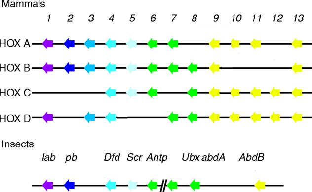
Sex combs reduced
The vertebrate Hox genes have been shown to confer regional identity along the anteroposterior axis
of the developing embryo, especially within the central nervous system (CNS) and the paraxial
mesoderm. The notochord has been shown to play vital roles in patterning adjacent tissues along both
the dorsoventral and mediolateral axes. However, the notochord's role in imparting anteroposterior
information to adjacent structures is less well understood, especially since the notochord shows no
morphological distinctions along the anteroposterior axis and is not generally described as a segmental
or compartmentalized structure. Four zebrafish hox genes (hoxb1, hoxb5, hoxc6
and hoxc8) are regionally expressed along the anteroposterior extent of the developing notochord.
Notochord expression for each gene is transient, but maintains a definite, gene-specific anterior limit
throughout its duration. The hox gene expression in the zebrafish notochord is spatially colinear with
those genes lying most 3' in the hox clusters having the most anterior limits. The expression patterns of
these hox cluster genes in the zebrafish are the most direct molecular evidence for a system of
anteroposterior regionalization of the notochord in any vertebrate studied to date (Prince, 1998).
To test whether the mouse Hox-1.3 gene is a cognate of the Drosophila Sex combs reduced
gene, a hsp 70-Hox-1.3 fusion gene hasbeen inserted into the Drosophila genome. Transgenic flies
display Scr-like homeotic transformations after ectopic expression of Hox-1.3 induced by heat
shock. In larvae, the thoracic segments T2 and T3 are transformed toward T1. In adults, head
structures are dramatically disrupted, including transformation of antenna towards leg.
Transformations are not the result of ectopic activation of the endogenous Scr gene. Rather,
Hox-1.3 appears to directly regulate Scr target genes, as demonstrated by the ectopic activation of
forkhead by Hox-1.3. The results suggest that mouse Hox-1.3 cannot only substitute functionally
for Drosophila Scr in the determination of external structures but also can participate in the
regulatory hierarchy of insect organogenesis (Zhao, 1993).
Two homeotic cluster (HOM-C) genes of C. elegans have antagonistic roles in the ability of vulval precursor cells (VPCs) to respond to the inductive signal from the anchor cell of the somatic gonad. The vulva develops from a subset of ectodermal vulval precursor cells distributed along the anteroposterior axis. Vulval patterning depends on both a localized inductive signal by the epidermal growth factor homolog LIN-3 (see Drosophila Vein for more information) and lateral signaling between inducing VPCs. One HOM-C gene, the Antennapedia homolog mab-5, is expressed in the posterior two VPCs. By examining the response of single VPCs to controlled doses of inductive signal in wild-type and in mab-5 mutant animals, it can be seen that mab-5 mutation increases the responsiveness of these two cells. Moreover, a gain-of-function allele of mab-5 that causes ectopic expression of MAB-5 in all VPCs reduces the sensitivity of all VPCs to inductive signal. Additional experiments suggest that another HOM-C gene, the Sex combs reduced homolog lin-39, is required for VPCs in wild-type animals to respond to activation of inductive signal. Genetic epistasis tests are consistent with models in which lin-39 acts downstream of the RAS pathway to regulate response to inductive signal. It is proposed that the spatial pattern of HOM-C gene expression may enhance the precision of vulval fate patterning (Clandinin, 1997).
The fate of ventral epidermal cells differs among nematode species. Nonvulval cells fuse with the
epidermis in Caenorhabditis elegans, whereas the homologous cells undergo apoptosis in Pristionchus
pacificus. The homeotic gene lin-39 is involved in the regulation of these epidermal cell fates. In
Caenorhabditis, lin-39 prevents cell fusion of potential vulval cells and specifies the vulva equivalence
group. Pristionchus vulvaless mutants that display apoptosis of the vulval precursor cells were
isolated, and point mutations in lin-39 were identified. Thus, the evolution of these epidermal cell fates
is driven by different intrinsic properties of homologous cells (Eizinger, 1997).
The normal Pbx1 homeodomain protein (Homolog of Drosophila Extradenticle), as well as its oncogenic
derivative, E2A-Pbx1, binds the DNA sequence ATCAATCAA cooperatively with the murine Hox-A5 (homolog of Drosophila Scr) and
Hox-B7, Hox-B8, and Hox-C8 (Antennapedia class) homeodomain proteins, which are themselves known oncoproteins, as well
as with the Hox-D4 (homolog of Deformed) homeodomain protein. Cooperative binding to ATCAATCAA required the
homeodomain-dependent DNA-binding activities of both Pbx1 and the Hox partner. In cotransfection
assays, Hox-B8 suppresses transactivation by E2A-Pbx1. These results suggest that (1) Pbx1 may
participate in the normal regulation of Hox target gene transcription in vivo and thereby contribute to
aspects of anterior-posterior patterning and structural development in vertebrates; (2) that E2A-Pbx1
could abrogate normal differentiation by altering the transcriptional regulation of Hox target genes in
conjunction with Hox proteins, and (3) that the oncogenic mechanism of certain Hox proteins may
require their physical interaction with Pbx1 as a cooperating, DNA-binding partner (Lu, 1995).
To uncover roles for the Hoxa-5 gene during embryogenesis, a focus has been placed on structural
and functional defects that can be identified in organ systems underlying the perinatal lethality in Hoxa-5 homozygous
mutants. Analysis of the mutant phenotype shows that Hoxa-5 is essential for normal organogenesis
and function of the respiratory tract. Larynx and tracheae are greatly disorganized in all homozygous specimens. The cricoid cartilage is always larger than the thyroid cartilage due to fusion with the first tracheal rings. The tracheal rings do not display a normal banding pattern and there tends to be a reduction in the number of rings formed. There is also a striking reduction in the diameter of the tracheal lumen. In homozygous newborn mutants, improper tracheal and lung
morphogenesis can lead to tracheal occlusion, and to respiratory distress associated with a marked
decrease in the production of surfactant proteins. Collectively, these defects likely underlie the
pronounced mortality of homozygous mutant pups. The loss of Hoxa-5 function results in
altered TTF-1, HNF-3 beta, and N-myc gene expression in the pulmonary epithelium. Since expression
of Hoxa-5 is confined to the mesenchymal component of the developing trachea and lung, the effects
observed in epithelial cells may result from a disruption of normal epithelial-mesenchymal interactions (Aubin, 1997).
The anteroposterior (A-P) patterning of the developing heart underlies atrial and ventricular lineage specification and heart
chamber morphogenesis. The posteriorization of cardiomyogenic phenotype with retinoic acid (RA) treatment of primitive
streak stage chicken embryos is suggestive of a role for the clustered homeobox (Hox) genes in early heart patterning. A screen for Hox genes expressed in chick heart
primordia and primitive heart led to the isolation of anterior genes of the Hox clusters expressed during cardiogenesis. Specific
hoxd-3, hoxa-4, and hoxd-4 transcripts are detected at the early stages of heart formation and full-length cDNA clones have been
isolated. Expression of hoxd-3 is detected in the heart forming region of embryos prior to heart tube formation. Expression
of hoxa-4, hoxd-3, and hoxb-5 is increased in cardiogenic tissue treated with RA in culture conditions that also produced
changes in positionally restricted cardiomyogenic phenotypes. Hox genes expressed in cardiac explants exhibit distinct
sensitivities to RA and ouabain treatment, when compared to genes, such as nkx-2.5, that are involved in cardiac commitment
and differentiation. These studies support a role for Hox genes in early heart patterning and suggest that positional information
in the cardiogenic region is established by regulatory mechanisms distinct from early heart lineage specification (Searcy, 1998).
The Hox genes cooperate in providing positional information needed for spatial and temporal patterning
of the vertebrate body axis. However, the biological mechanisms behind spatial Hox expression are
largely unknown. In transgenic mice, gene fusions between Hoxa5 (previously called Hox-1.3) 5'
flanking regions and the lacZ reporter gene show tissue- and time-specific expression in the brachial
spinal cord in day 11-13 embryos. A 604-bp regulatory region with enhancer properties directs this
spatially specific expression. Fine-detail mapping of the enhancer has identified several elements
involved in region-specific expression, including an element required for expression in the brachial
spinal cord. Factors in embryonic day 12.5 nuclear extracts bind this element in electrophoretic mobility
shift assays (EMSA) and protect three regions from DNase digestion. All three sites contain an
AAATAA sequence and mutations at these sites reduce or abolish binding. Furthermore, this element
binds specific individual embryonic proteins on a protein blot. The binding activity appears as a gradient
along the anterior-posterior axis with two- to three-fold higher levels observed in extracts from anterior
regions than from posterior regions. In parallel with the EMSA, the proteins on the protein blot also
show reduced binding to probes with mutations at the AAATAA sites. Most importantly, transgenic
mice carrying Hoxa5/lacZ fusions with the three AAATAA sites mutated either do not express the
transgene or have altered transgene expression. The brachial spinal cord element and its binding
proteins are likely to be involved in spatial expression of Hoxa5 during development (Nowling, 1999).
A short sequence element (L7ATE) within the proximal
promoter of a Purkinje cell-specific gene, pcp-2(L7), is required for the normal pattern of expression of
this gene in the cerebellum of transgenic mice. The presence of a series of TAAT sequence motifs in
this element suggests its interaction with homeodomain proteins. To extend these observations,
degenerate oligonucleotides were used to clone by reverse-transcriptase polymerase chain reaction
members of the mouse Hox gene family expressed in neonatal cerebellum but not forebrain. Two of
these, HoxB7 and HoxA5, are continuously expressed from the neonatal period into adult stages in
cerebellar Purkinje cells. These Hox proteins are shown to synergistically activate the L7 promoter by
cotransfection assay in vitro. In contrast, another homeodomain protein that is normally expressed in
Purkinje cells only during the embryonic period, En-2, has a negative effect on L7 gene expression.
These data suggest a biphasic, combinatorial control mechanism for the Purkinje cell-specific
expression of the pcp-2(L7) gene (Sanliouglu, 1998).
In Drosophila and mouse, Polycomb group genes are involved in the maintenance of homeotic gene
expression patterns throughout development. Skeletal phenotypes are found in mouse compound mutants
for two Polycomb group genes bmi1 (Drosophila homolog: Posterior sexcombs) and M33 (Drosophila homolog: Polycomb). Mice deficient for both bmi1 and M33 present
stronger homeotic transformations of the axial skeleton as compared to each single Polycomb group mutant,
indicating strong dosage interactions between those two genes. These skeletal transformations are
accompanied with an enhanced shift of the anterior limit of expression of several Hox genes in the somitic
mesoderm. These results demonstrate that in mice the Polycomb group genes act in synergy to control the
nested expression pattern of some Hox genes in somitic mesodermal tissues during development (Bel, 1998).
When Pc-G mutant mice are compared, loss of each Pc-G
gene shows a unique subset of affected Hox genes. For example, in M33 mutant mice, only anterior shifts for Hoxa3 can be detected and, in some cases, for Hoxc8. mel18 -/-, bmi1 -/- and rae28 -/- mice
present a more extensive overlap in affected Hox genes,
encompassing one prevertebrae anterior shifts of Hoxa5 and Hoxc8.
However, Hoxc6 and Hoxc5 are uniquely affected in bmi1 -/-
mice, while Hoxa7 and Hoxd4 are only affected in mel18 -/-
mice and Hoxb5 is unaffected in all those mutant mice. In M33 bmi1 double mutant mice, the
anterior limit of expression of at least two Hox genes, Hoxc9
and Hoxc8, is significantly more severely shifted as compared
to both single mutants, demonstrating the additive effect of
Pc-G products in maintaining the boundaries of selected Hox
genes. One striking observation is that deleting three gene doses
of these Pc-G genes does not increase the derepressive effect
of Hoxc8 or Hoxc9 expression; full deficiency of M33 and
bmi1 is required to induce extensive ectopic expression of
these two genes in mesodermal tissues. Nevertheless,
according to the differential expression of Hoxc8 and Hoxc9
in both single mutants, it seems that Hoxc8 is more sensitive
to M33 regulation since a one-segment anterior
shift is observed in M33 -/- bmi1 +/- (prevertebrae 10), whereas that shift is not visible
in bmi1 -/- M33 +/- (prevertebrae 11); reciprocally, Hoxc9 is more
sensitive to bmi1 regulation.
In contrast, some other Hox genes like
Hoxb1, Hoxd4 and Hoxd11 are not affected, either in single
or in double mutant mice. This suggests that several murine
multimeric Pc-G protein complexes of different composition
might exist that differ in their affinities for specific Hox
genes. Alternatively, since in mammals Pc-G genes exist as
highly related gene pairs (such as mel18/bmi1, Enx1/Enx2,
M33/MPc2, hPc1/hPc2 and Hph1/rae28/Hph2), a potential redundancy likely exists. This
suggests that the homologous gene complements part of the
function and thus maintains the boundaries of expression for
a subset of Hox genes. Analysis of double mutant mice for a
related pair such as mel18 and bmi1 should clarify the degree
of redundancy. Differential effects of Pc-G mutations on Hox
gene expression in different tissues like the somitic
mesoderm and the neural tube, also suggest that different Pc-G
complexes may regulate a specific subset of Hox genes in
a tissue-dependent manner (Bel, 1998).
Transgenic mice expressing the homeobox gene Hoxa5 under the control of Hoxb2 regulatory elements present a growth arrest during weeks two and three of postnatal development, resulting in proportionate
dwarfism. These mice present a liver phenotype illustrated by a 12-fold increase in liver insulin-like growth factor binding protein 1 (IGFBP1) mRNA and a 50% decrease in liver insulin-like growth factor 1 (IGF1) mRNA correlated with a 50% decrease in circulating IGF1. The Hoxa5 transgene is
expressed in the liver of these mice, leading to an overexpression of total (endogenous plus transgene) Hoxa5 mRNA in this tissue. Several cell lines were used to investigate a possible physiological interaction of Hoxa5 with the main regulator of IGFBP1 promoter activity, the Forkhead box transcription factor FKHR. In HepG2 cells, Hoxa5 has little effect by itself but inhibits the FKHR-dependent activation of the IGFBP1 promoter. In HuF cells, Hoxa5 cooperates with FKHR to dramatically enhance IGFBP1 promoter activity. This
context-dependent physiological interaction probably corresponds to the existence of a direct interaction between Hoxa5 and FKHR and
FoxA2/HNF3ß, as demonstrated by pull-down experiments achieved either in vitro or after cellular co-expression. In conclusion, it is
proposed that the impaired growth observed in this transgenic line relates to a liver phenotype best explained by a direct interaction between Hoxa5 and liver-specific Forkhead box transcription factors, in particular FKHR but also Foxa2/HNF3ß. Because Hoxa5 and homeogenes of the same paralog group are normally expressed in the liver, the present results raise the possibility that homeoproteins, in addition to their established role during early development, regulate systemic physiological functions. (Foucher, 2002).
The genetic control of gut regionalization relies on a hierarchy of molecular events in which the Hox gene family of transcription factors is suspected to be key participant. The role of Hox genes
in gut patterning has been examined using the Hoxa5-/- mice as a model. Hoxa5 is expressed in a dynamic fashion in the
mesenchymal component of the developing gut. Its loss of function results in gastric enzymatic anomalies
in Hoxa5-/- surviving mutants that are due to perturbed cell specification during stomach development. Histological, biochemical and molecular characterization of the mutant stomach phenotype may be compatible with a homeotic transformation of the gastric mucosa. As the loss of mesenchymal Hoxa5 function leads to gastric epithelial defects, Hoxa5 should exert its action by controlling molecules involved in mesenchymal-epithelial signaling. Indeed, in the absence of Hoxa5 function, there is alteration in the expression
of genes encoding for signaling molecules such as Sonic hedgehog (Shh), Indian hedgehog (Ihh), transforming growth factor ß family members and
fibroblast growth factor 10. These findings provide insight into the molecular controls of patterning events of the stomach,
supporting the notion that Hoxa5 acts in regionalization and specification of the stomach by setting up the proper domains of expression of signaling molecules (Aubin, 2002a).
It has been proposed that the original purpose of Hox genes was to pattern the gut, being co-opted afterwards to pattern other morphological structures such as the skeleton. One might therefore expect that Hox gene function in gut regional patterning will be highly conserved throughout evolution. In that regard, a parallel can be drawn between the anomalies encountered in the gut of Hoxa5–/– mice and those reported in Sex combs reduced Drosophila mutants. Scr is the Hoxa5 ortholog and its loss of function leads to the absence of the gastric cecae at the foregut-midgut boundary. scr is also expressed in the posterior part of the midgut, where it may play a role in the formation of the fourth midgut constriction. Both the gastric cecae and the fourth constriction correspond to functional frontiers separating the midgut from the rest of the digestive tract in Drosophila. Analogously, in the Hoxa5–/– mutant, morphological anomalies are encountered in the regions delimiting the midgut: the stomach and the proximal colon. Although the possibility that the Hoxa5 mutation could interfere with the expression of 5' located Hox genes that could result in colonic anomalies cannot be excluded, the similarity between Hoxa5 and scr expression patterns and function during gut development agrees with a conserved role for this paralog group in the delimitation of functional midgut boundaries (Aubin, 2002a).
Hoxa5 action in the establishment of Shh and Ihh gradients necessitates mesenchymally expressed intermediate(s). Bmps have been shown to be important regulators of glandular stomach development. Moreover in several species, a network exists between Hox, Bmp and Hh gut gene expression. For instance, ectopic Shh is able to induce Bmp4 expression in the chick hindgut and in the stomach. Although a complex situation prevails regarding the capacity of Shh to activate Bmp4 expression in foregut derivatives, it has been proposed that Hox genes influence the regionalized response to Shh. Even though the induction of Bmp4 by Shh in the stomach mesenchyme has not been directly addressed in the mouse, the change in the Bmp4 expression pattern observed in Hoxa5–/– stomachs is in agreement with this notion. It is also possible that Hoxa5 directly controls Bmp4 expression in the stomach. In the Drosophila midgut, the Ultrabithorax gene regulates at the transcriptional level the expression of the Bmp4 homolog decapentaplegic (Aubin, 2002a and references therein).
Hox and Pax transcription factors are master regulators of skeletal and organ morphogenesis. Some skeletal malformations
encountered in Hoxa5 mutants are shared by the undulated (un) mice, which bear a point mutation in the Pax1 gene. To
investigate whether Hoxa5 and Pax1 act in common pathways during skeletal development, Hoxa5;un
compound mutants were analyzed. Genetic studies show that Hoxa5 and Pax1 cooperate in the vertebral patterning of the
cervicothoracic transition region and in acromion morphogenesis. The dynamics of expression of Hoxa5 and Pax1 in the
pectoral girdle region suggest that both genes function in a complementary fashion during acromion formation. Whereas Pax1 is required for the recruitment of acromion precursor cells, Hoxa5 may provide regional cues essential for the correct formation of the acromion by ensuring Pax1 expression at the proper time and position during morphogenesis of the pectoral girdle. Hoxa5 also has a distinctive role in specifying the fate of perichondrial and chondrogenic cell lineages in a
Sox9-dependent way (Aubin, 2002b).
The regulatory interactions are described that cause anterior extension of the mouse 5' Hoxb expression domains from spinal cord levels to their definitive boundaries in the posterior hindbrain between embryonic day E10 and E11.5. This anterior expansion is retinoid dependent since it does not occur in mouse embryos deficient for the retinoic acid-synthesizing enzyme retinaldehyde dehydrogenase 2. A retinoic acid response element (RARE) was identified downstream of Hoxb5 and shown to be essential for expression of Hoxb5 and Hoxb8 reporter transgenes in the anterior neural tube. The spatio-temporal activity of this element overlaps with rostral extension of the expression domain of endogenous Hoxb5, Hoxb6 and Hoxb8 into the posterior hindbrain. The RARE and surrounding sequences are found at homologous positions in the human, mouse and zebrafish genome, which supports an evolutionarily conserved regulatory function (Oosterveen, 2003).
Best known as epigenetic repressors of developmental Hox gene transcription, Polycomb complexes alter chromatin structure by means of post-translational modification of histone tails. Depending on the cellular context, Polycomb complexes of diverse composition and function exhibit cooperative interaction or hierarchical interdependency at target loci. The present study interrogated the genetic, biochemical and molecular interaction of BMI1 [Drosophila homologs Psc and Su(z)2] and EED (Drosophila homolog; Esc), pivotal constituents of heterologous Polycomb complexes, in the regulation of vertebral identity during mouse development. Despite a significant overlap in dosage-sensitive homeotic phenotypes and co-repression of a similar set of Hox genes, genetic analysis implicated eed and Bmi1 in parallel pathways, which converge at the level of Hox gene regulation. Whereas EED and BMI1 formed separate biochemical entities with EzH2 and Ring1B, respectively, in mid-gestation embryos, YY1 engaged in both Polycomb complexes. Strikingly, methylated lysine 27 of histone H3 (H3-K27), a mediator of Polycomb complex recruitment to target genes, stably associated with the EED complex during the maintenance phase of Hox gene repression. Juxtaposed EED and BMI1 complexes, along with YY1 and methylated H3-K27, were detected in upstream regulatory regions of Hoxc8 and Hoxa5. The combined data suggest a model wherein epigenetic and genetic elements cooperatively recruit and retain juxtaposed Polycomb complexes in mammalian Hox gene clusters toward co-regulation of vertebral identity (Kim, 2006).
At least two PcG complexes with diverse composition and function in chromatin remodeling have been identified in mammals. The Polycomb repressive complex 1 (PRC1) involves the paralogous PcG proteins BMI1/MEL18, M33/PC2, RAE28, and RING1A. Evidence for PRC1-mediated chromatin modification derived from ubiquitylation at lysine 119 of histone H2A (H2A-K119). A second PcG complex, PRC2, encompasses EED, the histone methyltransferase EZH2, the zinc finger protein SUZ12, the histone-binding proteins RBAP46/RBAP48, and the histone deacetylase HDAC1. Several EED isoforms, generated by alternate translation start site usage of eed mRNA, differentially engage in PRC2-related complexes (PRC2/3/4), targeting the histone methyltransferase activity of EZH2 to H3-K27 or H1-K26. PcG complexes bind to cis-acting Polycomb response elements (PREs), which encompass several hundred base pairs and are necessary and sufficient for PcG-mediated repression of target genes. Whereas the function of several PREs has been delineated in Drosophila, similar elements await characterization in mammals (Kim, 2006 and references therein).
An antibody raised against residues 123-140 of the EED amino terminus
precipitated three distinct isoforms of approximately 50 and 75 kDA from E12.5
trunk, representing three of the four EED isoforms previously reported in 293 cells. In
addition to EZH2 and YY1, dimethylated H3-K27 co-immunoprecipitated with EED. Immunoprecipitation identified three BMI1 isoforms of approximately 39-41 kDA. BMI1 was found in a complex with RING1B, but not dimethylated H3-K27. Similar to the EED complex,
the BMI1 complex also contained YY1. It should be emphasized that all (co-)immunoprecipitating bands were detected by at least two antibodies against different epitopes. Strikingly, while dimethylated H3-K27 engaged in the EED complex,
trimethylated H3-K27 did not appear to associate with either the EED or the
BMI1 complex. Importantly, reciprocal co-immunoprecipitation detected EED and
BMI1 in separate protein complexes (Kim, 2006).
Ectopic expression in mutant embryos revealed Hoxc8 and
Hoxa5 as downstream targets of EED and BMI1 function. ChIP detected EED and
BMI1 binding immediately upstream of the Hoxc8 transcribed region
near putative promoter elements. The binding sites could not be separated, indicating close proximity of the complexes. EED and BMI1 binding also
clustered within a small fragment 1.5 kb upstream of the Hoxc8
transcription start site, suggesting long-range juxtaposition of heterologous PcG
complexes. Similar to EED and BMI1, YY1 localized to both regions. In support
of YY1 binding to Hox regulatory regions, inspection of the mouse genome
sequence revealed clusters of putative YY1 binding sites in
both regions a and b, including TGTCCATTAG and
CCCCCATTCC (region a), as well as ACACCATGGC,
TTTCCATTAG and TCCCCATAAA (region b). CCAT represents
the core of the YY1 consensus binding site, while flanking sequences exhibited
significant tolerance for multiple nucleotides. EED,
BMI1 and YY1 also co-localized approximately 1.5 kb upstream of the
transcription start site of Hoxa5. In addition to PcG binding, ChIP detected trimethylated H3-K27 throughout the regulatory regions of Hoxc8 and Hoxa5. Furthermore, dimethylated H3-K27 localized to region b of Hoxc8 (Kim, 2006).
Spatial regulation of EED and BMI1 binding to Hox regulatory regions was
evident from ChIP analysis of dissected anterior and posterior regions of
E12.5 trunk. In agreement with transcriptional silencing of Hoxc8 and
Hoxa5, EED and BMI1 binding was detected upstream of these loci in
anterior regions of the trunk. By contrast, EED and BMI1 binding was absent from posterior regions of the trunk, where Hoxc8 and Hoxa5 are transcribed.
These findings implicate PcG complexes in Hox gene repression in anterior
regions of the AP axis (Kim, 2006).
The combined interpretation of the co-immunoprecipitation and ChiP results
indicates that trimethylated H3-K27 did not form a complex with EED or BMI1,
despite co-localization of the three proteins in Hox regulatory regions. By
contrast, co-immunoprecipitation demonstrated physical association of the EED
complex with dimethylated H3-K27. In aggregate, the results support a model in
which EED- and BMI1-containing chromatin remodeling complexes exist as
separate, but juxtaposed, biochemical entities at Hox target loci (Kim, 2006).
How adjacent organ fields communicate during development is not understood. This study identified a mechanism in which signaling within the forelimb field restricts the potential of the neighboring heart field. In zebrafish embryos deficient in retinoic acid (RA) signaling, the pectoral fins (forelimbs) are lost while both chambers of the heart are enlarged. Evidence is provided that both of these phenotypes are due to RA signaling acting directly within the forelimb field. hoxb5b, an RA-responsive gene expressed within the forelimb field, is required to restrict the number of atrial cells arising from the adjacent heart field, although its function is dispensable for forelimb formation. Together, these data indicate nonautonomous influences downstream of RA signaling that act to limit individual chamber size. Therefore, these results offer new perspectives on the mechanisms regulating organ size and the possible causes of congenital syndromes affecting both the heart and forelimb (Waxman, 2008).
Retinoic acid (RA) signaling plays an important role in determining the anterior boundary of Hox gene expression in the neural tube during embryogenesis. In particular, RA signaling is implicated in a rostral expansion of the neural expression domain of 5' Hoxb genes (Hoxb9-Hoxb5) in mice. However, underlying mechanisms for this gene regulation have remained elusive due to the lack of RA responsive element (RARE) in the 5' half of the HoxB cluster. To identify cis-regulatory elements required for the rostral expansion, a recombineering technology was developed to serially label multiple genes with different reporters in a single bacterial artificial chromosome (BAC) vector containing the mouse HoxB cluster. This allowed simultaneous monitoring of the expression of multiple genes. In contrast to plasmid-based reporters, transgenic BAC reporters faithfully recapitulated endogenous gene expression patterns of the Hoxb genes including the rostral expansion. Combined inactivation of two RAREs, DE-RARE and ENE-RARE, in the BAC completely abolished the rostral expansion of the 5' Hoxb genes. Knock-out of endogenous DE-RARE lead to significantly reduced expression of multiple Hoxb genes and attenuated Hox gene response to exogenous RA treatment in utero. Regulatory potential of DE-RARE was further demonstrated by its ability to anteriorize 5' Hoxa gene expression in the neural tube when inserted into a HoxA BAC reporter. These data demonstrate that multiple RAREs cooperate to remotely regulate 5' Hoxb genes during CNS development, providing a new insight into the mechanisms for gene regulation within the Hox clusters (Ahn, 2014).
The duplication-degeneration-complementation (DDC) model predicts that subfunctionalization of duplicated genes is a common mechanism for their preservation. The additional Hox complexes of teleost fish constitute a good system in which to test this hypothesis. Zebrafish have two hoxb complexes, with two hoxb5 genes, hoxb5a and hoxb5b, the expression patterns of which suggest subfunctionalization of an ancestral hoxb5 gene. Conserved non-coding elements (CNEs) were characterized near the zebrafish hoxb5 genes. One CNE, J3, is retained only in the hoxb5a locus, whereas the others, J1 and J2, are present in both hoxb5 loci. When tested individually, the enhancer activity of individual CNEs, including J3, extensively overlapped and did not support a role in subfunctionalization. By contrast, reporter transgene constructs encompassing multiple CNEs were able to target reporter gene expression to unique domains of hoxb5a and hoxb5b expression. The deletion of J3 from the hoxb5a locus resulted in expression that approached that of hoxb5b, whereas its insertion in the hoxb5b locus increased reporter expression and rendered it more similar to that of hoxb5a. These results highlight the importance of interactions between CNEs in the execution of complementary subfunctions of duplicated genes (Jarinova, 2008).
back to Sex combs reduced Evolutionary homologs part 1/2 |
Home page: The Interactive Fly © 1995, 1996 Thomas B. Brody, Ph.D.
The Interactive Fly resides on the
Sex combs reduced:
Biological Overview
| Regulation
| Targets of Activity, Homeotic Effects, Post-Transcriptional Regulation and Protein Interactions
| Developmental Biology
| Effects of Mutation
| References
Society for Developmental Biology's Web server.