

Deep Homology?
| |
Deep Homology? |
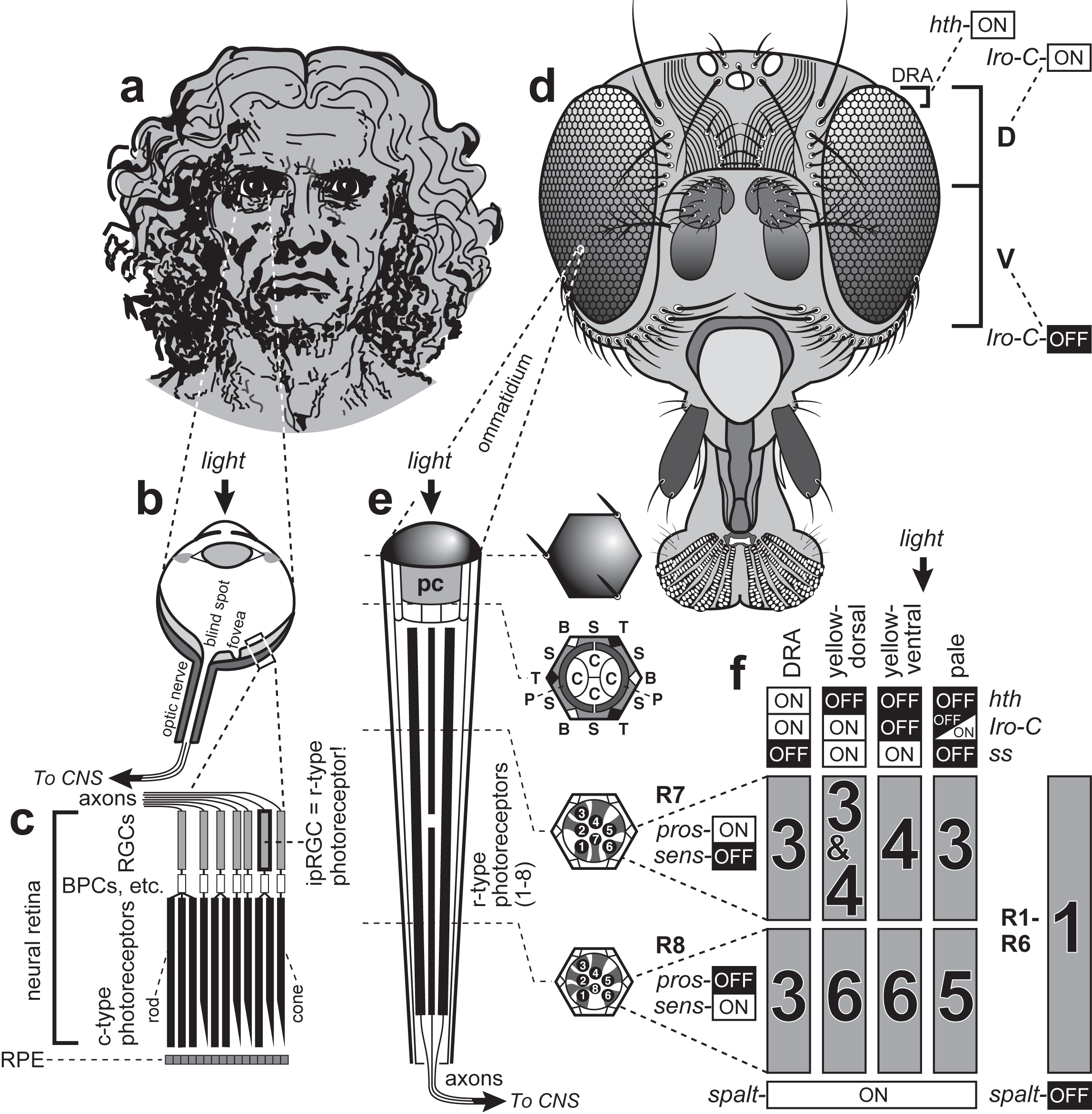 How eyes function in vertebrates (a-c) and flies (d-f). a. Vertebrates see the world through single-chamber "camera" eyes. The resolving power of our eyes is ~100 times better than a fly's, in part due to our huge size. b. Light rays are refracted by the cornea (outer hump) and lens (gray oval) onto PRCs in the neural retina (light gray layer). Photons that escape capture are absorbed by the retinal pigment epithelium (RPE; dark gray layer). Acuity is highest in the fovea but absent in the blind spot. c. Enlargement of boxed area in b. The optic nerve (at top) conveys axons from retinal ganglion cells (RGCs; front layer) to the central nervous system (CNS). About 1% of mammalian RGCs are intrinsically photosensitive (ipRGCs) insofar as they possess an r-type opsin and r-type transduction cascade. However, they lack stacked membranes and only work in bright light. After light traverses the RGCs, bipolar cells (BPCs), and other interneurons, it reaches the rod and cone (c-type) PRC layer. Each eye has ~7 million cones and ~120 million rods (≈ 20,000X more than the ~6000 PRCs per fly eye). Mammals use a combinatorial code (not shown) to assign identities to retinal cells, as do flies (f). d. Each compound eye has ~750 "ommatidial" facets. Flies also have three camera eyes (white ovals) atop the head that are too unfocused to form images. Also, two tiny remnants of larval eyes (not shown) regulate circadian rhythms. e. Each ommatidium is a tapered cylinder capped by a cuticular dome. After light rays enter the dome (≈ cornea) they pass through a pseudocone (pc) lens secreted by four "cone" cells (C in the cross section; no relation to human cone PRCs). Each ommatidium has eight r-type PRCs, six of which (R1-R6) span its height and form a trapezoid. The other two are shorter and are stacked one atop the other. Numbered circles in the lower two cross sections are rhabdomeres. A sleeve of primary (P), secondary (S) and tertiary (T) pigment cells encases R1-R8 and absorbs stray photons. At three of the six vertices a bristle (B) replaces a T-pigment cell. f. Flies use five opsins (Rh1, 3, 4, 5, and 6 in gray rectangles). R1-R6 cells use Rh1 ("Rh" stands for Rhodopsin), while R7 and R8 use Rh3, Rh4, Rh5, or Rh6 based on a binary "bar code" of multiple transcription factors. |
|
Introduction: cover image Body axes: figure 2 | figure 3 | figure 4 | figure 5 | figure 6 Nervous system: figure 7 | figure 8 Vision: figure 9 | figure 10 | figure 11 | figure 12 | figure 13 Touch and hearing: figure 14 | figure 15 Smell and taste: figure 16 Limbs: figure 17 Epilogue: figure 18 The Interactive Fly resides on the web server of the Society for Developmental Biology. |