

Deep Homology?
| |
Deep Homology? |
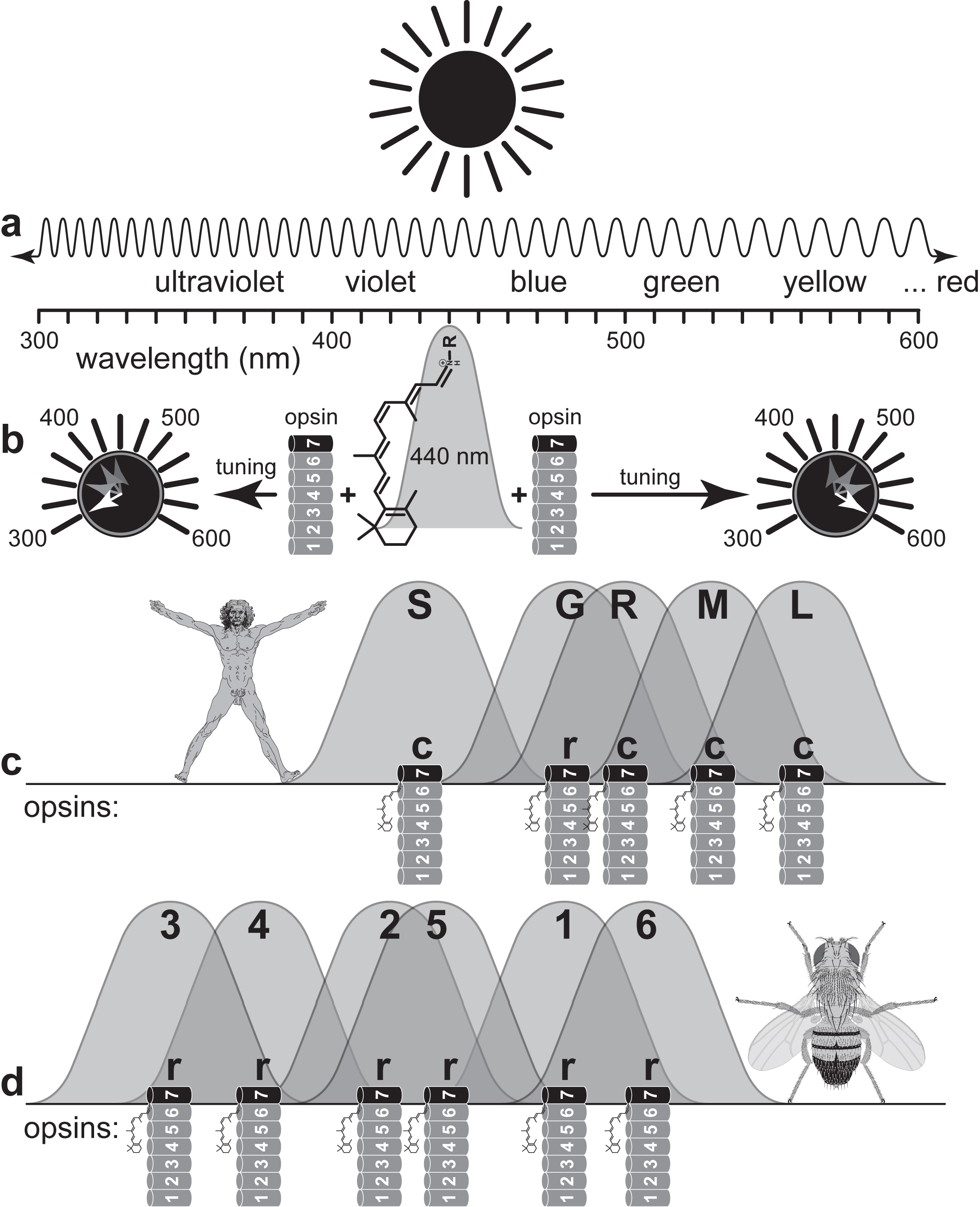 How opsin tunes 11-cis-retinal to different colors. a. Spectrum of ultraviolet and visible light. Across the scale are listed what we perceive as colors from violet to red, though red (~620-750 nm) is really beyond the right end, and not all labels are centered in their ranges. b. Peak wavelength (lmax = 440 nm) that is absorbed by "naked" 11-cis-retinal (the protonated Schiff's base unmoored from its opsin). When tucked into an opsin protein (helices drawn as cylinders), the 11-cis-retinal can be tuned to other wavelengths by charged or polar amino acid residues in its vicinity. Opsin thus acts like the knob of an AM/FM radio dial, allowing the species (≈ listener) to receive selected frequencies of light (≈ sound) relevant to its niche. c. Spectral sensitivities of our three (c-type) cone opsins, which absorb light at short (S), medium (M), and long (L) wavelengths, plus our (c-type) rod (R) opsin and the melanopsin pigment used by a subset of our (r-type) retinal ganglion (G) cells. Melanopsin works like the r-opsins in flies, but its role in mammals is to measure brightness rather than to help form images. N.B.: Absorbance curves are broader and less symmetric than depicted here (and in d), so humans can actually see light beyond 700 nm. Every opsin has a minor absorbance shoulder in the UV range (not shown). Most mammals lack UV-tuned PRCs, unlike honeybees and other insects, which use them to identify flowers. d. Spectral sensitivities of the six (r-type) fly opsins Rh1-Rh6 that are expressed in ocelli (Rh2) or in compound eyes (Rh1 and Rh3-6). The absence of opsins in the yellow-red range is due to screening pigments that give fly eyes their red color: flies use orange light (~570 nm) to convert all-trans retinal back into 11-cis retinal. Red-sensitive opsins are rare in nature, possibly because they are susceptible to thermal noise from infrared radiation. Mantis shrimps hold the "world record" for the most differently tuned PRCs (up to ~21) of any animal, which they achieve via pigments in their lenses. N.B.: The 11-cis-retinal chromophore is actually buried inside the protein, not on its surface as drawn (for clarity) here (and in c). |
|
Introduction: cover image Body axes: figure 2 | figure 3 | figure 4 | figure 5 | figure 6 Nervous system: figure 7 | figure 8 Vision: figure 9 | figure 10 | figure 11 | figure 12 | figure 13 Touch and hearing: figure 14 | figure 15 Smell and taste: figure 16 Limbs: figure 17 Epilogue: figure 18 The Interactive Fly resides on the web server of the Society for Developmental Biology. |