

Deep Homology?
| |
Deep Homology? |
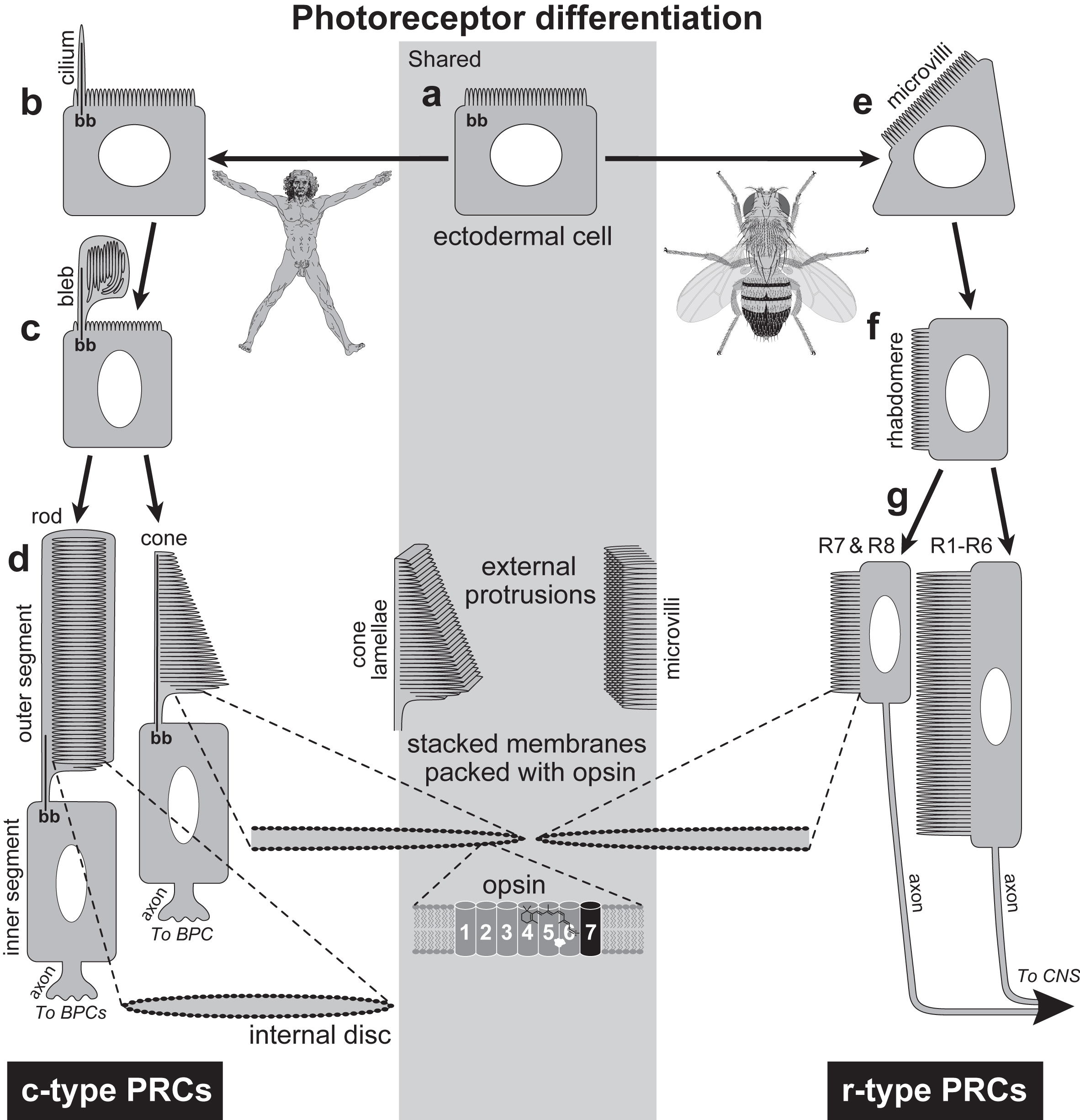 How photoreceptors differentiate. a. Structures in the shaded rectangle are common to humans and flies, whereas those outside are not. In both humans and flies, photoreceptor cells (PRCs) begin as ordinary ectodermal cells with a lawn of finger-shaped microvilli (actin-filled protrusions) on the apical surface. The oval is the nucleus, and "bb" denotes a basal body. Mature PRCs contain stacks of membranes packed with opsin at a density of ~4 X 10⁴ proteins per square micron. This combination of high surface area with high opsin density maximizes the chance that a photon will be absorbed as it passes through the cell. The photosensitive membranes of rod PRCs (flat vesicles) are internal, while those of cones (flat lamellae) and fly PRCs ("toothbrush" filopodia) are external. b-d. Development of ciliary (c-type) PRCs in mice and, by inference, humans. b. A cilium grows out from the basal body. c. The plasma membrane of the cilium inflates. This "outer segment" fills with flattened vesicles that are roughly parallel to the cilium. d. The outer segment elongates, vesicles reorient ~90° , and more are added. Rods and cones are named for the shapes of their outer segments. Vertebrate PRCs use an enigmatic synapse to innervate bipolar cell (BPC) interneurons. The ciliary axoneme spans the outer segment of cones but not rods. Photosensitive membranes of rods ("discs") have no connection with the plasma membrane, but those of cones ("lamellae") are contiguous with it. N.B.: PRCs are actually thinner than depicted, and they have more discs or lamellae. All views are cross sections except the central schematics. e-g. Development of rhabdomeric (r-type) PRCs in flies. e. The apical fringe of microvilli pivots by 90°, though not as neatly as shown here. f. Eventually, microvilli cover one flank to form the rhabdomere. g. The rhabdomeres of R1-R6 (monochromatic) PRCs elongate to ~80 mm with ~30,000 microvilli, while most R7 and R8 (color-detecting) PRCs reach only half this size. The shorter microvilli of R7 and R8 afford greater acuity but less sensitivity. R7 and R8 PRCs in the dorsal rim area are exceptions: they have larger rhabdomeres for sensing polarized UV light, which they use as a solar compass. Unlike our PRCs, fly PRCs send axons directly to the CNS, rather than via interneurons. One rhabdomeric lamella is enlarged to show how it is dotted with opsins, though it actually has a 10-fold lower density than vertebrate rods. |
|
Introduction: cover image Body axes: figure 2 | figure 3 | figure 4 | figure 5 | figure 6 Nervous system: figure 7 | figure 8 Vision: figure 9 | figure 10 | figure 11 | figure 12 | figure 13 Touch and hearing: figure 14 | figure 15 Smell and taste: figure 16 Limbs: figure 17 Epilogue: figure 18 The Interactive Fly resides on the web server of the Society for Developmental Biology. |