Select image to enlarge

Figure 4.5
Regenerative potency of wing-disc fragments and an early model.
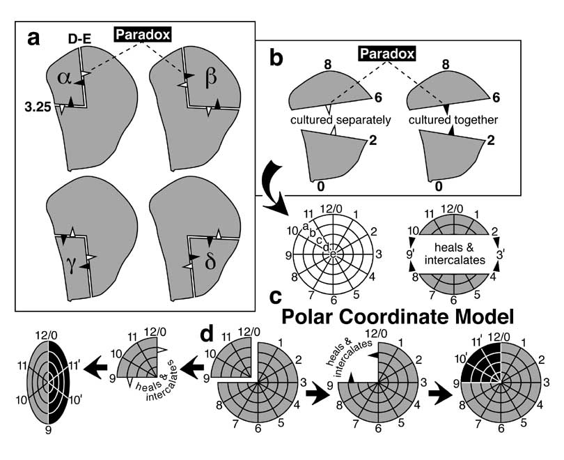
a. Fate map (abridged) of a mature right wing disc (notum, light shading; arc = suture; wing, dark shading), as per Bryant [524] except that dots are actual SOP sites [1925] and thick lines are pre-vein zones (I-V) [4189]. Vein I is the bristled part of the margin [1741] (thin dashed line = bristleless part). Thick dashed line is the A/P compartment boundary (cf. Fig. 4.4). Directions (A, anterior; P, posterior; D, dorsal; V, ventral) are given in the compass at right. In all bristle pairs except scutellars (SC; bounded area = scutellum), the P partner is on the right (cf. Fig. 3.4 for abbreviations). White trapezoid is a unique spot (cf. b).
b. Results of bisecting a disc lengthwise (B-F) or transversely (1-7) along folds (dashed lines) or elsewhere and letting each piece grow in isolation inside the body of a host adult [524]. As indicated in the key, an unfilled arrowhead means that the fragment to its rear duplicates (i.e., makes two copies of its fated structures as mirror images), while a solid arrowhead means that the fragment behind it regenerates (i.e., restores the whole). Note that every cut line has arrowheads of opposite type. Thus, the wing disc obeys a 'Reciprocity Rule': when one piece regenerates, the reciprocal fragment duplicates. The fact that regeneration proceeds away from D, E, 3, and 3.5 lines suggests that the disc uses x-y coordinates, but diagonal cuts (not drawn) showed that potency actually declines radially from the center piece (black trapezoid).
c. The Gradient of Developmental Capacity (GDC) Model was based on the findings shown in b [524]. It proposed that the center piece contains the peak of a conical gradient of regenerative potency. The gradient is schematized here as triangles (side view) above the columnar epithelium (inscribed with imaginary contour lines). When the disc is cut, the gradient is supposed to force cells at each edge to grow down its slope.
d. According to the GDC Model, any piece that includes the peak should regenerate, but the 'CF24' piece actually duplicates (as does BF16; not shown) [526]. This and other paradoxes (cf. Fig. 4.6) led to the GDC model being abandoned in favor of the Polar Coordinate Model [1775].
|
|