| Developmental and Signaling Pathways: The hierarchical dual-oscillator model of the Drosophila's circadian clock neuron network |
| Reference: Yao, Z. and Shafer, O. T. (2014). The Drosophila circadian clock is a variably coupled network of multiple peptidergic units. Science 343(6178): 1516-1520. PubMed ID: 24675961 |
|
|
|
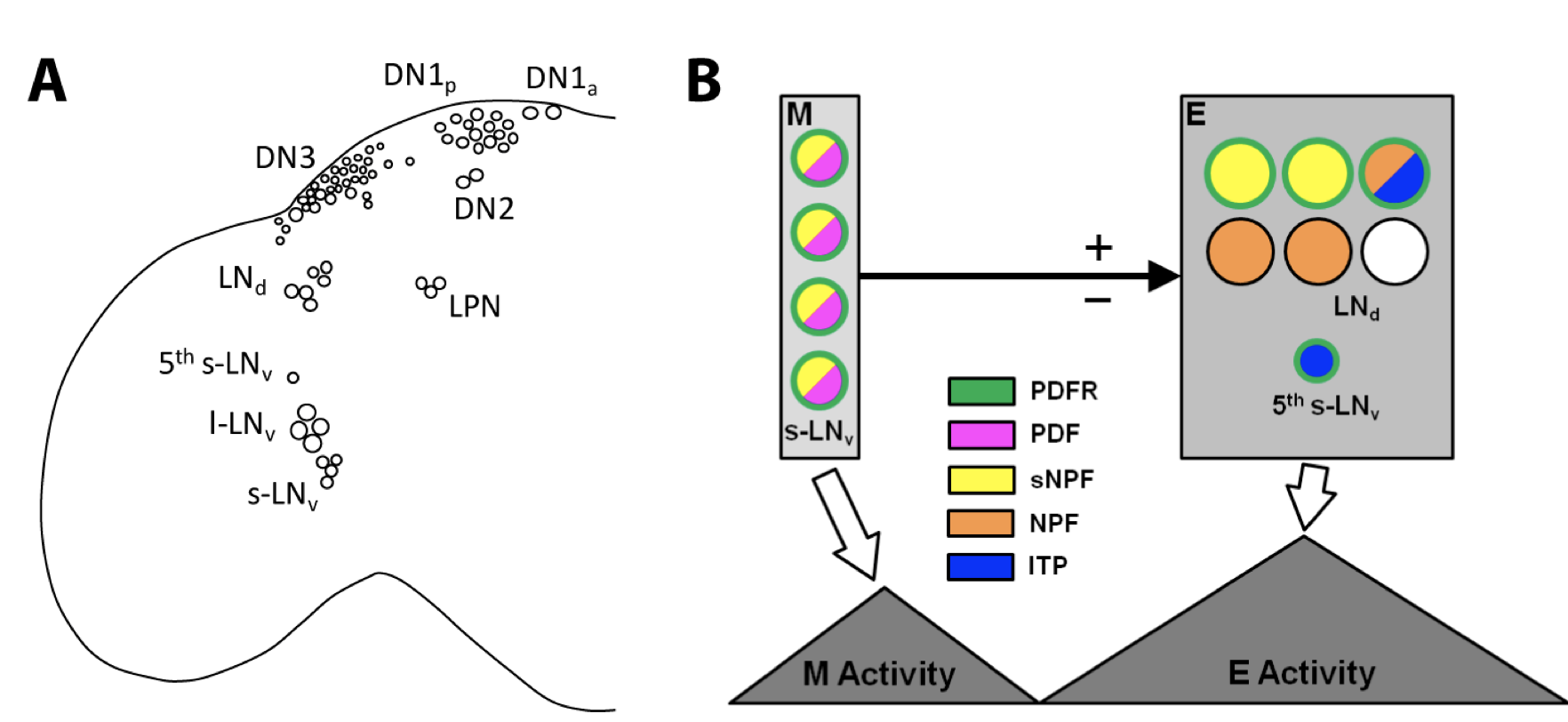
Legend: The hierarchical dual-oscillator model of the Drosophila's circadian clock neuron network (A) A somatic map of the clock neuron network of Drosophila. A single hemisphere (left) is shown and the various classes of clock neurons are labeled. The lateral neurons (LN) consist of large ventral lateral neurons (l-LNvs), small ventral lateral neurons (s-LNvs), a 5th small ventral lateral neuron (5th s-LNv), and dorsal lateral neurons (LNds). The dorsal neurons (DN) contain DN1as (anterior), DN1ps (posterior), DN2s, and DN3s. LPN: lateral posterior neurons. The l-LNvs and s-LNvs express pigment-dispersing factor (PDF); all other clock neurons are PDF negative. (B) The dual-oscillator model of the lateral clock neuron network and its control of activity rhythms. The LNds and 5th s- LNv (evening (E) oscillator) control evening activity. The s-LNvs (morning (M) oscillator) control morning activity and reset the evening oscillator through daily advances (+) or delays (-), thereby maintaining clock network synchrony under constant conditions. The neuropeptides expressed by the lateral clock neurons are shown. PDF, pigment-dispersing factor; sNPF, short neuropeptide F; NPF, neuropeptide F; ITP, ion transport peptide. Only subsets of the evening oscillator neurons express PDF receptor (PDFR). |
back to Graphic Pathways
Home page: The Interactive Fly © 1995, 1996 Thomas B. Brody, Ph.D.
The Interactive Fly resides on the
Society for Developmental Biology's Web server.