Select image to enlarge in new window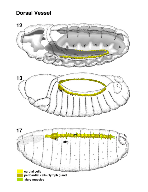 | Dorsal Vessel pages 46-47
From
stage 13 onward, the primordium of the dorsal vessel moves dorsally along
with the overlying ectoderm (Campos-Ortega and Hartenstein 1985; Hartenstein
and Jan 1992; see Bate, this volume). The two rows of cells express different
characteristics: Cells of the dorsal row adopt a cuboidal shape and become
perfectly aligned. These cells are the cardioblasts (car) that
form the dorsal vessel proper. The lateral cells keep their rounded shape and
remain more irregularly arranged. They give rise to the pericardial cells
(per), large cells that flank the dorsal vessel on either side.
Close to the anterior end of the developing dorsal vessel, a cluster of
mesodermally derived cells that are confluent with the pericardial cells
form the primordium of the lymph glands (lg).
|
Select image to enlarge in new window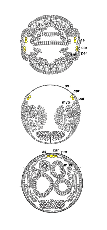 |