Select image to enlarge in new window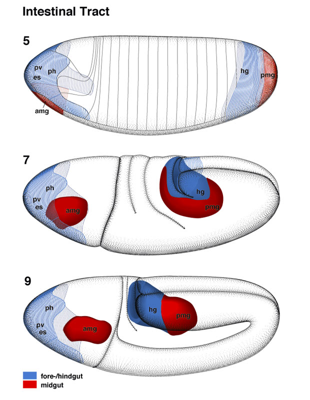 | Intestinal Tract pages 28-29 | 30-31 | 32-33 | 34 | 35
|
Select image to enlarge in new window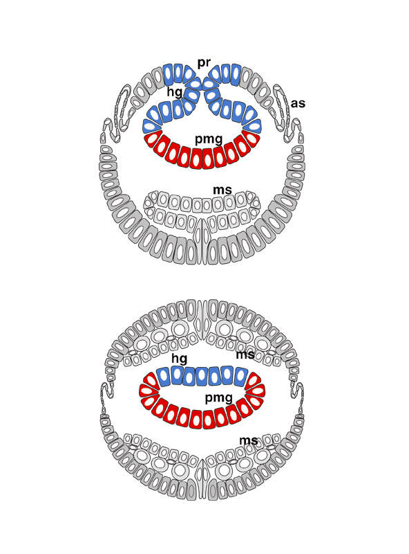 |
The early development of the primordia of the foregut and the hindgut
is also quite different. The foregut primordium does not start invaginating
before stage 10; after that, invagination is a slow process that ends only in
late embryogenesis [stages 15-17]. On the other hand, the hindgut primordium invaginates
right after the posterior midgut rudiment [stages 7-9]. In the stage 7 embryo
depicted, both the posterior midgut rudiment (pmg) and the
already invaginated part of the hindgut primordium (hg) form a pouch
called the amnioproctodeal invagination (or proctodeum, pr).
During stage 9, the remaining parts of the hindgut primordium enter into the embryo, leading to an increase in length of the amnioproctodeal invagination. At the same time, this structure is pushed anteriorly by the elongating germ band. Proliferation of the gut primordia takes place during stages 8-10. All cells of the primordium of the foregut and the midgut primordia divide three times in a parasynchronous way (Hartenstein and Campos-Ortega 1985). The hindgut primordium also performs three complete divisions; after that, some parts of it (e.g., what will become the Malpighian tubules) divide for a fourth time. (as) Amnioserosa; (ms) mesoderm. |