Atlas of Drosophila Development by Volker Hartenstein Table of Contents
Select image to enlarge in new window
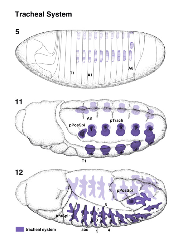
Tracheal System pages 18-19 | 20-21 | 22 | 23

The larval tracheal tree originates from a series of placodes that, projected on the blastoderm (stage 5), map to the dorsal ectoderm. Segments T2 through A8 each gives rise to one pair of tracheal placodes (Poulson 1950; Campos-Ortega and Hartenstein 1985; see Manning and Krasnow, this volume).

Tracheal placodes start invaginating during late stage 10. At stage 11, they form spherical vesicles that open to the outside right behind the developing segment furrows (tp). These external openings (abdominal spiracles, abs) of the tracheal pits obliterate around stage 14. At the position of the former openings, cords of cells remain that form permanent connections between the epidermis and the lateral segmental tracheae (Hartenstein and Jan 1992). Cells of these cords form the tracheal histoblasts that, during larval and pupal stages, produce much of the adult tracheae (see below). The posterior spiracle develops as follows: In addition to the tracheal pit in the anterior part of A8, the placode of the posterior spiracle (ps) appears in the dorsal-posterior part of A8. During late stage 11, both structures fuse to generate a single opening, the posterior spiracle.

Select image to enlarge in new window

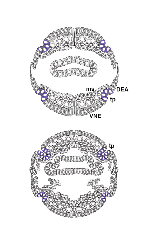
During stage 12, the invaginated tracheal pits elongate and form a dorsal and a ventral stem. The ventral stem bifurcates into a posterior branch (4) and an anterior branch (5), The dorsal stem gives (1) off two side branches: one toward the anterior (2) and the other toward the interior of the embryo (3), (A1, A8) Abdominal segment 1, 8; (as) anterior spiracle; (DEA) dorsal ectoderm; (ms) mesoderm; (T1) thoracic segment 1; (VNE) ventral neurogenic region.


Atlas of Drosophila Development

SDB home page

The Interactive Fly resides on the
Society for Developmental Biology's Web server.