Select image to enlarge in new window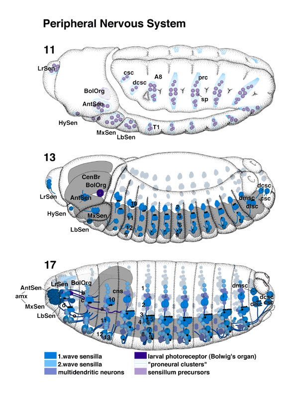 |
Select image to enlarge in new window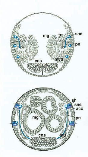 |
| PNS pages 12-13 | page 14 | page 15
The precursor cells of the larval sensilla can be first
identified in the ectoderm of a stage 11 embryo (Ghysen and O'Kane 1989;
Bodmer et al. 1989; see Jan and Jan, this volume). These cells develop from
specialized parts of the ectoderm ('proneural clusters'; Ghysen and
Dambly-Chaudière 1989). Within the proneural clusters (prc), cell-cell
interactions take place to select the sensillum precursors (sp) from
the epidermal precursors. These interactions seem to be similar to those
involved in determining the neuroblasts at an earlier stage. During stages 11
and 12, sensillum precursors perform two or three divisions to produce the
different cells that make up the sensilla (Bodmer et al. 1989). Typically, a
sensillum contains one bipolar sensory neuron (sne), three
accessory cells (acc) that form sheaths around the dendrite, and
a sheath cell associated with the axon (Hartenstein 1988). Some sensilla have
more than one neuron (e.g., basiconical sensilla, which have three neurons).
Two major classes of sensilla exist: the external sensilla (mechanoreceptors
and chemoreceptor) and chordotonal organs (stretch receptors). In the external
sensilla, the accessory cells remain in the epidermis and produce a specialized
cuticular apparatus involved in receiving the stimulus. In the chordotonal
organs, neurons and accessory cells come to lie subepidermally. A special type
of sensillum is the larval photoreceptor (Bolwig's organ, (bo), which consists of 12 modified bipolar sensory neurons and no accessory cells (Steller
et al. 1987; Green et al. 1993). Figure legend for enlarged view: (A8) Abdominal segment 8; (amx) antennomaxillary complex; (cns) central nervous system; (csc) caudal sensory cone; (dcsc) dorsal-caudal sensory cone; (dlsc) dorsal-lateral sensory cone; (dmsc) dorsal-medial sensory cone; (do) dorsal organ; (lis) labial sensory complex ('hypophysis'); (lrs) labral sensory complex ('epiphysis'); (mg) midgut; (mu) somatic musculature; (myo) myoblasts; (sh) sensory hairs; (T1) thoracic segment 1; (to) terminal organ; (tr) trachea; 1 dorsal campaniform sensilla (singly innervated); 2 dorsal trichoid sensillum (doubly innervated); 3 lateral trichoid sensilla (singly innervated); 4 lateral campaniform sensilla (singly innervated); 5 lateral pentascolopidial chordotonal organ; 6 lateral monoscolopidial chordotonal organ; 7 ventral campaniform sensillum 5 (doubly innervated); 8 ventral campaniform sensilla (singly innervated); 9 ventral chordotonal organ; 10 dorsal triscolopidial chordotonal organ; 11 lateral basiconical sensillum; 12 ventral basiconical sensillum; 13 Keilin's organ; (a) Bolwig's nerve; (b) labral nerve; (c) antennal nerve; (d) maxillary nerve; (e) labial nerve. |
|