Select image to enlarge
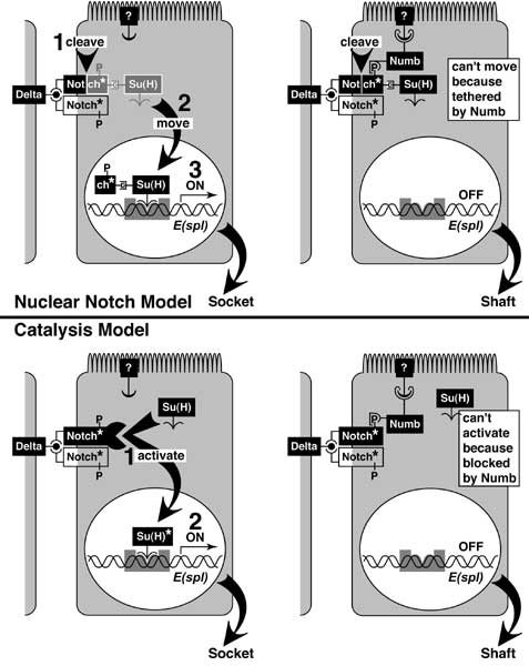
Figure 2.2
Models for Notch signaling and its blockage by Numb.
Black rectangles are proteins, and connecting 'wires' are binding sites. Contact with Delta ligand on a neighbor's surface activates (asterisk) the Notch receptor, possibly by dimerization (partner outlined) [3022]. Cells that lack Numb (left) can relay the signal to its nucleus, while those that express Numb (right) cannot.
The models differ in how Numb stops the signal. In the Nuclear Notch Model (above) [1307, 1448, 1651, 2299, 4027, 4244, 4542] Numb stops Notch from leaving its roost (ghost image) by anchoring it to the membrane [2267] via an unknown linker ('?' = possibly Partner of Numb). In the Catalysis Model (below) [112, 132, 1131, 3022, 4244], Numb blocks an active site for Su(H) activation (covalent modification?).
Numb is shown binding Notch at a phosphotyrosine (P), but Numb's PTB domain is unusual and may not need a phosphate [2530, 4789], and Notch is only known to have phosphoserines [2209]. Notch resides in the apicolateral membrane [184, 1203, 1448, 2070]. The cell's apex is carpeted with microvilli. Su(H) can activate transcription (right-angle arrow) of E(spl) (a.k.a. 'm8'; cf. Fig. 2.4) by binding its promoter (gray rectangle), but E(spl) may not dictate bristle cell fates, nor is Su(H) needed for signal relay in neurons or sheath cells (see text). Estimates are that a signal at the membrane takes ~20-90 mins. to cause detectable changes in target gene expression [184]. See also App. 7.
|
|