The Interactive Fly
Drosophila neuroblasts
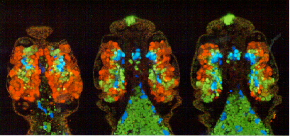
Drosophila cephalic lobe neuroblasts [image © 1998 Ward Odenwald]
 Drosophila cephalic lobe (brain) neuroblasts
and their offspring stained for three transcription factors, Hunchback, Pdm-1 and Castor. Three serial confocal immunofluorescent views from dorsal (left) to ventral (right) of embryos triple-labeled for Hb (green), Pdm-1, and (green) Cas (red). Hb is expressed in early sublineages, Cas is expressed in late sublineages and Pdm-1 is expressed in intermediate sublineages. Note the more lateral position of the Cas-positive cells relative to the more internal Pdm-1 and Hb sublineages. Few cells are found that express more than one of these proteins. The most anterior staining marks progenitors of the larval eye known as Bolwig's organ. Staining of the intestinal yolk cells is non-specific. In addition to its CNS expression, Hb is also found in a subset of cells lining the gut.
Drosophila cephalic lobe (brain) neuroblasts
and their offspring stained for three transcription factors, Hunchback, Pdm-1 and Castor. Three serial confocal immunofluorescent views from dorsal (left) to ventral (right) of embryos triple-labeled for Hb (green), Pdm-1, and (green) Cas (red). Hb is expressed in early sublineages, Cas is expressed in late sublineages and Pdm-1 is expressed in intermediate sublineages. Note the more lateral position of the Cas-positive cells relative to the more internal Pdm-1 and Hb sublineages. Few cells are found that express more than one of these proteins. The most anterior staining marks progenitors of the larval eye known as Bolwig's organ. Staining of the intestinal yolk cells is non-specific. In addition to its CNS expression, Hb is also found in a subset of cells lining the gut.
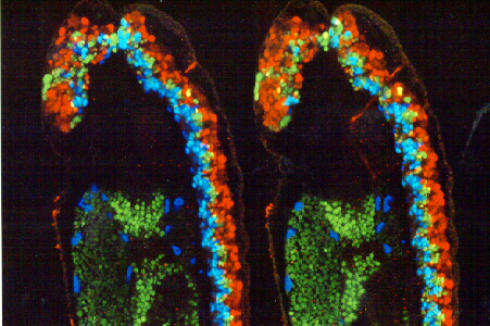
Lateral views of Drosophila CNS [image © 1998 Ward Odenwald]
 Lateral views of Drosophila CNS stained for Hunchback, Pdm-1 and Castor. Two serial confocal immunofluorescent views of embryos triple-labeled for Hb (green), Pdm-1, and (green) Cas (red). Hb is expressed in early sublineages, Cas is expressed in late sublineages and Pdm-1 is expressed in intermediate sublineages. Note the more ventral-ventral/lateral position of the Cas-positive cells relative to the more internal Pdm-1 and dorsally positioned Hb sublineages. Staining of the intestinal yolk cells (green) is non-specific. Hb is also expressed in a subset of cells lining the gut.
date revised: 15 June 98
Home page: The Interactive
Fly © 1995, 1996 Thomas B. Brody, Ph.D.
The Interactive Fly resides on the
Society for Developmental Biology's Web server.
 Drosophila cephalic lobe (brain) neuroblasts
and their offspring stained for three transcription factors, Hunchback, Pdm-1 and Castor. Three serial confocal immunofluorescent views from dorsal (left) to ventral (right) of embryos triple-labeled for Hb (green), Pdm-1, and (green) Cas (red). Hb is expressed in early sublineages, Cas is expressed in late sublineages and Pdm-1 is expressed in intermediate sublineages. Note the more lateral position of the Cas-positive cells relative to the more internal Pdm-1 and Hb sublineages. Few cells are found that express more than one of these proteins. The most anterior staining marks progenitors of the larval eye known as Bolwig's organ. Staining of the intestinal yolk cells is non-specific. In addition to its CNS expression, Hb is also found in a subset of cells lining the gut.
Drosophila cephalic lobe (brain) neuroblasts
and their offspring stained for three transcription factors, Hunchback, Pdm-1 and Castor. Three serial confocal immunofluorescent views from dorsal (left) to ventral (right) of embryos triple-labeled for Hb (green), Pdm-1, and (green) Cas (red). Hb is expressed in early sublineages, Cas is expressed in late sublineages and Pdm-1 is expressed in intermediate sublineages. Note the more lateral position of the Cas-positive cells relative to the more internal Pdm-1 and Hb sublineages. Few cells are found that express more than one of these proteins. The most anterior staining marks progenitors of the larval eye known as Bolwig's organ. Staining of the intestinal yolk cells is non-specific. In addition to its CNS expression, Hb is also found in a subset of cells lining the gut.
 Lateral views of Drosophila CNS stained for Hunchback, Pdm-1 and Castor. Two serial confocal immunofluorescent views of embryos triple-labeled for Hb (green), Pdm-1, and (green) Cas (red). Hb is expressed in early sublineages, Cas is expressed in late sublineages and Pdm-1 is expressed in intermediate sublineages. Note the more ventral-ventral/lateral position of the Cas-positive cells relative to the more internal Pdm-1 and dorsally positioned Hb sublineages. Staining of the intestinal yolk cells (green) is non-specific. Hb is also expressed in a subset of cells lining the gut.