Select image to enlarge
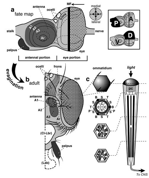
Figure 7.1
The eye disc, its adult derivatives, and the cellular components of an ommatidium.
a, b. Fate map (abridged) of an eye-antenna disc (a) and its cuticular derivatives (b). Abbreviations: A1-A3 (antennal segments 1-3), Ar (arista), Fr (frons; parallel grooves ≈ a human fingerprint), MF (morphogenetic furrow, black bar; arrow shows direction of movement), n (nerve = optic stalk), Oc (ocelli; oval = lateral ocellus; half-oval = median ocellus), Palp (palpus).
a. A left eye-antenna disc at maturity. In size and shape the eye part resembles a wing pouch, and the antenna part looks like a half-size leg disc. Dots are photoreceptor clusters. The disc's hind half is cup-shaped (concave-side down) with two flaps (dashed lines on underside). The antennal part (medium shading) telescopes toward the viewer during eversion. Directions (compass at right) are prospective (a) or actual (b) axes of the adult. The vectors are confusing because eye discs rotate ~180° after they arise -- inverting their A-P and D-V axes (inset box) [4146]. Thus, the P compartment (≈ en-ON domain) winds up anterior [2931], and the true D compartment winds up ventral (compare the fringe-ON domains in Figs. 6.8 and 7.4) and is conventionally called the 'V' compartment (as shown). This inversion explains (1) why the dpp-ON and wg-ON sectors of the antenna are upside-down relative to those of the leg (compare Figs. 7.3 and 5.8) [2631, 4277] and perhaps also (2) why disabling the Dpp pathway (via dppLOF or MadLOF) removes the D half of the leg but the V half of the eye [723, 930, 1812, 4648] (but see [2752]). (N.B.: The antennal D-V axis is mislabeled in [1037].) The A/P boundary is established by late 2nd instar (~72 h AEL) [2931, 2933], and the same is true for antennae transformed into legs [1635, 2926, 2933], while the D/V boundary arises in early 1st instar [189]. These times contrast with the blastodermal onset of A/P restrictions in thoracic discs [4651], but they resemble the timing for the D/V line in the wing disc [354]. See Fig. 6.9 for the origin of the eye vs. antenna dichotomy.
b. Left half of a head (frontal aspect). Unlike the compound eyes, ocelli do not focus images, but rather may detect moving shadows [293]. Blank areas show where structures made by other discs would insert: 'Cl-Lbr' (clypeus and labrum) from clypeolabral disc and 'Lab' (labellum) from labial disc. Interommatidial bristles are omitted. Reshaping during evagination is complicated (not shown) [1311, 1777, 2864, 2865, 3516].
c. One facet from the lattice is shown in frontal view (left). At right is a side view of the entire conical ommatidium -- a simple eye [2909, 3230]. Cross-sections are sketched (left) at 3 levels (dashed lines). Abbreviations: B (bristle), C (cone cell; no relation to 'cone' photoreceptors in vertebrates); P, S, T are primary, secondary, and tertiary pigment cells; 1-8 are photoreceptors R1-R8. Nuclei are omitted. In the upper section, 4 C cells are embraced by 2 P cells, which are bordered by 6 S cells, with 3 B and 3 T cells at alternating vertices. Adjacent ommatidia share S, B, and T cells (Fig. 7.2). Light is refracted by the cornea (dark shading) and underlying pseudocone (light shading) [1720, 4364]. The cornea is a cuticular secretion of the cone and primary pigment cells, while the pseudocone (pc) is a gel secreted by cone cells alone [602, 2034]. Photons are then transduced by the 8 rhabdomeres (black bars at right = numbered circles at left). Rhabdomeres are compact arrays of photosensitive microvilli [715, 844, 1817, 2323, 3568, 4458] on R1-R8 cells (shaded; intercellular spaces are exaggerated) [1270, 2594, 3350, 4270]. R7's rhabdomere lies atop R8's. Both are smaller than the other six, which nearly span the height of the ommatidium. Note (lower sections) that the cytoplasmic stalks (and hence rhabdomere gratings) of R7 and R8 are orthogonal [1318, 4355] -- a trick for detecting polarized light [1720, 4398]. The transduction cascade culminates in electrical signals [374, 2908, 2909, 3229] that are sent to the CNS via axons (below) [1390, 2801]. Stray photons that might ricochet to adjacent ommatidia (and hence degrade the image) are absorbed by the intervening pigment cells [1048]. These pigment cells thus serve as insulating walls [3369].
Panel a is redrawn from [1777] with details added for ocelli [3663] and photoreceptors (Fig. 1b of [724]), b is adapted from [1777] with territories ascribed to different cephalic discs as per [1408], and c is simplified from [602, 1048, 1239, 1726, 4715]. See [3540] for a 3-dimensional rendition.
N.B.: The arista's branches may arise from hairs (vs. bristles) [3362]. Unlike the 3rd antennal segment which senses odors [2221], the arista appears to sense temperature and humidity [1260].
|
|