

Deep Homology?
| |
Deep Homology? |
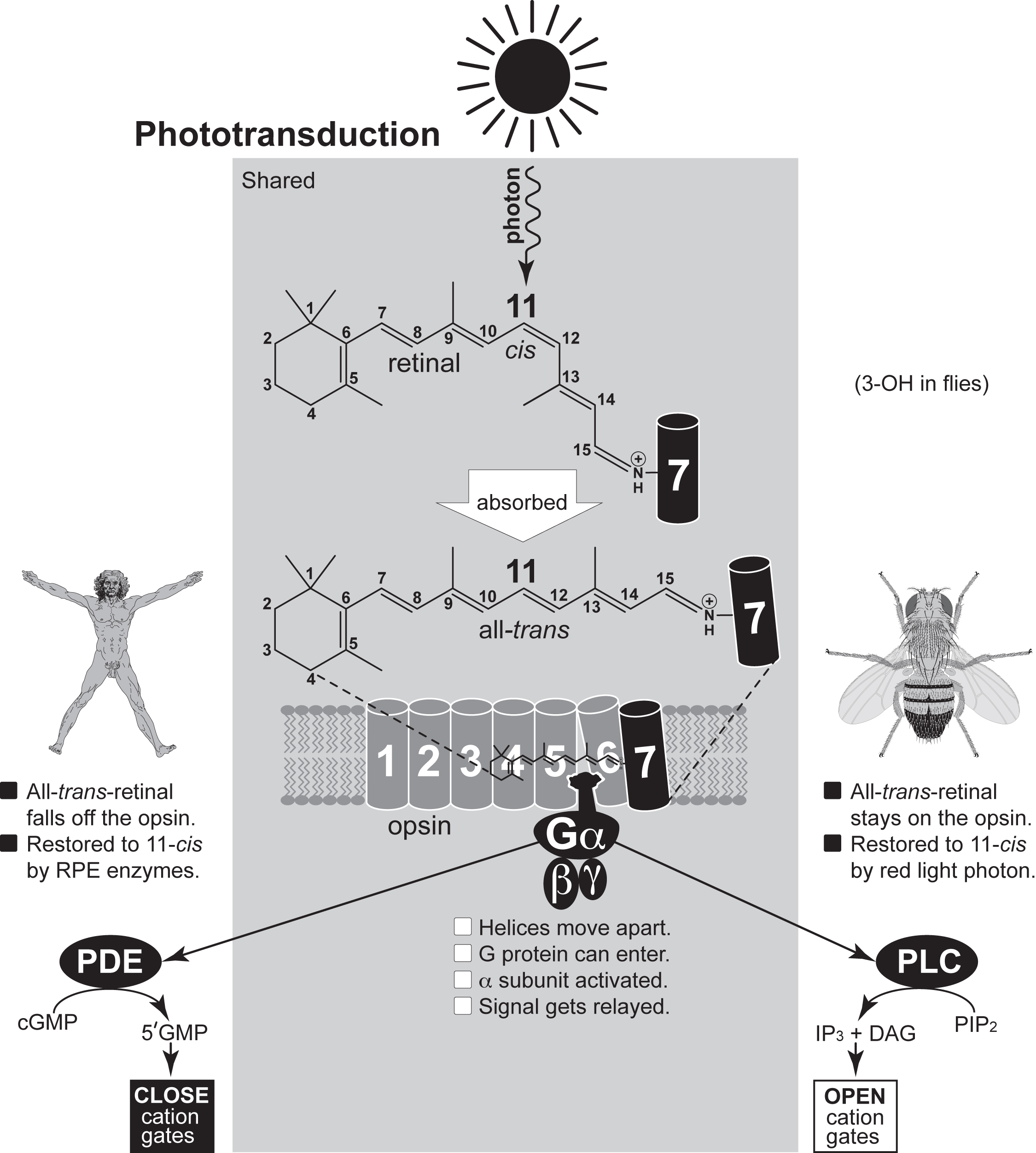 How photoreceptors transduce light into neuronal signals. Features depicted in the shaded rectangle are common to humans and flies, whereas those outside are not. The same chromophore is used by all animal eyes. That molecule is 11-cis retinal or a variant thereof. One minor variant, which is found in flies, has a hydroxyl group on its C-3 carbon. The 11-cis retinal is bound covalently to a lysine in the 7th transmembrane helix (black cylinder) of an "opsin" protein. When a photon is absorbed, the kink at C-11 straightens to yield all-trans isomer, and opsin's 6th helix shifts to unveil a binding site for the Ga subunit of a trimeric G-protein, though that site actually involves helices 2 and 3 as well (not shown). All-trans retinal leaves the opsin in vertebrates but usually stays attached in flies. It is converted back to the 11-cis isomer enzymatically by the retinal pigment epithelium (RPE) in vertebrates, but the reversion in Drosophila is primarily achieved non-enzymatically by subsequent absorption of a second (~570 nm) photon -- which helps explain why their eyes are red. Recently, flies were found to possess an auxiliary enzymatic pathway for restoring the 11-cis retinal, and that pathway operates in pigment cells like the RPE of vertebrates. In flies the Ga signal is relayed to phospholipase C (PLC), which converts phosphatidylinositol-4,5-bisphosphate (PIP2) into inositol-1,3,5-trisphosphate (IP3) and diacylglycerol, resulting in the opening of cation gates. In vertebrates, on the other hand, the Ga signal is relayed to phosphodiesterase (PDE), which converts cGMP to 5'GMP, resulting in the closure of cation gates. Different subclasses of Ga protein are used by c- versus r-opsins (not shown). The helices of opsin are drawn aligned, but they actually form an irregular bundle. The cavity between helices 5 and 6 is the Ga binding site. Gray ovals with zigzag lines are membrane phospholipids. Omitted are opsin's N- and C-terminal tails and inter-helix loops. Parts are not drawn to scale. |
|
Introduction: cover image Body axes: figure 2 | figure 3 | figure 4 | figure 5 | figure 6 Nervous system: figure 7 | figure 8 Vision: figure 9 | figure 10 | figure 11 | figure 12 | figure 13 Touch and hearing: figure 14 | figure 15 Smell and taste: figure 16 Limbs: figure 17 Epilogue: figure 18 The Interactive Fly resides on the web server of the Society for Developmental Biology. |