|
| DB Cinema Main Page
|
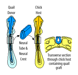
Quail-Chick Chimeras |
| Nicole Le Douarin pioneered the use of quail-chick chimeras to study the developmental fate of cells in the bird embryo. The videotape Nicole Le Douarin gave us permission to digitize is titled, "Quail-Chick Chimeras in Development of the Nervous System and Immune System" and it was made in 1987. These digital video sequences and still images come from the first part of her videotape. |
|
Click on the "thumbnail" image or the QuickTime link to watch a sequence. You need QuickTime to watch these sequences; all sequences are silent. |
|
|
 |
Sequence showing
newly hatched quail-chick chimeras; white feathers are chick and dark, pigmented
feathers are quail. QuickTime (477 K) |
|
| Nicole Le Douarin discovered that Feulgen stain will distinguish chick cells from quail cells. (The Feulgen stain, stains DNA.) Feulgen stained quail cells have interphase nuclei with condensed nucleolar-associated heterochromatin that's not found in chick interphase nuclei. To the right is an image of a Feulgen stained section showing quail cells on the left and chick cells on the right. | 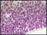 |
|
 |
Sequence showing
the injection of India ink beneath host chick embryo to help see the almost
transparent embryo in ovo. QuickTime (958 K) |
|
 |
Sequence showing
the preparation of the chick host; removing a portion of host's neural tube
and neural crest.
QuickTime (1.3 MB) |
|
 |
Sequence showing
the removal and "cleaning off" of donor quail neural tube and
neural crest. QuickTime (1.4 MB) |
|
 |
Sequence showing
transplantation and grafting of donor quail neural tube and neural crest
into the chick host; at the end of this sequence, you see the host chick
embryo 5 hours later with its healed in graft.
QuickTime (1.5 MB) |
|
| Feulgen stained transverse section through the host chick embryo containing the grafted quail nerual tube and neural crest. Host one day after receiving quail graft. The top of this section is dorsal; the right edge of this image shows a lateral half of the donor quail neural tube. Donor quail neural crest are adjacent to the dorsal quail neural tube. | 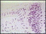 |
|
| Newly hatched quail-chick chimera. Donor quail feathers (at the level of the graft) are dark and host chick feathers are light. This newly hatched chimera has normal sensory and motor activity throughout its body. | 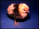 |
|
| Young quail-chick chimera with wings extended to better show pigmented quail feathers at the level of the graft. | 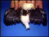 |
|
| When the quail-chick chimera is about two months old, the host chick rejects the quail graft; the chimera develops a neurologic syndrome that causes paralysis of the limbs. | 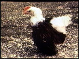 |
|
| We want to thank Pascal Meunier for translating the French dialogue of Nicole Le Douarin's videotape. | ||
|
||
| DB Cinema Main Page | ||
| We welcome your comments and suggestions for the Developmental Biology Cinema. | |
 SOCIETY
FOR DEVELOPMENTAL BIOLOGY SOCIETY
FOR DEVELOPMENTAL BIOLOGY
SDB Webmaster |
|