Wolff, T. and Ready, D. F. (1993). Pattern formation in the Drosophila retina. In: The Development of Drosophila melanogaster. Cold Spring Harbor Laboratory Press. Vol. 2 Pp. 1277-1325

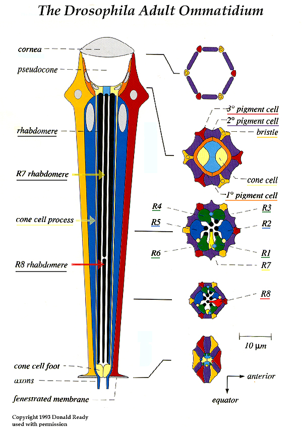
To play the animation (below: 1940Kb), an exploded view of the figure on the left, you will need to have, or to install or upgrade (free) to Quicktime 4 for either Windows or Macintosh. You can also go directly to this site or copy/paste this URL into your Quicktime movie player: http://www.sdbonline.org/fly/virtomm.mov
The animation image is enlarged when played through the Quicktime movie player.

The adult compound eye presents a regular hexagonal array of approximately 750 facets, the lenses of the unit eyes, or ommatidia. An adult ommatidium is a precise 19-cell assembly of 8 photoreceptors and 11 accessory cells.

The photoreceptors (R cells) are of three distinct types: the R1-R6 class, which lie in a ring surrounding two central receptors; R7, the distal, or outer, central cell; and R8, the proximal, or inner, central cell. Genetic and morphological criteria subdivide the outer receptors into three groups: R1/6, R3/4 and R2/5. Each R cell contributes a distinct pattern element to the photoreceptor group.

Overlying the photoreceptors is a quartet of cone cells. Two primary pigment cells, which surround the cone cells are mirror-image twins that meet on the vertical midline of the ommatidium. A mesh of shared pigment cells surrounds each ommatidial core of photoreceptors, cone cells, and primary pigment cells in an exact, honeycomb-like matrix.

Secondary pigment cells lie between two ommatidia, and tertiaries are shared among three ommatidia at a vertex. Rhabdomeres are the rhodopsin-located apical surfaces of a photoreceptor, accommodating more than 90% of the photoreceptor's plasma membrane in a closely packed stack of about 60,000 microvilli.

The small mechanosensory bristles of the eye are products of a typical four cell complement of neuron, glia and two support cells. Eye bristles are developmentally distinct from ommatidia and project their sensory axons into the brain. The corneal lens is a chitinous extracellular secretion of the four underlying cone cells and the two primary pigment cells (Wolff, 1993).