Select image to enlarge in new window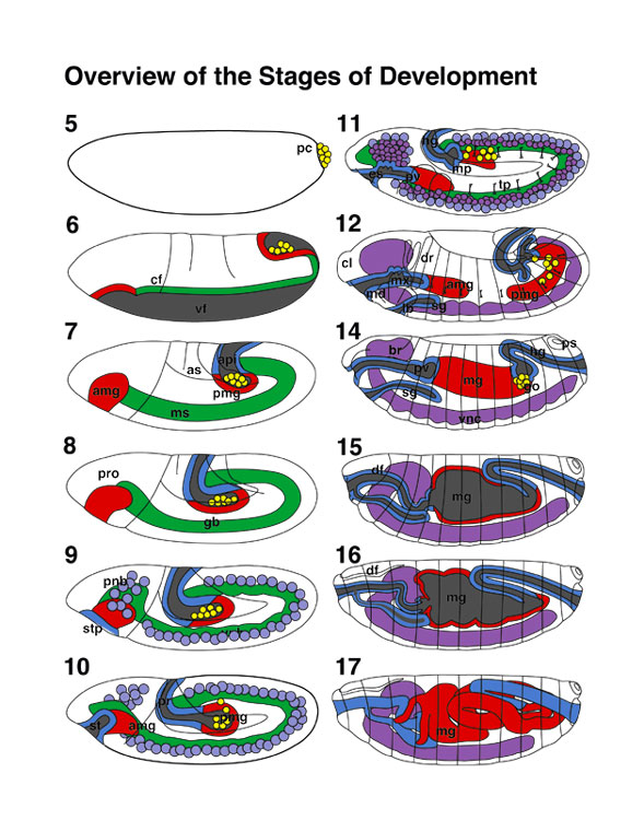 |
Stages of Embryonic Development page 52
All embryos are in lateral view (anterior to the left). Endoderm, midgut; mesoderm; central nervous
system; foregut, hindgut and pole
cells in yellow. |
| Atlas of Drosophila Development by Volker Hartenstein | Table of Contents |
| Select image to enlarge in new window |
Stages of Embryonic Development page 52
All embryos are in lateral view (anterior to the left). Endoderm, midgut; mesoderm; central nervous
system; foregut, hindgut and pole
cells in yellow. |
The Interactive Fly resides on the
Society for Developmental Biology's Web server.