Select image to enlarge in new window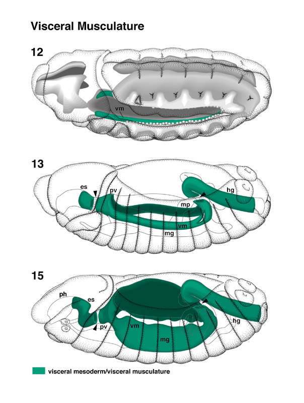 | Visceral Musculature pages 42-43
During early stage 12, the visceral mesoderm (vm) on either side of the embryo forms a band of cells that are located dorsally and internally of the somatic mesoderm (Poulson 1950; see Bate, this volume). During stage 12 and 13, the visceral mesoderm becomes attached to the midgut primordium (mg), which, by this stage, is also represented by two bilateral symmetrically arranged plates of cells. Farther anterior and posterior, visceral mesoderm also flanks the esophagus (es) and hindgut (hg), respectively. These terminal parts of visceral mesoderm are separated from the midgut visceral mesoderm by wide gaps (indicated by arrowheads). Thus, there exist segments of the developing foregut and hindgut that have no visceral mesoderm attached (Hartenstein and Jan 1992). The segment of the foregut that lacks visceral mesoderm becomes the inner part of the proventriculus (pv); the part of the hindgut without visceral mesoderm corresponds to the narrow segment where the Malpighian tubules (mp) open into the hindgut (see Skaer, this volume). During later stages [14-16], the cells of the visceral mesoderm spread in the transversal axis eventually to encircle the entire gut. (fb) Fat body; (mus) somatic musculature; (myo) myoblasts; (ph) pharynx; (sm) somatic mesoderm. |
Select image to enlarge in new window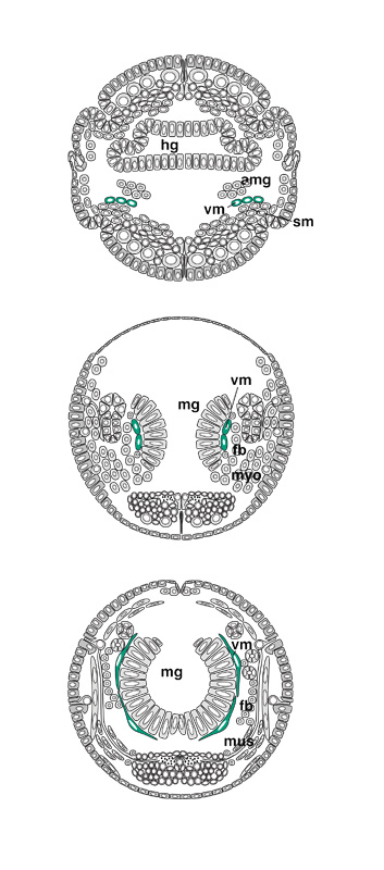 |