|
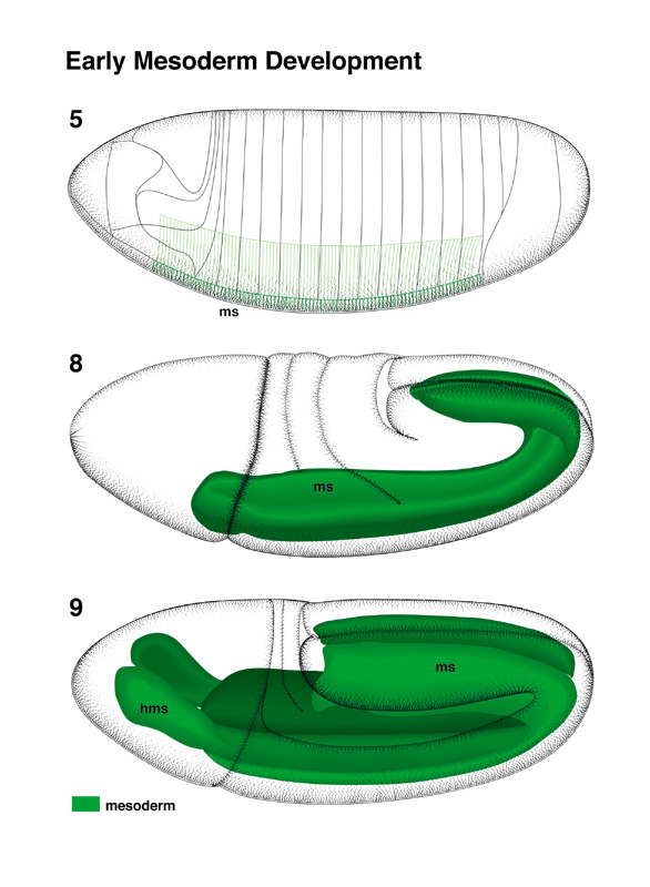
In the blastoderm [stage 5], the prospective mesoderm
occupies a midventral stripe of about 18 cell diameters in width. This region
invaginates during gastrulation [stages 6-7] and gives rise to the
mesoderm (ms; Poulson 1950; see Chasan and Anderson; Costa et al.; Bate; all this volume).
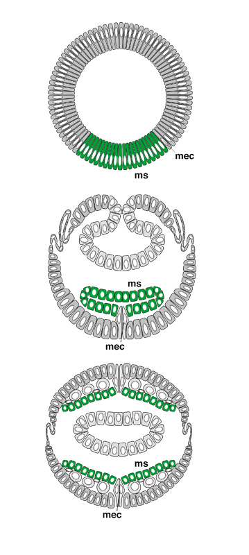
In the stage 8 embryo, the mesoderm forms a
dorsal-ventrally flattened tube that is connected to the ventral ectoderm via
two rows of cells, called mesectoderm (mec; see Goodman and Doe,
this volume).
At the transition to stage 9, mesoderm cells
rearrange and form a monolayer. In the ventral midline, this monolayer still
contacts the mesectoderm. Mesoderm cells remain in the monolayered configuration
until late stage 10 when they split up into different organ primordia. During
stages 8-11, the mesoderm undergoes three waves of mitoses (Hartenstein
and Campos-Ortega 1985). Another mitosis is seen in most, if not all, mesoderm
cells during stage 12 (see Bate, this volume). The anterior part of the
mesoderm (head mesoderm, hms) forms two vertical plates flanking the
anterior midgut rudiment and, from stage 10 onward, the stomodeum. These
plates lose their contact to the mesoderm of the trunk during later stages and
move into the anterior tip of the head where they give rise to the musculature
of the head and macrophages.
From early stage 12 onward, the mesoderm splits up
into separate cell masses that give rise to the somatic musculature, visceral
musculature, dorsal vessel, and fat body. The development of these
organs is described separately in the following sections (also see Bate, this volume). |