| Intestinal Tract pages 28-29 | 30-31 | 32-33 | 34 | 35 | |
Select image to enlarge in new window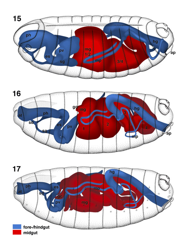 |
Select image to enlarge in new window |
|
By stage 15, the presumptive midgut (mg) has closed
ventrally and dorsally. A constriction in its posterior third gives the midgut
a heart-like shape. Anteriorly and posteriorly, the midgut lumen has become
continuous with the lumen of the presumptive proventriculus (pv) and
hindgut (hg), respectively. The different parts of the foregut have
progressed in their differentiation (see Skaer, this volume). The pharynx (ph) forms a wide, vertically flattened structure; the esophagus (es) represents
a round tube that is bent like an S. A circular constriction separates the
presumptive proventriculus into an anterior and posterior chamber.
The posterior chamber opens into the midgut. During later development (stage
16), the anterior chamber invaginates into the posterior chamber. The
salivary duct (sd) has formed. Its external opening has reached the
ventral lip of the stomodeum.
|
|