Select image to enlarge in new window
 | Epidermis pages 24-25 | 26 | 27
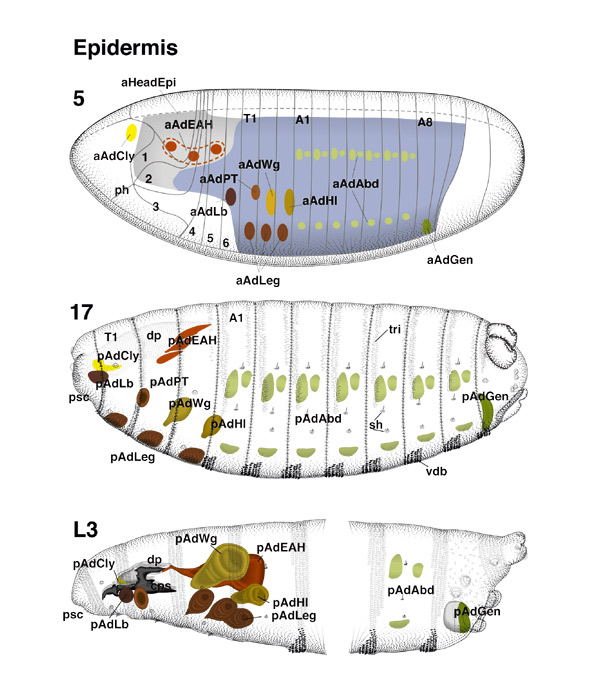
In the fate map [stage 5], the blastoderm region that will
form the larval epidermis is shaded blue. The major part of this region (i.e.,
the ventral neurogenic region) also gives rise to the precursors of the ventral
nerve cord (see Martinez Arias, this volume). The Drosophila larva is
acephalic; i.e., most of the head involutes to form the rostral part of the
alimentary tract (pharynx [ph] and atrium) and the dorsal pouch (dp, shaded gray). Only small parts of the head segments, which bear the sensory
antennomaxillary complex, are exposed to the outside at the anterior tip of the
larva (pseudocephalon, psc). The rest of the bodywall is formed
by the thoracic and abdominal segments.
The drawing of the stage 17 embryo
shows the main cuticle specializations formed by the larval cuticle. Prominent
among these are the ventral denticle belts (vdb), the dorsal
trichomes
(tri), sensory structures (sh), and the
cephalopharyngeal skeleton (cps; see drawing of L3) formed by the
epidermis of the involuted head. The primordia of the adult epidermis, which
are set apart from the larval epidermis during midembryogenesis (stages 13-15;
Bate and Martinez Arias 1990; Cohen et al. 1991; see Cohen, this volume), are
represented in the fate map as differently colored ovals.
By early stage 17,
some of these primordia have invaginated as imaginal discs connected to the
larval epidermis only by a thin peripodial stalk (leg discs, ld; wing
disc, wd; haltere disc, hd). Some other primordia (genital
disc, gd; labial disc, lbd; eye-antennal disc, ead) invaginate
late in embryogenesis, shortly before hatching. The primordia of the abdomen
(abdominal histoblasts, hib), prothorax (pd), and
labrum (clypeolabral disc [bud], clb) remain permanently in the
larval epidermis.
The third larval instar [L3] depicts the spatial
arrangement of the imaginal discs and histoblasts at late larval stages. All
discs have grown in size and cell number; after puparium formation, there are
but one to two more rounds of division before the epidermal cells become
postmitotic. In contrast, the abdominal histoblasts have not grown
substantially; for them, proliferation takes place during the pupal phase. (Al, A8) Abdominal segments 1, 8; (T1) thoracic segment 1;
(1-6) head segments 1-6.
|