back to lateral views on page 8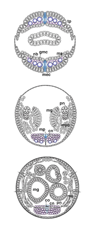
| CNS pages 6-7 | 8 | 9 | 10-11 Shortly after their segregation, neuroblasts start dividing with a perpendicularly oriented spindle. During stages 9-13, neuroblasts (nb) undergo eight waves of mitosis (Hartenstein et al. 1987). Their progeny, called ganglion mother cells (gmc), are placed between the neuroblasts and the mesoderm (ms). Each ganglion mother cell performs one equal division yielding two neurons. Ganglion mother cells and neurons form an irregular layer of increasing thickness on top of the neuroblasts. With the onset of overt segmentation during stage 12, deep indentations appear in the flanks of the developing CNS. Throughout stages 9 and 10, the mesectoderm (mec) formed a double row of cells between the neuroblast layers of either side. During stage 11, the mesectoderm loses contact with the outer surface. Segmentally repeated swellings mark the ap\-pearance of neuronal precursors (the so-called midline neuronal precursors, median neuroblasts, and midline glial cells, mp) that originate from the mesectoderm. In a stage-11 embryo, the cells that will form the optic lobe are still in the head ectoderm where they occupy a dorsal-lateral position behind the developing brain (optic lobe placode, op). During stage 12, these cells invaginate. Unlike all other neuroblasts, the optic lobe precursors maintain their epithelial characteristics. From stage 13 onward, they form a vesicle (ol) that is attached to the basal surface of the brain hemispheres. Neuronal differentiation begins at stage 13 (see Goodman and Doe, this volume). A population of identifiable neurons lays down a scaffold of fibers on the dorsal surface of the CNS. Later-appearing axons fasciculate along the pioneer tracts. Longitudinal fibers form the connectives (cn); transversal fibers form two commissures (co) in each segment that cross the midline while in contact with the midline glial cells, descendants of the mesectoderm. Axons that leave the CNS form an anterior fascicle (af; also called intersegmental nerve) and a posterior fascicle (pf; segmental nerve). Ingrowing sensory axons (pn) fasciculate with both of these tracts. Starting at stage 14, the ventral nerve cord condenses. At stage 17, the deep indentations in the lateral margins of the ventral nerve cord (vg) that had separated individual neuromeres have disappeared. The CNS becomes invested with a sheet of perineurial cells. (br) Brain; (mg) midgut; (mu) somatic musculature; (myo) myoblasts; (pn) peripheral nerve; (tp) tracheal pit. |