Select image to enlarge in a new window.
FATEMAP OF THE BLASTODERM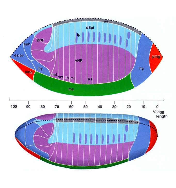
| Fatemap of the Blastoderm page 5 Shown are composite fate maps of the blastoderm. Map positions are based on Hartenstein et al. (1985) and Jürgens et al. (1986).
Top:
The fate map is projected onto a planimetric reconstruction of the blastoderm. In this type of reconstruction, one half of the curved blastoderm is flattened in order to depict true distances between blastodermal positions. The upper margin of the drawing (dashed line) represents the dorsal midline and the lower margin represents the ventral midline. The scale shows distances in percent egg length, with 0% being at the posterior tip.
Bottom:The fate map is projected onto a blastoderm shown in the standard dorsal-lateral view used for all drawings in this Atlas. Thick dashed line
Dorsal midline. The anlagen of different tissues are illustrated in different colors used throughout the figures in this Atlas.
Abdominal segment 1; amg anterior midgut
rudiment (endoderm);
as amnioserosa;
dEpi dorsal epidermis;
eph epipharynx;
es esophagus;
hg hindgut;
hy hypopharynx;
lb labium;
md mandible;
ms
mesoderm;
mx maxilla;
pmg posterior midgut rudiment (endoderm);
pNR procephalic neurogenic region;
pv proventriculus;
vNR ventral neurogenic region;
T1 thoracic segment 1;
tp tracheal placodes.
|