Select image to enlarge in new window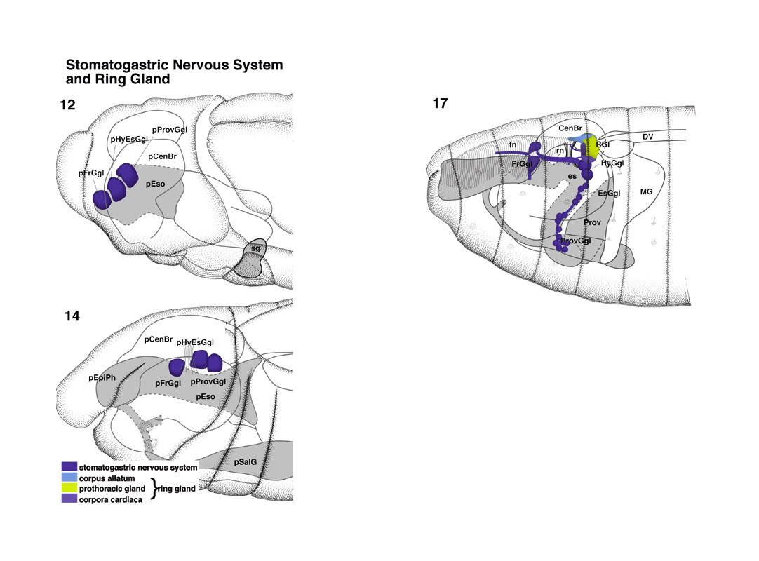 Stomatogastric Nervous System and Ring Gland pages 16-17
During early stage 12, the primordia of the stomatogastric
nervous system (stp) evaginate as three unpaired, linearly
arranged pockets from the roof of the esophagus primordium (es; Poulson
1950). During stage 13, these pockets pinch off the esophagus epithelium and
form closed vesicles with an inner lumen; up until stage 14, they stay in
contact with the esophagus and move posteriorly. During late stage 14, the
primordia of the stomatogastric nervous system lose their epithelial characteristics.
Cells that were included in the anterior-most pocket migrate anteriorly
(Campos-Ortega and Hartenstein 1985) and end up as the frontal ganglion (fg),
which in Drosophila is a paired structure attached to the anterior
surface of the brain (br). The middle and posterior pockets give
rise to at least two different neural structures: the hypocerebral ganglion (hcg) and a group of neurons that migrate along the esophagus and proventriculus (pv) and might correspond to the ventricular ganglion (vgl) of
other insects. At stage 17, many neurons of the stomatogastric nervous system
have differentiated and formed axons. Neurons of the frontal ganglion project
axons to the brain and to the pharynx musculature (frontal nerve,fn).
Axons of the hypocerebral ganglion send their axons to the frontal ganglion
(recurrent nerve, rn). |