| CNS pages 6-7 | 8-9 | 10-11 Unlike most other larval organs, the CNS persists into the adult stage. Recent evidence (for review, see Truman 1990; Truman et al., this volume) suggests that most motor neurons and large interneurons of the insect adult nervous system are of embryonic
origin. To this set of embryonically born neurons, a large number of neurons are added during larval and early pupal stages. The neuroblasts that generate these postembryonic neurons seem to be the same as those that had produced the larval CNS in the embryo (Prokop and Technau 1991).
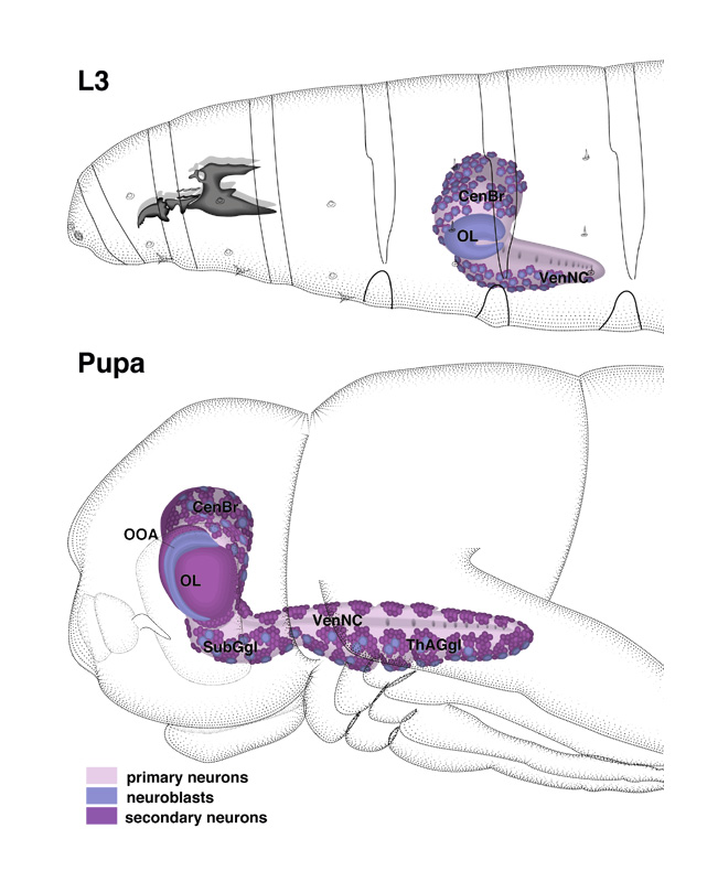
In the first larval instar, these neuroblasts reappear at the outer surface of the CNS ( see enlarged view of L3 ). In some parts of the CNS (e.g., thoracic segments), they closely match the number of neuroblasts present in the embryo (Truman and Bate 1988). In other regions (e.g., posterior abdominal segments), the number of postembryonic neuroblasts is very small. Each neuroblast resumes its proliferatory activity. As before in the embryo, neuroblasts divide in a stem cell mode with a perpendicularly oriented spindle. Their progeny, the presumptive adult neurons, remain undifferentiated until pupal stages. The optic lobe (ol), which started out as a small vesicle attached to the basal surface of the early larval brain, proliferates and gives rise to the outer and inner optic anlagen (outer optic anlage, ooa
on drawing of pupa). These structures are curved epithelial sheets that cap the lateral aspect of the brain hemispheres throughout larval life. The outer optic anlage forms the lamina and part of the medulla; the inner optic anlage gives rise to the remaining part of the medulla, the lobula, and the lobula plate (Hertweck 1931; Hofbauer and Campos-Ortega 1990; see Meinertzhagen and Hanson in Bate and Arias, 1993).
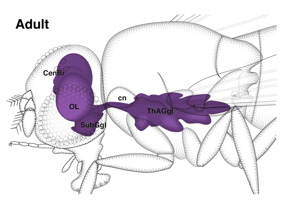
The gross anatomy of the CNS changes markedly during late postembryonic development (Hertweck 1931). The larval brain hemispheres (br) to which the massive optic lobes and several central brain structures are added, become the supraesophageal ganglion (seg) of the adult brain. Another part of the adult brain is the subesophageal ganglion (sub). This structure develops from the neuromeres of the three gnathal segments that, in the larva, had formed part of the ventral ganglion (vg).
The three thoracic neuromeres of the larval ventral nerve cord, which had grown massively by postembryonic neuroblast proliferation, form the major part of the adult thoracico-abdominal ganglion (tha). The fused abdominal neuromeres remain small; they form an unpaired 'cone' attached to the third thoracic neuromere. (cn) Cervical connective. |