|
|
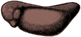
Another Way To Look at Serially Sectioned Frog and Chick EmbryosWhat we show here is the first part of a project we call "4-D Embryology--Embryos in Three Dimensions and Their Changes Over Time." We captured digital images of all the sections of serially sectioned embryos. We then used the sequence of images of an embryo to make a QuickTime digital movie. After we made movies of several serially sectioned frog and chick embryos, we started using them in our developmental biology labs. Students like watching and using these QuickTime movies to learn about developing embryos, and we want to share these movies with other students of developmental biology. What you can see here are small "postage-stamp" versions of these movies of serially sectioned frog and chick embryos. All show sequential cross sections going anterior-to-posterior (head-to-tail).
 Get Quicktime Get Quicktime
Why We Look At Serial Sections of EmbryosWhen we study embryology, we need to visualize the changes that occur in developing embryos. Direct observation is the ideal method for visualizing developing embryos. Everyone enjoys watching living embryos develop or watching time-lapse movies of developing embryos. Some embryos remain transparent throughout their development and we can see what's going on inside and outside at the same time. Most embryos are not transparent; once they become more than a few cell layers thick, we only see what's happening on the outside.
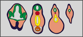
To see what's going on inside, we typically cannot dissect an embryo because it's too small. We have to look at serial sections of embryos. Serial sections are where we slice an embryo as if it was a sausage. We affix each section, in order, on a microscope slide and stain the sections before we look at them with the microscope. We then "read" these two-dimensional sections to try to visualize the three-dimensional organization of all the layers and parts of an embryo at one stage of development.
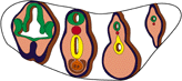
The above three figures are modified versions of Fig. A, p.3 of Schoenwolf, G.C. 1995, Laboratory Studies of Vertebrate and Invertebrate Embryos. Guide and Atlas of Descriptive and Experimental Development. 7th Edition, Prentice Hall, New Jersey.
How We Make the MoviesTo capture images of sections to make into movies, we use a compound microscope with a video camera attached to a computer (Macintosh) with a video board.Within an image editing application (Photoshop), we capture an image of each section of a serially sectioned embryo.We also use Photoshop to align images before we make the movies. To make the movie, we used the QuickTime "Convert to Movie" application.
What Else Is Being Done in the "4-D Embryology" Project"Flying" through a QuickTime movie of a serially sectioned embryo gives us a hint of the three-dimensional (3-D) spatial arrangement of embryonic structures. Now we are taking the 2-D images we used to make the movies to reconstruct an embryo into its natural three dimensions. What you see below are some "slices" through a reconstructed 7 mm frog embryo.
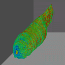 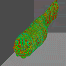 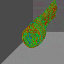 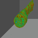
When we have embryos reconstructed at consecutive stages of development, we'll be able to look at the spatial arrangement of the cells, tissues, and organs as they appear over time--the fourth dimension of embryology.
Contributed by Laurie Iten |
||||
|
Page Modified:
|
News | About SDB | Membership | Meetings | Jobs | Education | Interactive Fly | Publications | Virtual Library | © Society for Developmental Biology | ||